By definition, all members of the Na+:neurotransmitter family (SNF) are Na+ dependent (Nelson and Lill, 1994), including the γ-aminobutyric acid (GABA) transporter from Manduca sexta (Mbungu et al., 1995). Reflecting the adaptation of M. sexta to its K+-rich plant diet, KAAT1 (K+ -coupled amino acid transporter; Castagna et al., 1998) is unique among cloned SNF transporters in using K+as well as
Na+ as a co-substrate for neutral amino acid uptake. Apparently, the substrate binding site of the superfamily was altered during evolution to accept L-, α-amino acids rather than
γ-amino acids, while the Na+ recognition site was altered to
accept both K+and Na+. KAAT1 is 30% identical to the GABA transporter, GAT1, which is the prototype of the superfamily (Guastella et al., 1990), the KAAT1 residues that diverge from
doi:10.1242/jeb.00065
KAAT1, a K+-coupled, neutral amino acid transporter
from larval insect midgut, differs from other members of the Na+:neurotransmitter transporter family (SNF) in two
important ways: (1) it transports nutrient L-, α-amino acids, rather than neurotransmitters such as γ-aminobutyric acid (GABA), and (2) it accepts K+ as well as Na+ as a
co-substrate. To determine whether the restoration of KAAT1 residues to their GABA transporter GAT1 cation-binding equivalents might abolish its K+but not its Na+recognition
site, we constructed a multiple mutant in which nine divergent KAAT1 residues were mutated back to the conserved form of the superfamily. To investigate the amino-acid-binding site, we constructed several single mutants that had been identified in GAT1. Wild-type (WT) or mutant cRNA was injected into Xenopus oocytes and the effects of external amino acids and ions upon labeled leucine uptake and substrate-induced currents were examined.
The multiple mutant exhibited no amino-acid-induced currents, indicating that one or more of the mutated residues are crucial for function. W75L and R76E mutations in the first transmembrane helix of KAAT1 led to results equivalent to those observed in the corresponding mutants of GAT1; namely, substrate (leucine) uptake and substrate-evoked net inward current were severely curtailed. The KAAT1 A523S mutant, which corresponds to a serotonin transporter mutant that is thought to render Li+ equivalent to Na+ as a co-transported ion, functioned
no differently to WT.
The effects of mutation Y147F in the third transmembrane helix of KAAT1 were dramatically different from the equivalent mutation, Y140F, in GAT1. Although kinetic characteristics, expression levels and plasma membrane localization were all similar in Y147F and WT, the Y147F mutant exhibited a sevenfold increase in labeled leucine uptake by Xenopus oocytes in Na+
buffer. This increase is in sharp contrast to the complete loss of uptake activity in the GAT1 Y140F mutant. KAAT1 Y147F also differed from WT in cation selectivity and substrate spectrum, as revealed by amino-acid-induced net inward currents that were measured with a two-electrode voltage clamp.
Amino-acid-independent currents induced by Li+ and
Na+ chloride salts were observed in both WT and the
Y147F mutant. The Li+-induced current was 30% higher
in Y147F than in WT, whereas no substrate-independent K+-induced currents above control levels were detected
either in WT or Y147F. These results suggest that transport of K+, the physiological co-substrate in insect
midgut, is tightly coupled to that of amino acids in KAAT1, in contrast to the independence of cation and amino acid transport in the closely related cation amino acid transporter channel, CAATCH1.
Key words: CAATCH1, KAAT1, GAT1, potassium, sodium, amino acid, transporter, leakage current.
Summary
Introduction
K
+amino acid transporter KAAT1 mutant Y147F has increased transport
activity and altered substrate selectivity
Zhilin Liu
1,3, Bruce R. Stevens
2, Daniel H. Feldman
2,*, Matthias A. Hediger
3and
William R. Harvey
1,2,†1The Whitney Laboratory, University of Florida, St Augustine, FL 32080, USA, 2Department of Physiology and Functional Genomics, University of Florida College of Medicine, Gainesville, FL 32610, USA and 3Harvard
Institutes of Medicine, Harvard Medical School, Boston, MA 02115, USA
*Present address: Research Department, Shriners Hospital for Children of Northern California, Sacramento, CA 95817, USA.
†Author for correspondence (e-mail: [email protected])
the conserved family sequence being scattered throughout the 12 transmembrane domains. These diverged residues are likely to account for the unique features of KAAT1, including its K+ acceptance.
Amino acid residues in highly conserved regions within the 12 transmembrane domains of SNF transporters have been investigated intensively, especially in GAT1. All charged and aromatic residues of GAT1 have been mutated to determine if they react in a critical way with Na+, Cl–or amino or carboxyl groups of GABA. In an effort to abolish the K+, but not the Na+, recognition site of KAAT1, nine residues were mutated back to the superfamily’s conserved form in a multiple mutant. In addition, noting that mutating serine 545 to alanine (S545A) in the serotonin transporter, SERT, renders Li+equal to Na+as a co-substrate (Sur et al., 1997), we mutated the corresponding KAAT1 residue, alanine 523 to serine (A523S) and examined its effects on cation selectivity.
Finally, we noted that, among 10 tryptophan residues, 12 tyrosine residues and five charged residues that were mutated in GAT1, only tryptophan 68, arginine 69, and tyrosine 140, which are conserved in the SNF family, were functionally critical. A single substitution in any of these highly conserved residues resulted in a complete loss of GABA transport activity
(Pantanowitz et al., 1993; Kleinberger-Doron and Kanner, 1994; Bismuth et al., 1997). Residues that correspond to these three critical residues in GAT1 are also conserved in KAAT1. These three residues were mutated to yield W75L, R76E and Y147F (Fig. 1). The results of these mutations provide fresh insight into binding sites for ions and amino acids in SNF transporters.
Materials and methods
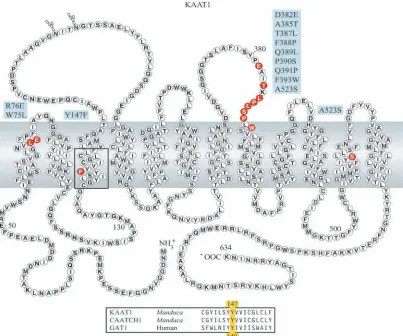
Location of mutants in the postulated KAAT1 membrane topology
The nine KAAT1 residues that were restored to the SNF transporter equivalent in a multiple mutant were D382E, A385T, T387L, F388P, Q389L, P390S, Q391P, F393W and A523S. Four single amino acid substitution mutants, W75L, R76E, Y147F and A523S, were also made. The locations of these mutations in the postulated KAAT1 membrane topology are shown in Fig. 1.
[image:2.612.102.505.73.409.2]Mutagenic primers were synthesized by Midland Certified Reagent Co. (Midland, TX, USA). The ‘domain swapping’ method was applied to eliminate the occasional involvement of undesired random mutations. After an EcoRI site of the pSport vector was removed, the resulting structure has four unique sites that are located in the KAAT1 coding region from nucleotides 1 to 1902. They are EcoRI 175, NcoI 500, XhoI 1366 and StuI 1738, which divide the entire coding region into four domains. Mutants were verified by sequencing, and the mutation-containing domain, flanked by two unique sites, was cut and pasted into the corresponding domain of wild-type KAAT1. Capped cRNA transcripts were synthesized using the mMESSAGE mMACHINE™ kit (Ambion, Austin, TX, USA). The quality of the transcripts was routinely monitored on formaldehyde denaturing agarose gels (Lehrach et al., 1977).
Radiolabeled amino acid uptake
[3H]L-leucine uptake was measured 2–3 days after injection with 100 ng of cRNA. Oocytes (10 per assay) were exposed to 1.9×10⁵Bq ml–1 [3H]L-leucine (Amersham, Piscataway, NJ, USA) at 22°C for 30 min or 60 min. Data were collected and analyzed as previously described (Stevens, 2001; Castagna et al., 1998; Feldman et al., 2000).
Electrophysiology
Electrical determinations were carried out as described earlier (Feldman et al., 2000). Briefly, injected oocytes were superperfused (22°C) with modified ND96 media, containing 98 mmol l–1 NaCl, 2 mmol l–1 KCl, 1 mmol l–1 MgCl
2, 1 mmol l–1 CaCl
2, 10 mmol l–1 Taps/NMG+ buffer {3-[tris(hydroxymethyl)methyl] amino propanesulphonic acid/N-methyl glucamine, pH 8.0}, using a peristaltic pump. In some experiments, Na+ was completely replaced by K+, Li+ or NMG+. In experiments designed to determine anionic specificity, Cl– was completely replaced by gluconate–. Transmembrane currents were measured in intact oocytes using a two-electrode voltage clamp (Warner model OC725-B, Hamden, CT, USA) with agar-bridged bath electrodes. Current/voltage relations were generated using voltage steps or ramps (36 mV s–1, 1.8 mV per point) between –150 mV to +30 mV from a holding potential of –60 mV. The protocol to measure steady-state currents minimized or eliminated the rapidly decaying transient currents that were previously reported for KAAT1 (Bossi et al., 1999). Substrate-dependent, current/voltage data were obtained by subtracting control current values (i.e. those in the absence of substrate) from those in the presence of substrate.
Immunofluorescent microscopy
Stage V–VI Xenopus oocytes were injected with 50–100 ng of Y147F or wild-type (WT) cRNA or water. Injected oocytes were incubated in Barth’s solution (88 mmol l−1NaCl, 1 mmol l−1 KCl, 2.4 mmol l−1 NaHCO
3, 15 mmol l−1 Hepes, 0.32 mmol l−1 CaNO3, 0.4 mmol l−1CaCl2and 0.81 mmol l−1MgSO4) at 18°C for 3 days. After incubation in 20% sucrose containing phosphate-buffered saline (PBS) overnight, the oocytes were
[image:3.612.333.553.73.596.2]2×10⁻³mg ml–1 affinity-purified antibody at room temperature for 30 min, washed three times with PBS, then incubated with secondary, tetramethylrhodamine isothiocyanate-labeled, goat-anti-rabbit immunoglobulin G (Sigma) for 30 min and again washed three times with PBS. Images were observed with a fluorescence microscope (Axioplan, Zeiss, Germany).
Results
Immunofluorescence and western blots of KAAT1 WT and Y147F
The injected RNA for both KAAT1 WT and the mutant was expressed and the transporter proteins were localized in the oocyte plasma membrane, as shown in confocal micrographs of sections labeled with fluorescent antibody to KAAT1 (Fig. 2). The expression was confirmed in western blots (data not shown). [3H]L-leucine uptake by oocytes expressing mutants of KAAT1
Several mutations have been shown to affect transporter function in GAT1 (see reviews by Bismuth et al., 1997; Kanner, 1994). RNA from a multiple mutant of KAAT1 that included nine of these mutations was expressed in Xenopus oocytes. Critical residues of the mammalian GABA transporter (GAT1) and serotonin transporter (SERT) that have been identified previously (Sur et al., 1997) were mutated singly in KAAT1. Uptake of [3H]L-leucine was measured for 30 min in oocytes that had been injected previously with cRNA. The multiple mutant had a much lower uptake rate than WT (data not shown). Mutations W75L and R76E of KAAT1 abolished transporter-mediated uptake (Fig. 3), as they do in GAT1. The mutation S523A had no discernible effect (data not shown). In sharp contrast to these results, [3H]L-leucine uptake was sevenfold higher in the Y147F mutant than in KAAT1 WT (Fig. 3).
Amino acid modulation of currents in KAAT1 Y147F- and WT-injected oocytes
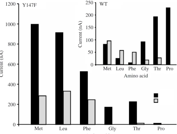
The effects of amino acid substrates on KAAT1 WT currents are different in K+than in Na+media, and the difference is pH dependent (Castagna et al., 1998). Currents with K+are much lower than those with Na+for almost all amino acids, despite K+ being the predominant cation in the midgut lumen in vivo. For WT at pH 8 in K+ buffer, the selectivity sequence is Met>Leu>Phe>Thr>GlyⰇPro (Fig. 4, inset),
whereas in Na+ buffer it is
Pro>Thr>Gly=Met>Leu>Phe (Fig. 4, inset). By contrast, for the Y147F mutant in K+buffer the sequence is Leu>Met>Phe with Thr, Gly and Pro being barely detectable, whereas in Na+buffer it is Met>Leu>Phe>Thr>Gly>Pro (Fig. 4). Confirming the results from tracers, the transporter net currents with both K+ and Na+ were usually greater in Y147F than in WT; thus, the change in substrate-evoked net inward currents measured in Y147F ranged from 10 nA to 1000 nA (Fig. 4), whereas for WT they ranged from 10 nA to 225 nA (Fig. 4, inset). For methionine in Na+medium, the Y147F value was
W75L R76E Y147F WT Control
200 400 600 800
0
[
3H]
L
-l
euci
n
e up
take (
n
m
ol
oo
cy
te
–
1 3
0
mi
n
–
[image:4.612.74.265.74.276.2]1)
Fig. 3. Uptake of [3H]L-leucine by KAAT1 wild-type (WT) and
several mutants (Y147F, R76E and W75L). Accumulated
radioactivity was measured in oocytes after 30 min exposure to Na+
media containing 0.2 mmol l–1 [3H]L-leucine. Oocytes had been
injected 2–3 days earlier with cRNA encoding W75L, R76E, Y147F, WT or water control.
200 400 600 800
0
Met 1000
1200
Curr
en
t (nA)
Leu Phe Gly Thr Pro
Amino acid 50 100 150 200
0
Curr
en
t (nA)
Met Leu Phe Gly Thr Pro
Amino acid 250
Y147F WT
Fig. 4. Typical amino acid selectivity spectrum for KAAT1 Y147F mutant and wild type (WT; insert)
in K+ and Na+ media. Data represent net inward
currents evoked by each amino acid in the presence
[image:4.612.46.356.499.734.2]1000 nA compared with <100 nA for WT. Proline uptake in Na+ was a notable exception; the Y147F value was 10 nA compared with 225 nA for WT.
Kinetics of [3H]L-leucine uptake by KAAT1 WT and Y147F
The concentration dependence of labeled leucine uptake in Na+medium was measured in oocytes incubated with [3H]L -leucine for Y147F and WT (Fig. 5A; WT data are enlarged in Fig. 5B). The Vmwas much higher in Y147F than in WT but the Kmwas the same in mutant and WT.
Activation of leucine-associated currents by leucine and K+
The L-leucine dependence of the current that flows into oocytes clamped at –60 mV was measured in the presence of 100 mmol l–1K+ at pH 7.6 and 25°C. For Y147F, the V
max was
755 nA, the apparent Km was 3.0 mmol l–1 L-leucine and the apparent Hill coefficient (η) was 1.0 (Fig. 6A; Table 1). The WT Vmax (734 nA) was nearly the same value (Fig. 6B; Table 1). Thus, paradoxically, the maximal leucine-associated current was unchanged by the mutation, whereas, as discussed above, the Vmaxfor [3H ]L-leucine uptake was increased by the mutation. The ability of K+ to activate L-leucine-evoked current was measured in oocytes that were clamped at –60 mV in the presence of 0.2 mmol l–1 L-leucine at pH 7.6 and 25°C. For Y147F, the change in net inward current Vmax was 1103 nA, the Km was 93 mmol l–1K+and ηwas 1.9 (Fig. 7A; Table 1). In WT, values were estimated by non-linear regression analysis of the obtained [K+] value up to the physiologically plausible [K+] limit of 100 mmol l–1. In this case, the estimated V
max(1136 nA) was similar to that of Y147F, the apparent Km(245 mmol l–1) was somewhat higher and the apparent Hill coefficient (η=1.2) was less than that of the mutant (Fig. 7B; Table 1).
Amino-acid-independent Li+and Na+currents in Y147F- and
WT-injected oocytes
GAT1 exhibits GABA-independent, chloride-enhanced, Cs+ -and Li+-activated currents (Mager et al., 1996). Similarly, Li+ -Table 1. Activation of KAAT1 currents in Xenopus oocytes by
leucine and K+
Activator Vmax Apparent Hill
apparent Km (nA; net coefficient
Activator KAAT1 (mmol l–1) inward current) (η)
Leucine Y147F 3.0±1.0 755±100 1.0±0.1
Leucine WT 2.7±1.4 734±109 0.7±0.1
K+ Y147F 93.4±5 1103±55.7 1.9±0.1
K+ WT 245±60 1125±229 1.2±0.1
[Leu] (mmol l–1)
200 300 400 500
0
[
3H
]
L
-leu
cin
e u
ptak
e (
n
mol oo
cyt
e
–1 3
0
m
in
–1)
4 6 8 16 18 20
Y147F
WT 100
10 12 14
0 0.05 0.10 0.15 0.20 0.25 0.30 0.35
A
B
0 0.1 0.2 0.3 0.4 0.5 0.6 0.7
WT
[Leu] (µmol l–1)
200 300 400 500
0
C
u
rr
ent
(
nA
)
0 400 500 600
Y147F
WT 100
100 200 300
0 5000 10000 15000
A
B
0 1000 2000 3000 4000
Fig. 5. [3H]L-leucine uptake by KAAT1 (A) Y147F mutant and (B)
[image:5.612.60.289.74.373.2]wild type (WT) in K+media.
Fig. 6. Typical activation of L-leucine-associated net inward currents
as a function of leucine concentration in 100 mmol l–1K+-containing
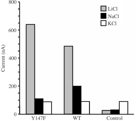
[image:5.612.334.553.76.407.2] [image:5.612.49.299.655.740.2]and Na+-activated currents, which were greater than those in controls, were exhibited in both KAAT1 Y147F and WT. However, no K+currents were observed in oocytes expressing Y147F or WT in the absence of amino acids (Fig. 8). Thus, even though KAAT1 is <40% identical to GAT1 (whereas other neurotransmitter transporters are >80% identical to each other), it appears to retain many aspects of the molecular mechanism for co-transport that are observed in GAT1. The Cl–-dependent Li+current is approximately 30% higher in the KAAT1 mutant than in WT (Fig. 8). The Li+ current appears to vary directly with the transport activity in both KAAT1 and GAT1; thus, both Li+current and amino acid transport are increased in the KAAT1 mutant, whereas the Li+ current decreases by 30% when GABA transport is lost in the GAT1 mutant.
Discussion
Nutrient vs neurotransmitter transporters
Many neurotransmitter amino acid transporters, such as the glutamate and GABA transporters, are well characterized, whereas nutrient amino acid transporters are still being discovered and cloned. The first invertebrate nutrient amino
acid transporter to be cloned, KAAT1, is a System B type co-transporter (Stevens, 1992) that accepts either K+or Na+ and requires Cl–. It is 30% identical to the GABA transporter GAT1 and clearly belongs to the SNF family (Castagna et al., 1998). A second invertebrate nutrient amino acid transporter, CAATCH1 (which is 92% identical to KAAT1), is activated by K+or Na+, although it does not require Cl–and was initially thought to be a co-transporter (Feldman et al., 2000). However, Quick and Stevens (2001) showed that, although K+ or Na+ and amino acids are mutually dependent co-activators, their transport activities are not thermodynamically coupled, i.e. CAATCH1 is not a co-transporter.
KAAT1 is a classical nutrient, neutral amino acid transporter Our hypothesis that KAAT1 is a System B type co-transporter (Stevens, 1992) is supported by the interaction between lysine and KAAT1 at pH values of 7.5 and 10. Classical studies on whole cells (Christensen, 1984) and membrane vesicles (Liu and Harvey, 1996) had shown that the electrical charge on an amino acid plays a critical role in its interaction with transporters, but the co-existence of heterogeneous transporters in vivo complicated the analysis. The overexpression of the cloned KAAT1 transporter in Xenopus eggs largely eliminated the complication. At pH 7.5, where lysine is mainly cationic, few lysine-evoked inward currents beyond controls were observed; by contrast, at pH 10, where lysine is mainly zwitterionic, lysine-associated current was substantial (Fig. 9, upper panel). The same result was obtained by measuring lysine-induced currents at the two pH values (Fig. 9, lower panel).
Amino-acid- and cation-binding sites
The most striking effects of the KAAT1 mutation Y147F are the enhanced leucine uptake and the altered pattern of
amino-[K+] (mmol l–1)
200 300 400 500
0
Current
(
nA
)
0 200 250 300
Y147F
WT 100
50 100 150
0 20 40 60
A
B
60080 100
WT Control
200 400 600 800
0
C
u
rr
ent
(
n
A)
Y147F
[image:6.612.321.554.75.283.2]LiCl NaCl KCl
Fig. 7. Typical activation of L-leucine-associated net inward current
as a function of K+concentration in 500µmol l–1leucine-containing
medium. (A) KAAT1 Y147F mutant and (B) wild type (WT).
Fig. 8. Typical Li+-, K+- and Na+-induced currents in mutant Y147F
[image:6.612.50.286.76.407.2]acid-evoked currents in voltage-clamped oocytes (Fig. 4). These effects contrast sharply to the complete loss of transport activity in the equivalent position 140 of GAT1. Noting that Y140 is conserved in all SNF transporters and that the mutation at position 140 from tyrosine to phenylalanine is essentially the loss of a hydroxyl group, Bismuth et al. (1997) suggested that Y140 may be the amino-binding residue of GAT1. They speculated that an H-bond may form between the oxygen atom of tyrosine 140 and an amine hydrogen of GABA. KAAT1 Y147 is equivalent to GAT1 Y140 because the amino terminus of KAAT1 is seven amino acids longer than that of GAT1. However, if leucine acts on KAAT1, as GABA does on GAT1, then our finding of a sevenfold increase in transporter activity in KAAT1 Y147F would be contrary to this hypothesis – there is no longer a tyrosine oxygen atom present to bind to the amino hydrogen of leucine – yet the transport is increased dramatically (Figs 5, 10). Despite the increased [3H]L-leucine transport (Figs 3, 5) and leucine-associated net inward current in the Y147F mutant over WT (Figs 4–8), the impact on proline-associated net inward current is similar to that observed for GABA in GAT1 and the primary substrates of other SNF transporters. Proline transport in KAAT1 exclusively requires Na+in the manner that GABA requires Na+ in GAT1. Notably,
the mutant loses the capability for proline-evoked currents (Fig. 4).
Nevertheless, the two dramatic effects – loss of GABA transport in the GAT1 mutant, Y140F, and gain of leucine transport in the KAAT1 mutant, Y147F – support the hypothesis that this site is crucial for solute transport. As Na+ is a substrate for all SNF transporters, perhaps the binding of Na+(or K+) at KAAT1 Y147 or the regulation of amino acid or ion binding at a distant site, is dependent on events at KAAT1 Y147. The presence of electronegative cysteine residues at positions C151 and C154, near Y147, in KAAT1 (Fig. 1, lower box) but not at the corresponding locations of GAT1 are consistent with this view. The corresponding Y147 site is also crucial in CAATCH1, which has both transporter and channel functions (Feldman et al., 2000) but is not a co-transporter (Quick and Stevens, 2001). The mutation Y147F also enhanced transporter activity and altered the amino acid current-inducing spectrum in CAATCH1, as well as abolishing its channel function (Stevens et al., 2002).
Neurotransmitter vs nutrient amino acid transporters A comparison of the neurotransmitter transporter GAT1 with the two nutrient amino acid transporters, KAAT1 and CAATCH1, demonstrates some fundamental differences between nutrient and neurotransmitter amino acid transporters (Fig. 10).
GAT1, KAAT1 and CAATCH1 are similar in that they are all activated by alkaline metal cations. GAT1 and KAAT1, but not CAATCH1, are also activated by Cl–. GAT1 has 599 amino acid residues, whereas KAAT1 has 634 residues and CAATCH1 has 633 residues, with moderately high sequence identity (30%) between GAT1 and the nutrient transporters. All three transporters have 12 transmembrane domains and fit a common template, with minimal insertions or deletions of residues (Fig. 1; see also Nelson and Lill, 1994). All three transporters are energized directly by H+V-ATPase-generated voltages and possibly indirectly by K+ or Na+ gradients that are secondary to the primary proton transport (Moriyama et al., 1992; Nelson and Harvey, 1999).
The three types of transporter differ in several important ways:
[image:7.612.60.291.71.340.2](1) GAT1 was first cloned from rat brain (Guastella et al., 1990) and later from a caterpillar embryo, where it is expressed in neural tissues (Mbungu et al., 1995), whereas KAAT1 (Castagna et al., 1998) and CAATCH1 (Feldman et al., 2000) were first cloned from caterpillar midgut. GAT1 accepts a single substrate, GABA, which has a γ-amino group, whereas KAAT1 and CAATCH1 recognize several structurally different neutral amino acids that have α-amino groups. Binding of L-, α-amino acids to KAAT1 and CAATCH1 appears to have loose restrictions, whereas binding of GABA to GAT1 has tight restrictions that allow only minute changes in transporter structure. This difference may reflect the physiological functions of KAAT1 and CAATCH1, which mediate nutrient amino acid uptake in the highly variable lumen of caterpillar midgut, whereas GAT1 mediates Fig. 9. KAAT1 wild type (WT) uptake at pH 7.5 and pH 10. Upper
panel: bars A and B, 0.2 mmol l–1[3H]-L-leucine at pH 7.5 and pH 10,
respectively; C and D, 0.2 mmol l–1 [3H]-L-lysine uptake at pH 7.5
and pH 10, respectively; E, 0.2 mmol l–1 [3H]-L-leucine uptake with
10 mmol l–1 lysine; F, 0.2 mmol l–1 [3H]-L-lysine with 10.0 mmol l–1
leucine; G and H, 10 mmol l–1[3H]-L-leucine and 10 mmol l–1 [3H]-L
-lysine uptake, respectively, by water-injected oocytes. Lower panel:
currents measured in KAAT1 cRNA-injected oocytes in Na+-free K+
medium with lysine added (0.2 mmol l–1) and then removed.
A B C D E F G H
50 100 150 200
0
Lys Con Leu Con Lys in Leu Leu in
Lys 10 Lys 7.5 Lys 10 Leu 7.5 Leu
+Lys –Lys
50 nA pH 7.5
pH 10.0
[
3H
]
L
-am
ino
ac
id
u
p
ta
k
e
(p
m
o
l
oo
cy
te
–
1 3
0
m
in
–
neurotransmitter re-uptake in the constant cerebrospinal fluid that is separated from surrounding tissues by the blood–brain barrier.
(2) GAT1 has a high affinity for GABA (Km=7.3µmol l–1) and a low Vmax (24 pmol oocyte–1h–1; Guastella et al., 1990), apparently adaptations to its role in removing neurotransmitter from the synaptic cleft. By contrast, KAAT1 has a low affinity for neutral amino acids, such as leucine (Km=2.7 mmol l–1), and a high Vmax relative to other substrates (734 nA; Table 1), apparently adaptations to its role in moving large quantities of amino acids from lumen to cells.
(3) Approximately 70% of the residues in KAAT1 are different from those in GAT1 and other SNF transporters. KAAT1 does not transport GABA, and GAT1 does not transport leucine (Z. Liu and D. Trotti, unpublished data).
(4) GAT1 is Na+ coupled, whereas KAAT1 is K+coupled
under physiological conditions (although Na+ can substitute for K+in vitro).
(5) GAT1 and CAATCH1 have both transporter and channel properties, whereas KAAT1 lacks a channel property.
(6) GAT1 Y140F has no transport activity, whereas KAAT1 Y147F (the corresponding residue) has much higher transport activity and altered substrate specificity compared with WT.
Implications of oocyte current data for living insects Neither the Na+nor Li+currents reported here in oocytes are likely to have immediately obvious physiological significance for caterpillars because the [Na+] is extremely low in these plant-eating insects (Harvey et al., 1975) and Li+ is not detectable. However, an Na+ channel may be important in mosquito larvae that have moderately high [Na+] in the hemolymph (Edwards, 1982).
Transmembrane currents in Xenopus oocytes that are overexpressing xenic transporters are commonly thought to be carried by ions that are co-transported with an organic substrate. For example, Gaustella et al. (1990) initially interpreted inward currents measured in GAT1-injected oocytes as the co-transport of Na+, Cl–and GABA into the oocytes, and this interpretation has been experimentally proven by Loo et al. (2000). Similarly, currents measured in KAAT1-injected oocytes (Castagna et al., 1997) and CAATCH1-injected oocytes (Feldman et al., 2000) were interpreted as the co-transport of Na+ and amino acids into the oocytes. However, Quick and Stevens (2001) showed that 3H-labeled amino acid uptake has no stoichiometric relationship to inward currents measured simultaneously with a two-electrode voltage clamp in the same CAATCH1-expressing oocyte. They concluded that amino acid uptake and ion fluxes are not coupled in the oocytes. This finding was unexpected because the magnitude of the inward current depends upon the amino acid that is present (Feldman et al., 2000).
The implication that cation flux and amino acid transport are not coupled in living caterpillar midgut, from which 140
Y140F WT
A
B
AA (+)
OH
147 AA (+)
OH
147 Pro (+)
OH
140 AA
147 AA
147 Met (+)
Ions
H2O
Na+ Cl–
GABA
Y147F WT
Y147F WT
Met (–) Pro (–)
C
Cl– Cl–Na+
Cl–
[3H]Leu
Na+
Cl–
Na+
[3H]Pro
Na+
Na+ Met
GAT1
KAAT1
CAATCH1
(?)
Na+ saline
[image:8.612.43.317.75.519.2][3H]Leu
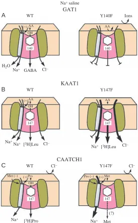
Fig. 10. Comparison of cation and amino-acid-associated net
inward currents and transport occurring in Na+ medium by
Xenopus oocytes injected with full-length cRNA from wild type
(WT) and mutants of nutrient and neurotransmitter transporters.
(A) In GAT1, WT Na+ and Cl– activate the uptake of
γ-aminobutyric acid (GABA) but not α-amino acids, and a water
channel is present. The transport is curtailed completely in the
equivalent mutant Y140F. (B) In KAAT1, WT Na+ and Cl–
activate the uptake of leucine. The co-transport of the amino acids and ions is enhanced more than sevenfold in the mutant
Y147F. (C) In CAATCH1 WT, there is no Cl–activation and
Na+fluxes are thermodynamically uncoupled from the transport
of proline and other neutral amino acids. In CAATCH1 Y147F
mutant, methionine, but not proline, activates the Na+channel
CAATCH1 was cloned, is even more puzzling, because the only driving force for amino acid uptake in vivo is the transapical membrane voltage, which is thought to drive cation-coupled amino acid uptake (reviewed by Castagna et al., 1997; Harvey et al., 1998). If cation flux and amino acid uptake are not coupled then the apically localized, electrogenic H+ V-ATPase would have no obvious function, and the source of energy for driving the amino acid uptake would be unknown. Three solutions to this puzzle come to mind. First, as the lipid composition of plasma membranes in oocytes is different from that in posterior midgut columnar cells, transport and uptake may be uncoupled in CAATCH1-expressing oocytes but not in midgut. As a precedent, Ca+ transport and Ca2+ -ATPase activity become uncoupled when ‘boundary lipids’ are removed from the membrane containing the Ca2+ pump (Hidalgo, 1982). Second, CAATCH1 may possess a coupling ‘slip’ (Gerencser and Stevens, 1994), i.e. the stoichiometric coupling of amino acid uptake and ionic fluxes may vary, from say 1:1 to zero. Such a slip is well documented in the coupling of ATP hydrolysis to proton transport by H+ V-ATPases (Moriyama and Nelson, 1988). Finally, perhaps CAATCH1 has other functions than cation:amino acid co-transport. For example, it may function as a channel that mediates fluxes that alkalinize the midgut.
On the other hand, KAAT1 appears to be a cation:neutral amino acid co-transporter, as suggested by Castagna et al. (1998). Thus, leucine induces large increases in K+and Na+ currents in KAAT1-expressing oocytes (Bossi et al., 1999). Moreover, K+ and Na+, but not Li+, currents coincide with leucine uptake, suggesting to Bossi et al. (2000) that there are two populations of transporters present, one in which amino acid uptake and cation flux are coupled and another in which they are not. Leucine uptake was maximal near the physiological pH of 10 (Vincenti et al., 2000). Acidic pH led to complete inhibition of coupled currents in the presence of either Na+ or K+ (Peres and Bossi, 2000); these results suggested that: “the operation of the transporter is maximal in the physiologically alkaline native environment”. The dependence of ionic current upon amino acid identity (Fig. 4) and the absence of K+current in both Y147F and WT in the absence of amino acids (Fig. 7) are consistent with this view. Taken together, these studies suggest that KAAT1 functions as a co-transporter in living insects, where it couples the fluxes of the predominant cation, K+, to the uptake of neutral amino acids.
Conclusions
(1) The tyrosine residue at position 147 in KAAT1 and CAATCH1, and at position 140 in GAT-1, plays a key role in amino acid uptake because mutation from tyrosine to phenylalanine has dramatic (although opposite) effects in the two types of transporter.
(2) Y140-OH of GAT1 WT is thought to be involved in γ -amino group binding, because its absence in F140 of GAT1 Y140F results in loss of activity. However, Y147-OH, the equivalent residue in KAAT1 and CAATCH1 WT, cannot play
the same role in amino acid binding because its absence in the mutant Y147F leads to greatly enhanced activity.
(3) The contrasting effects of the mutation Y147 in KAAT1 and CAATCH1 with the mutation Y140F in GAT1 are convenient diagnostic tools for classifying nutrient vs neurotransmitter amino acid transporters.
(4) The requirement of amino acids for K+currents in both mutant and WT of KAAT1 suggests that the transport of K+ and amino acids is tightly coupled by KAAT1 in sharp contrast to their independence in CAATCH1 (Quick and Stevens, 2001).
This research was supported in part by grants NIH RO1 AI30464 (W.R.H.), American Heart Association 50975 (B.R.S.) and NIH RO1 DK43171 (M.A.H.). We thank Dr S. Ursberger for the immunohistochemical images of Fig. 2. We thank Mrs Lynn M. Milstead and Mr Daniel J. Harvey for assistance with the illustrations.
References
Bismuth, Y., Kavanaugh, M. P. and Kanner, B. I. (1997). Tyrosine 140 of
the gamma-aminobutyric acid transporter GAT-1 plays a critical role in neurotransmitter recognition. J. Biol. Chem. 272, 16096-16102.
Bossi, E., Sacchi, V. F. and Peres, A. (1999). Ionic selectivity of the coupled
and uncoupled currents carried by the amino acid transporter KAAT1.
Pflugers Arch. 438, 788-796.
Bossi, E., Vincenti, S., Sacchi, V. F. and Peres, A. (2000). Simultaneous
measurements of ionic currents and leucine uptake at the amino acid cotransporter KAAT1 expressed in Xenopus laevis oocytes. Biochim.
Biophys. Acta 1495, 34-39.
Castagna, M., Shayakul, C., Trotti, D., Sacchi, F., Harvey, W. R. and Hediger, M. A. (1997). Molecular characteristics of mammalian and insect
amino acid transporters: implications for amino acid homeostasis. J. Exp.
Biol. 200, 269-286.
Castagna, M., Shayakul, C., Trotti, D., Sacchi, V. F., Harvey, W. R. and Hediger, M. A. (1998). Cloning and characterization of KAAT1, a
potassium-coupled amino acid transporter. Proc. Natl. Acad. Sci. USA 95, 5395-5400.
Christensen, H. N. (1984). Organic ion transport during seven decades. The
amino acids. Biochim. Biophys. Acta 779, 255-269.
Edwards, H. A. (1982). Ion concentration and activity in the haemolymph of Aedes aegypti larvae. J. Exp. Biol. 101, 143-151.
Feldman, D. H., Harvey, W. R. and Stevens, B. R. (2000). A novel
electrogenic amino acid transporter is activated by K+or Na+, is alkaline
pH-dependent, and is Cl–independent. J. Biol. Chem. 275, 24518-24526.
Gerencser, G. A. and Stevens, B. R. (1994). Thermodynamics of symport and
antiport catalyzed by cloned or native transporters. J. Exp. Biol. 196, 59-75.
Guastella, J., Nelson, N., Nelson, H., Czycyk, L., Keynan, S., Miedel, M. C., Davidson, N., Lester, H. A. and Kanner, B. I. (1990). Cloning and
expression of a rat brain GABA transporter. Science 249, 1303-1306.
Harvey, W. R., Maddrell, S. H. P., Telfer, W. H. and Wieczorek, H. (1998).
H+ V-ATPases energize animal plasma membranes for secretion and
absorption of ions and fluids. Am. Zool. 38, 426-441.
Harvey, W. R., Wood, J. L., Quatrale, R. P. and Jungreis, A. M. (1975).
Cation distributions across the larval and pupal midgut of the Lepidopteran,
Hyalophora cecropia, in vivo. J. Exp. Biol. 63, 321-330.
Hidalgo, C. (1982). Lipid–protein interactions and calcium transport in
sarcoplasmic reticulum Ann. N. Y. Acad. Sci. 402, 561-562.
Kanner, B. I. (1994). Sodium-coupled neurotransmitter transport: structure,
function and regulation. J. Exp. Biol. 96, 237-249.
Kleinberger-Doron, N. and Kanner, B. I. (1994). Identification of
tryptophan residues critical for the function and targeting of the gamma-aminobutyric acid transporter (subtype A). J. Biol. Chem. 269, 3063-3067.
Lehrach, H., Diamond, D., Wozney, J. M. and Boedtker, H. (1977). RNA
Liu, Z. and Harvey, W. R. (1996). Cationic lysine uptake by System R+
and zwitterionic lysine uptake by System B in brush border membrane vesicles from larval Manduca sexta midgut. Biochim. Biophys. Acta 1282, 32-38.
Loo, D. D., Eskandari, S., Boorer, K. J., Sarkar, H. K. and Wright, E. M.
(2000). Role of Cl–in electrogenic Na+-coupled cotransporters GAT1 and
SGLT1. J. Biol. Chem. 275, 37414-37422.
Mager, S., Kleinberger-Doron, N., Keshet, G. I., Davidson, N., Kanner, B. I. and Lester, H. A. (1996). Ion binding and permeation at the GABA
transporter GAT1. J. Neurosci. 16, 5405-5414.
Mbungu, D., Ross, L. S. and Gill, S. S. (1995). Cloning, functional
expression, and pharmacology of a GABA transporter from Manduca sexta.
Arch. Biochem. Biophys. 318, 489-497.
Moriyama, Y., Maeda, M. and Futai, M. (1992). The role of V-ATPase in
neuronal and endocrine systems. J. Exp. Biol. 172, 171-178.
Moriyama, Y. and Nelson, N. (1988). The vacuolar H+-ATPase, a proton
pump controlled by a slip. Prog. Clin. Biol. Res. 273, 387-394.
Nelson, N. and Harvey, W. R. (1999). Vacuolar and plasma membrane
proton-adenosinetriphosphatases. Physiol. Rev. 79, 361-385.
Nelson, N. and Lill, H. (1994). Porters and neurotransmitter transporters. J. Exp. Biol. 196, 213-228.
Pantanowitz, S., Bendahan, A. and Kanner, B. I. (1993). Only one of the
charged amino acids located in the transmembrane alpha-helices of the
gamma-aminobutyric acid transporter (subtype A) is essential for its activity. J. Biol. Chem. 268, 3222-3225.
Peres, A. and Bossi, E. (2000). Effects of pH on the uncoupled, coupled and
pre-steady-state currents at the amino acid transporter KAAT1 expressed in
Xenopus oocytes. J. Physiol. 525, 83-89.
Quick, M. and Stevens, B. R. (2001). Amino acid transporter CAATCH1 is
also an amino acid-gated cation channel. J. Biol. Chem. 276, 33413-33418.
Stevens, B. R. (1992). Amino acid transport in intestine. In Mammalian Amino Acid Transport: Mechanisms and Control (ed. M. S. Kilberg and D.
Haussinger), pp. 149-164. New York: Plenum.
Stevens, B. R. (2001). Theory and methods in nutrient membrane transport.
In Surgical Research (ed. W. W. Souba and D. W. Wilmore), pp. 845-856. San Diego: Academic Press.
Stevens, B. R., Feldman, D. H., Liu, Z. and Harvey, W. R. (2002).
Conserved tyrosine-147 plays a critical role in ligand-gated current of the epithelial cation/amino acid transporter/channel (CAATCH1). J. Exp. Biol.
205, 2545-2553.
Sur, C., Betz, H. and Schloss, P. (1997). A single serine residue controls the
cation dependence of substrate transport by the rat serotonin transporter.
Proc. Natl. Acad. Sci. USA 94, 7639-7644.
Vincenti, S., Castagna, M., Peres, A. and Sacchi, V. F. (2000). Substrate

![Fig. 5. [3wild type (WT) in KH]L-leucine uptake by KAAT1 (A) Y147F mutant and (B)+ media.](https://thumb-us.123doks.com/thumbv2/123dok_us/1134264.633841/5.612.60.289.74.373/fig-wild-type-leucine-uptake-kaat-mutant-media.webp)