Adenosine 5′-triphosphate (ATP) synthesis by oxidative phosphorylation or photophosphorylation is a multi-step, membrane-located process that provides the bulk of cellular energy in eukaryotes and many prokaryotes. Most of the ATP synthesis in these cells is catalyzed by the enzyme, F1Fo-ATP
synthase, also called F1Fo-ATPase (F-ATPase), which in its
simplest, bacterial, form is composed of eight subunits (α3:β3:γ:δ:ε:a:b2:c9–12). The archaeal A1Ao-ATP synthase
(A-ATPase) has ten subunits (A3:B3:C:D:E:F:G:H:I:Kx), the
actual subunit stoichiometry being unknown. The term ATPase reflects the fact that the F- and A-enzymes are reversible and can act as proton (or Na+)-pumping complexes. The F- and the
A-ATPases transform energy from a gradient of ions across the membrane to synthesize ATP (Mitchell, 1961; Dimroth, 1997; Müller et al., 1999). Conversely, the free energy of ATP hydrolysis can be coupled to proton (or Na+) translocation and
generate an ion-motive force (IMF), as in the genetically related vacuolar-type, H+-translocating ATPases (V-ATPases).
The V-ATPases, consisting of at least twelve distinct subunits (A3:B3:C:D:E:F:Gy:Hz:a:d:e:c6), generate IMFs that are used
for ligand trafficking, signaling, nutrient uptake and diverse activities in endomembranes and plasma membranes of animal cells (Wieczorek et al., 1999).
A-, V- and F-ATPases consist of a mosaic of globular
structural units, including domain and secondary structures, which also serve as functional units. Morphologically each of these enzymes has three components: a membrane-bound sector, Ao/Fo/Vo, which contains the ion channel, a central
connecting stalk, and an approximately spherical assembly, A1/F1/V1, which contains the catalytic sites (Schäfer et al.,
1999; Leslie and Walker, 2000; Forgac, 2000). Side-view projections of the F1Fo- (Wilkens and Capaldi, 1998) and
V1Vo-ATPases (Boekema et al., 1997) show a second stalk
(stator) as a fourth distinct feature extending from the Foor Vo
portion. In the case of the Escherichia coli F1 moiety the
central stalk is composed of γec and εec, which are the equivalent of δmin mitochondrial F1Fo, and the stator is formed
by the δecand b subunits (Pedersen et al., 2000). The bacterial
δsubunit (δec) bears homology to one of the mitochondrial Fo
subunits called OSCP (Table 1). The mitochondrial F1 ε
subunit (εm) has no counterpart in the bacterial F1Foenzyme.
The central element of the F1 complex, subunit γ, has been
shown to move relative to the α3β3 complex during ATP hydrolysis (Capaldi et al., 1996; Junge et al., 1997; Masaike et al., 2000). This rearrangement is proposed to drive the motion of a ring of c9–14subunits (Fillingame, 1996; Stock et al., 1999;
Seelert et al., 2000; Stahlberg et al., 2001) in the Fodomain
(Sambongi et al., 1999; Pänke et al., 2000; Tsunoda et al.,
JEB3339
Ion-translocating ATPases, such as the F1Fo-, V1Vo- and archaeal A1Ao enzymes, are essential cellular energy converters which transduce the chemical energy of ATP hydrolysis into transmembrane ionic electrochemical potential differences. Based on subunit composition and primary structures of the subunits, these types of ATPases are related through evolution; however, they differ with respect to function. Recent work has focused on the three-dimensional structural relationships of the major, nucleotide-binding subunits A and B of the A1/V1 -ATPases and the corresponding β and α subunits of the
F1-ATPase, and the location of the coupling subunits within the stalk that provide the physical linkage between the regions of ATP hydrolysis and ion transduction. This review focuses on the structural homologies and diversities of A1-, F1- and V1-ATPases, in particular on significant differences between the stalk regions of these families of enzymes.
Key words: A1Ao-ATPase, archaea-type ATPase, F1Fo-ATPase, H+ translocating vacuolar-type ATPase, V1-ATPase, small-angle X-ray scattering, Escherichia coli, Manduca sexta, Methanosarcina mazei.
Summary
REVIEW
STRUCTURE–FUNCTION RELATIONSHIPS OF A-, F- AND V-ATPases
GERHARD GRÜBER1,*, HELMUT WIECZOREK2, WILLIAM R. HARVEY3 ANDVOLKER MÜLLER4
1FR 2.5 Biophysik, Universität des Saarlandes, D-66421 Homburg, Germany,2Department of Biology,
University of Osnabrück, D-49069 Osnabrück, Germany, 3Whitney Laboratory, University of Florida, St Augustine,
FL 32080, USA and 4Lehrstuhl für Mikrobiologie der Ludwig-Maximilians-Universität München, D-80638,
München, Germany
*Author for correspondence (e-mail: ggrueber@ med-rz.uni-saarland.de)
Accepted 9 May 2001
2001), each containing two transmembrane helices (Rastogi and Girvin, 1999).
Based on their subunit composition and primary sequences, the A-type (archaeal) enzymes are more closely related through evolution to V-type than to F-type ATPases (Iwabe et al., 1989; Ihara et al., 1992; Müller et al., 1999). Three interdigitating copies of the nucleotide-binding subunits A and B of the A1/V1-ATPases and subunits β and α of the F1-ATPase,
respectively, exhibit more than 25 % primary sequence identity (Nelson, 1992). The minor subunits C, D, E, F, G and C, D, E, F, G, H of the A1and V1-ATPases, respectively, form the
stalk and are proposed, by analogy to F-ATPases, to be involved, either directly or indirectly, in conversion of energy into controlled motion (Müller et al., 1999; Grüber et al., 2000a). However, the minor stalk subunits of A1, F1and V1
-ATPases show much less similarity than the headpiece subunits, suggesting that there are differences between the three classes of enzyme (Müller et al., 1999). One fundamental distinction is the reversible dissociation of the V1from the Vo
complex as an in vivo regulatory mechanism for the control of V-ATPase activity (Wieczorek et al., 2000). By contrast, the A1/F1and Ao/Fosectors form stable associated complexes in
the cell. Moreover, A- and F-ATPases, unlike V-ATPases, share the ability to synthesize ATP. Nevertheless, lineage profiles based on primary sequence reveal that A- and V-ATPases are more closely related to each other than to F-ATPases (Schäfer et al., 1999). Despite the fact that A-ATPases display chimeric properties of V- and F-A-ATPases the structure/function relationships of these enzymes remain a mystery. This review will focus on recent advances in
elucidating structural and functional relationships of A1/V1
-and F1-ATPases.
F1-ATPase: structure and subunit function
Structure description of the F1headpiece
Over the past decade, a tremendous amount of structural information about the F1Fo-ATPase has been obtained using
electron microscopy (reviewed in Gogol, 1994; Böttcher and Gräber, 2000), macromolecular crystallography (Abrahams et al., 1994; Shirakihara et al., 1997; Bianchet et al., 1998; Hausrath et al., 1999; Stock et al., 1999; Groth and Pohl, 2001) and nuclear magnetic resonance (NMR) spectroscopy (Wilkens et al., 1995; Wilkens et al., 1997; Rastogi and Girvin, 2000). Significant insights into the molecular mechanism of ATP hydrolysis came from the X-ray structure of the bovine heart α3β3γcomplex of the F1-ATPase (MF1; Abrahams et al.,
1994). The crystallographic model describes the three alternating α and β subunits as being arranged hexagonally, surrounding a solvent-filled cavity which is traversed by a part of the γsubunit. This coiled-coil structure of the γsubunit is asymmetrically located in the shaft relative to the axis of the
α3β3 complex and protrudes from it by about 30 Å into the stalk region. A third short α-helix of the γsubunit is inclined at a 45 ° angle to the coiled-coil domain at the bottom of the F1as it merges with the stalk that connects the F1and Foparts.
A key feature of the structural model is its asymmetry, particularly in the nucleotide occupancy and conformations of the catalytic β subunits. One β subunit, designated βTP, is ligated with MgAMP-PNP and linked to the short α-helix of the γsubunit via the C-terminal domain. A second βsubunit,
βDP, has MgADP bound and the third catalytic site, βE, is free of nucleotides and Mg2+. The three non-catalytic α
subunits are ligated with MgAMP·PNP and adopt similar conformations, although one α subunit, which contributes to
βTPand is therefore denoted αTP, displays a small rigid body rotation of the top domain relative to the other two domains (Abrahams et al., 1994).
Arrangement of the stalk subunits
Neither the structural model of the bovine F1-ATPase
(Abrahams et al., 1994) nor the subsequently determined structure of rat liver F1-ATPase (Bianchet et al., 1998), which
includes almost all of the residues of the α and β subunits, include either the small subunits δmand εm, or approximately half of the γsubunit residues. Subsequently, crystals containing all five subunits of the Escherichia coli F1-ATPase
(α3:β3:γ:δec:εec) and the α3β3γεec complex of the same organism have been obtained and diffracted to a resolution of 6.4 Å and 4.4 Å, respectively (Grüber et al., 1997; Hausrath et al., 1999). Besides the αand βsubunits and the known part of the coiled-coil α-helices of the MF1the electron-density map
[image:2.612.42.293.95.373.2]at 4.4 Å extends 12 and 20 residues, respectively, thereby adding 15 Å to the length of the N-terminal α-helix and 23 Å to the C-terminal helix of the γsubunit (Hausrath et al., 1999). This structure reveals that γextends from the α3β3hexagon far
Table 1. Listing of similar ATPase gene products in the A-,
F-and V-ATPases
M. mazei Gö1 M. sexta Bovine E. coli
A1AO V1VO F1FO F1FO
A A β β
B B α α
C C − −
− D − −
D E γ γ
E − OSCP δec
F F δm εec
− G − −
− H − −
− − εm −
I a a+b a+b
K c c c
- d − −
− e − −
H − − −
G − − −
− − d −
− − e −
− − f −
− − g −
− − F6 −
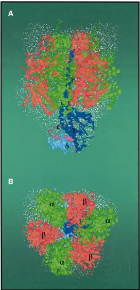
enough to traverse the full length of the central stalk, in agreement with the refined crystal structure at 2.4 Å resolution of the γec-εeccomplex from E. coli (Rodgers and Wilce, 2000) and the complete bovine F1-ATPase (Fig. 1; Gibbons et al., 2000). In these
structural models the γsubunit is arranged in six α -helices and five β-stranded β-sheets (Rodgers and Wilce, 2000; Gibbons et al., 2000). The bottom of γ is in contact with the external loops of c subunits (Watts et al., 1995; Watts et al., 1996); the entire γ subunit makes up a coupling domain, which couples ATP hydrolysis with ion pumping (Capaldi et al., 1996; Junge et al., 1997). Adjacent to the ‘bottom’ part of subunit γ an additional density has been observed in the map obtained from crystals of the
α3β3γδεc10subcomplex of yeast mitochondrial ATP
synthase at a 3.9 Å resolution (Stock et al., 1999) and the bovine F1 (Gibbons et al., 2000). In these
densities the structure of the εecsubunit (Wilkens et al., 1995; Uhlin et al., 1997), the counterpart of the yeast (δye; Giraud and Velours, 1994) and bovine δ subunit (Fig. 1; δm), has been modeled. Like subunit
εec, the subunits δye and δm are composed of a C-terminal helix–loop–helix structure and an N-terminal 10-stranded β-sandwich structure. However, the modeling of the εec subunit indicates that the C terminus of the polypeptide is turned away from the bottom domain of the catalytic β subunit. This domain is believed to be involved in the coupling of catalytic-site events (Capaldi et al., 1996; Grüber and Capaldi, 1996) along with γand εecacting as a rotor (reviewed in Junge et al., 1997; Masaike et al., 2000; Tsunoda et al., 2001). This structural feature is in conflict with the model of the γec-εec subcomplex (Rodgers and Wilce, 2000), in which the εec is located in close proximity to βvia its C-terminal α -helix and with its β-sandwich barrel turned toward the bottom of γ. As shown by cryo-electron microscopy (Gogol, 1994) and biochemical studies (Mendel-Hartwig and Capaldi, 1991; Wilkens and Capaldi, 1998), the εecsubunit can exist in different states in the complex depending upon whether ATP, MgATP or MgADP is bound to the enzyme (Mendel-Hartwig and Capaldi, 1991; Wilkens and Capaldi, 1998). Using E. coli F1 mutants with cysteine
[image:3.612.278.566.71.667.2]substitutions in the C termini of the α, β and εec subunits it has been shown that in the ATP-conformation, when the γand εecsubunits are linked to α subunits, the high-affinity site is completely closed with nucleotide unable to get in or out. In contrast, in the ADP-conformation, when the small subunits are linked to βsubunits, there is nucleotide exchange in and out from the high catalytic site (Grüber and Capaldi, 1996). Therefore, the question arises as to whether the arrangement of the equivalent to the bacterial εsubunit in the yeast and bovine F1Fo
complexes reflects a trapped state during its nucleotide-dependent movement.
A model-independent approach, based upon the multipole expansion method using spherical harmonics (Stuhrmann, 1970), has been developed to complement crystallographic studies of the quaternary structure of macromolecules such as the F1-ATPase from E. coli from solution X-ray scattering
data. Application of this approach has led to a low-resolution (32 Å) structure of the F1complex under nearly physiological
and saturating nucleotide conditions (Svergun et al., 1998a; Svergun et al., 1998b). The hydrated F1-ATPase (Fig. 1,
Fig. 5) is a compact molecule with a headpiece of approximately 108 Å from top to bottom and 110 Å wide. However, the overall structure is asymmetric due to the stem (stalk) that is approximately 42 Å in length and 53 Å in cross section (Svergun et al., 1998b; Grüber, 2000). These dimensions are consistent with recent data regarding the central stalk in the 2.4 Å resolution structure of the bovine F1-ATPase
(α3β3γδmεm) with a length of 47 Å and 51 Å × 41 Å in cross section (Gibbons et al., 2000), showing the regions of the γ, δm and εmsubunits are exposed at the foot of the stalk, and thereby in close contact with Fo subunits, which will consequently
facilitate the mechanistic linkage of ATP hydrolysis to ion pumping (Fig. 1).
Structure and mechanism of the V1-ATPase
Structural aspects of the V1complex
The idea that molecules now known to be V-ATPases may structurally resemble F-ATPases was suggested by early micrographs of insect plasma membranes (Gupta and Berridge, 1966; Anderson and Harvey, 1966), which showed repeating, spike-like units supporting globular structures. The spike-with-globule structures are widely distributed on transporting plasma membranes and were designated ‘portasomes’ (Harvey et al., 1981). Meanwhile, negatively stained membranes from bovine chromaffin granules (Schmidt et al., 1982) and the vacuolar membranes of Neurospora crassa (Bowman et al., 1989) were shown to contain similar structures, and sequencing of the genomic DNAs encoding V-ATPase subunits (Bowman et al., 1988) demonstrated beyond doubt that the particles are V1-ATPases. The regulatory mechanism
of reversible disassembly of the V1 and Vo complex, first
shown in the Manduca sexta midgut (Sumner et al., 1995) and also later in yeast (Kane, 1995), suggested that a study of the dissociated V1 complex could provide valuable information
about the structural features of this enzyme. The recovery of disassembled V1particles from the cytoplasm in high yield and
purity (Gräf et al., 1996) made the structural description possible.
Two major advances have been made toward elucidating the quaternary structure of V1during the past two years. First, the
gross structure of the M. sexta midgut V1-ATPase was
investigated by SAXS (Svergun et al., 1998b). The enzyme is highly elongated with a maximal length of about 220 Å. The solution scattering data define a hydrated complex with a
headpiece approximately 145 Å in diameter and a stalk approximately 110 Å in length (Fig. 5). Second, image processing of electron micrographs of negatively stained V-ATPases from Clostridium fervidus (Boekema et al., 1998) and V1-ATPase from M. sexta (Radermacher et al., 1999) yielded
two-dimensional structures at a resolution of 18 Å and 24 Å, respectively. A comparison of the independently identified structures (Boekema et al., 1998; Svergun et al., 1998b; Radermacher et al., 1999) revealed that the headpiece consists of a pseudo-hexagonal arrangement of six masses, surrounding a seventh mass. These six masses, which are assumed to consist of the major subunits A and B, are arranged in an alternating manner (Boekema et al., 1998; Svergun et al., 1998b). The first three-dimensional reconstruction of the V1
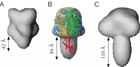
complex was determined at 32 Å resolution from negatively stained preparations of the M. sexta V1-ATPase (Fig. 2)
(Grüber et al., 2000a). A striking feature of the reconstruction is the presence of six elongated lobes, approximately 20 Å in diameter and 90 Å in length, which are parallel to the threefold axis (Fig. 2B). These lobes, which represent the alternating three copies each of subunits A and B, can be traced for most of the length of the V1-ATPase. The hexagonal barrel of
subunits A and B encloses a core of approximately 40 Å. In this model the V1 complex is barrel-shaped, being
approximately 110 Å high and 135 Å wide. At both ends of the hexagonal barrel extensions can be observed. The extensions on one side (Fig. 2A) are consistent with published two-dimensional average images of the V1Vo-ATPase from bovine
brain clathrin-coated vesicles, where elongated features (Fig. 2F–H; as, ce) can be seen at the very top of the V1domain
(Wilkens et al., 1999). The extensions on the opposite side can be attributed to traces of the stalk, e.g. the extension visible in Fig. 2B,C. The correspondence of dimensions of the hexagonal domain as determined by SAXS (see above) and electron microscopy indicate that the stalk is not completely resolved in the three-dimensional reconstruction, presumably due to absorption and drying of the V1particle on the carbon film.
However, a striking fact is that the shape and interdigitation of the A3B3subunits, located around the periphery of the barrel,
are in agreement with the three-dimensional model of the related F1-ATPase, derived from two- (Capaldi et al., 1992;
Gogol, 1994) and three-dimensional crystals of this complex (Abrahams et al., 1994; Bianchet et al., 1998; Hausrath et al., 1999), in which the alternating subunits αand βinterdigitate for the full length surrounding the γsubunit.
Topology and conformational rearrangements of the V1 -ATPase
E being modified most slowly. The rapid and slow cleavage of subunits D and E, respectively, is an important finding since both have been proposed as structural and functional homologues of the γ subunit of F-ATPases (Bowman et al., 1995; Nelson et al., 1995; Xu and Forgac, 2000). The observation that the D subunit is cleaved immediately into small peptides by trypsin is surprising; it implies that this polypeptide is exposed in the enzyme and does not support its putative role as a γhomologue (Grüber et al., 2000a).
Copper chloride-mediated disulfide formation yielded further insight into the proximity of the M. sexta V1subunits
to each other and into their functional relationships. When the enzyme was incubated with 2 mmol l−1CaADP on ice before
Cu2+treatment, two bands with apparent molecular masses of
120 and 110 kDa, consisting of the subunits A,E,F and B,H, respectively, were obtained. A B,H product did not occur when cross-linking was conducted in 2 mmol l−1 CaATP on ice to
slow down ATP hydrolysis, implying that subunit H moves away from B to the A,B interface (Fig. 3). This interpretation is consistent with the model of the yeast V1-ATPase, in which
subunit H was located at an interface of the nucleotide-binding subunits A and B (Tomashek et al., 1997). Moreover, in the presence of CaATP, two new bands with apparent molecular masses of 42 and 44 kDa, and composed of the subunits E,G and E,F, respectively, were observed. A homologous cross-linked product consisting of subunits E and G was also been generated using dimethyl sulfoxide (Thomashek et al., 1997) and disuccinimidyl glutarate (Xu et al., 1999) as cross-linking reagents.
Taken together, the trypsin cleavage and the cross-linking
data imply that one region of subunit E is shielded by the smaller subunits F and G (Fig. 3). In addition, the disulfide bonding of the catalytic A subunits with subunit E in the presence of CaADP indicates that these subunits are near neighbors. The close proximity of subunit E to subunits A, F and G would allow stalk subunit E to couple events in catalytic subunit A via stalk subunits F and G (Grüber et al., 2000a) in the V1 portion of the V1Vo-ATPase to events in the
ion-translocating Voportion (Fig. 3; Tomashek et al., 1997).
Redox modulation as a regulation of V-ATPases There is abundant evidence that V-ATPase activity is modulated by disulfide-bond formation (Feng and Forgac, 1992; Forgac, 2000). A mechanism of reversible disulfide-bond formation between cysteine residues (Cys254 and
Cys532) of the catalytic A subunit was proposed to regulate
the V-ATPase in vivo (Oluwatosin and Kane, 1995; Forgac, 2000). A mechanism was proposed in which disulfide bond formation is quickly followed by dissociation of the V1and
Vo complexes (Dschida and Bowman, 1995), implying that
nucleotide-binding and hydrolysis in the A subunit of the V1
domain have to be linked by the stalk region to ion translocation in the membrane-bound Vo domain. Recently
[image:5.612.54.565.75.329.2]SAXS experiments showed that reducing the V1-ATPase of M. sexta leads to significant changes in the overall dimensions of the complex. The radius of gyration of the oxidized and reduced enzyme are 62±6 Å and 58±6 Å, respectively, whereas the maximum dimension of both complexes remains constant at 220±10 Å (Grüber et al., 2000b). The shapes of both complexes (Fig. 4) were determined ab initio at a
Fig. 2. Surface representation of the three-dimensional reconstruction of the V1-ATPase from Manduca sexta determined from negatively stained specimens (Grüber et al., 2000a). An asymmetric (as), and a more centrally located (ce) extension can be seen above a subunit AB-interface. ec, protuberance. Bar, 100 Å.
ec
cc as
A
E
B
F
C
G
D
resolution of 27 Å by a simulated annealing procedure, based on a representation of the structure in terms of dummy atoms (Svergun, 1999). Both low-resolution structures have a characteristic mushroom-like shape with a central stalk of significant length, similar to the identified structures of the V1-ATPase from Clostridium fervidus (Boekema et al., 1998)
and M. sexta (Svergun et al., 1998), using electron microscopy and SAXS, respectively. Comparison of the oxidized and reduced models indicates that the main conformational changes upon reduction take place in both the crown-like region at the very top of the globular headpiece, where the major subunits A and B are located, and in the elongated stalk. Both regions evolve into an arrow-like shape after reduction (Grüber et al., 2000b). Based on homology of
the subunits A and B to the related F-ATPase subunits βand
α, respectively (Nelson, 1992), whose N termini form a β -barrel domain in a crown-like fashion (Bakhtiari et al., 1999), the conformational changes at the top of the V1-ATPase are
presumably due to rearrangements in the N termini of the A and B subunits. As shown more recently by three-dimensional reconstructions of the related F1Fo-ATPase from E. coli, a
crown-like shape, which is missing in the absence of the nucleotide (Böttcher et al., 2000), evolves upon binding of the non-cleavable nucleotide analogue AMP-PNP into the catalytic β subunit. The appearance of the crown has been attributed to rearrangements in the N-terminal domains of the
α and β subunits, located at the very top of F1. Moreover,
when AMP-PNP or ADP are bound to the catalytic site, subunit β assumes its closed conformation, in which the adenine-binding pocket moves into close proximity with the phosphate-binding domain, the P-loop; it moves away when the binding-site is empty (open conformation), as shown by the crystallographic model of the α3β3γsubcomplex of bovine heart mitochondrial F1-ATPase (Abrahams et al., 1994).
There is a striking similarity between the crown structure of the E. coli F1Fo-ATPase (Böttcher et al., 2000) that evolves
after binding of AMP-PNP (closed conformation) and the crown-like feature that is observed in the oxidized V1-ATPase
(Grüber et al., 2000b). In this state, the catalytic A subunit is proposed to be in a closed conformation (Forgac, 2000), and alters into a wedge-like shape after reduction of V1. In
summary, the structural changes in the headpiece, upon reduction of the enzyme, correspond with alterations of the protuberance of the stalk into a wedge-like feature, which enables the enzyme to transmit the activating movements that take place in the V1headpiece to the Vocomplex.
The archaeal A1-ATPase
Like F1 and V1 complexes of F- and V-ATPases, the A1
complex of archaeal ATPases possesses a pseudo-hexagonal arrangement of the major subunits A and B, as proposed from two-dimensional images of the thermoacidophilic archaea Sulfolobus acidocaldarius and Methanosarcina mazei Gö1 (Lübben et al., 1988; Wilms et al., 1996). However, in contrast to the related F1- and V1-ATPases described above, little is
known about the overall structure of the enzyme. This information gap is largely due to the instability of the isolated complexes (Wilms et al., 1996; V. Müller, unpublished). A new avenue of research was opened by the cloning and sequencing of the A1Ao-ATPase encoding genes from
methanogenic archaea. In M. mazei Gö1, the genes encoding the ATPase are clustered on the chromosome and comprise an operon. A fragment containing ahaE, ahaC, ahaF, ahaB, ahaA and ahaG was cloned in an overexpression vector and transformed into the F1Fo-ATPase negative mutant E. coli
DK8, which produced an A1-ATPase upon induction of gene
expression. This A1complex is made up of the five different
[image:6.612.49.288.76.229.2]subunits A, B, C, D and F, with apparent molecular masses of 64, 51, 41, 24 and 11 kDa, respectively, as estimated from the
Fig. 4. Low-resolution models of the oxidized (left) and reduced V1 -ATPase (right) from M. sexta (Grüber et al., 2000b).
30 Å
y
[image:6.612.49.285.533.711.2]x z
Fig. 3. Model of the subunit arrangement in the V1-ATPase from M.
sexta and its nucleotide-dependent rearrangement, based on the
combination of the solution-scattering X-ray data, the three-dimensional reconstruction (dark grey) and biochemical studies (Grüber et al., 2000a). Subunits C–H are placed within the envelope of the stalk of V1 from M. sexta as determined by SAXS data (Svergun et al., 1998b).
CaATP
amino acid sequences (Wilms et al., 1996); subunit E was not produced in E. coli (T. Lemker and V. Müller, unpublished). Based on SAXS data, the A3B3CDF-complex (Fig. 5)
comprises a headpiece approximately 94 Å long and 92 Å wide (Grüber et al., 2001). Superposition of the low-resolution structure of the A1complex with the atomic model of the α3β3γ
complex of the related F1-ATPase from E. coli (Hausrath et al.,
1999) reveals a striking similarity, especially with respect to the disposition of the nucleotide binding subunits αand β, the homologs of subunits B and A, respectively (Fig. 5B; Grüber et al., 2001). This structural similarity lends support to the view that A- and F-ATPases share a common catalytic mechanism for ATP synthesis. The overall structure of the hydrated particle is asymmetric because of the stalk, which is approximately 84 Å long and 60 Å in diameter. The shape and length of the stalk strongly resemble those in the stalk domain of the related V1-ATPase from M. sexta (see Fig. 5) (Svergun
et al., 1998b; Grüber et al., 2000b). A comparison of the overall structures of the A1- and V1-ATPases (Svergun et al., 1998b;
Grüber et al., 2001) with the low resolution (Svergun et al., 1998a; Svergun et al., 1998b) and atomic models of the E. coli (see Fig. 5; Hausrath et al., 1999) and bovine heart F1-ATPase
(Gibbons et al., 2000) identified major differences between these molecules. In particular, the F1-ATPase differs in shape
from the other two enzymes and has a significantly shorter stalk, being approximately 40–45 Å long and 50–53 Å wide (Gibbons et al., 2000; Grüber, 2000). These differences are consistent with the proposed evolutionary linkage of A1- and
V1-ATPases, which are thought to have evolved from common
ancestral genes (Iwabe et al., 1989; Ihara et al., 1992). Tryptic digestion studies of the A3B3CDF complex have shown that
the subunits C and F are exposed in the complex, whereas subunit D is well protected from the effect of trypsin (Grüber et al., 2001). The shielding of subunit D from trypsin is an important finding since this subunit has been proposed as the structural and functional homolog of the γ subunit of F-ATPases (Müller et al., 1999; Grüber et al., 2001). In experiments where CuCl2 was added after preincubation of
MgATP, the cross-linked product A-D was formed, which was
absent in the presence of MgADP+Pi(Ü. Coskun, J.
Godovac-Zimmermann, T. Lemker, V. Müller and G. Grüber, unpublished data). The disulfide bond that forms between catalytic subunit A and subunit D in the presence of MgATP indicates that these subunits are near neighbors. Furthermore, the absence of an A-D product, when MgADP+Piare bound,
also indicates a rearrangement of these subunits due to nucleotide binding. Taken together, the shielding of A1subunit
D from trypsin and the nucleotide-dependent cross-linking of A to D are reminiscent of the shielding of the V1subunit E
from the protease and the substrate-dependent proximity of E to the catalytic A subunits (Grüber et al., 2000a), which suggests in turn that A1subunit D and V1subunit E may have
similar functional roles.
Conclusions and future perspectives
Phylogenetic studies show that A- and V-ATPases evolved from a common ancestor (Iwabe et al., 1989; Ihara et al., 1992). The evolutionary relationship of both enzymes, and their relationship to F-ATP synthases, were confirmed by comparing the low-resolution structure of the A1- F1- and V1-ATPases,
which revealed that a knob-and-stalk-like shape is common to all three complexes. The stalk domains of the more closely related A1 and V1 are remarkably similar in shape and
dimensions, and are different in these respects from the F1
-ATPase. Despite the differences in structure, A1Aoand F1Fo
enzymes function as ATP synthases in cells whereas the V1Vo
-ATPase works as an ATP-driven ion pump. The elucidation of the structural basis for this functional difference is a challenge for future studies.
Further evidence for a closer relationship between A- and V-ATPases than either has to F-ATPase is found by comparing Aoand Voto Fodomains. Unlike the Fodomain (see above)
the Voand Aodomains contain only two membrane integral
subunits, a (I in A1Ao-ATPases) and c (the so called
proteolipid). There is a surprising variation in the number of proteolipid subunits: two additional proteolipid subunits, c′and c′′, both of which have homology to subunit c (Stevens and Forgac, 1997) are found in some V-ATPases, the F-ATPase from the bacterium Acetobacterium woodii contains two 8-kDa proteolipid subunits, c2 and c3, and the A-ATPase from
the archaeon Archeoglobus fulgidus contains two 8 kDa proteolipids (Müller et al., 1999; Müller et al., 2001). Moreover, there is an astonishing variability in the size of the proteolipids in archaeal A-ATPases with two, four or six transmembrane helices and a variable number of conserved ionizable groups per monomer (Müller et al., 1999). A proteolipid with four transmembrane helices but only one ionizable group has recently been described in the bacterial F1Fo-ATPase from A. woodii (Aufurth et al., 2000). Regarding
these remarkably complex machines there still remains the question of how the energy coupling between the proposed ion-gradient-driven motion within the Ao and Vo domains is
coupled by their stalks to the reversal of the ATP-driven motion in A1and V1domains.
42 Å
8
4 Å
110
Å
[image:7.612.52.298.72.189.2]A
B
C
This research was supported by grants from the Deutsche Forschungsgemeinschaft to V.M. (Mu801/10), H.W. (Wi 698 and SFB 431) and G.G. (GR 1475/6-1), respectively, and from the National Institutes of Health to W.R.H. (AI 22444).
References
Abrahams, J. P., Leslie, A. G. W., Lutter, R. and Walker, J. E. (1994).
Structure at 2.8 Å resolution of F1-ATPase from bovine heart mitochondria. Nature 370, 621–628.
Anderson, E. and Harvey, W. R. (1966). Active transport by the Cecropia
midgut II. Fine structure of the midgut epithelium. J. Cell Biol. 31, 107–134.
Aufurth, S., Schägger, H. and Müller, V. (2000). Identification of subunits a, b, and c1from Acetobacterium woodii Na+-F1Fo-ATPase: subunits c1, c2,
and c3constitute a mixed oligomer. J. Biol. Chem. 275, 33297–33301. Bakhtiari, N., Lai-Zhang, J., Yao, B. and Mueller, D. M. (1999).
Structure/Function of the β-Barrel Domain of F1-ATPases in the Yeast Saccharomyces cereviisiae. J. Biol. Chem. 265, 960–966.
Bernstein, F. C., Koetzle, T. F., Williams, G. J. B., Meyer, E. G., Jr., Brice, M. D., Rodgers, J. R., Kennard, O., Shimanouchi, T. and Tasumi, M.
(1977). The Protein Data Bank: a computer-based archival file for macromolecular structures. J. Mol. Biol. 112, 535–542.
Bianchet, M. A, Hullihen, J. Pedersen, P. L. and Amzel, M. L. (1998). The
2.8 Å structure of rat liver F1-ATPase: Configuration of a critical
intermediate in ATP synthesis/hydrolysis. Proc. Natl. Acad. Sci. USA 95, 11065–11070.
Boekema, E. J., Ubbink-Kok, T., Lolkema, J. S., Brisson, A. and Konings, W. N. (1998). Structure of V-type-ATPase from Clostridium fervidus by
electron microscopy. Photosyn. Res. 57, 267–273.
Böttcher, B. and Gräber, P. (2000). The structure of the H+-ATP synthase
from chloroplasts and its subcomplexes as revealed by electron microscopy.
Biochim. Biophys. Acta 1458, 404–416.
Böttcher, B., Schwarz, L. and Gräber, P. (2000). Direct visualization of
conformational changes in EF(0)F(1) by electron microscopy. J. Mol. Biol.
281, 757–762.
Bowman, E. J., Tenny, K. and Bowman, B. J. (1988). Isolation of genes
encoding the Neurospora vacuolar ATPase. Analysis of vma-1 encoding the 67 kDa subunit reveals homology to other ATPases. J. Biol. Chem. 263, 13994–14001.
Bowman, B. J., Dschida, W. J., Harris, T. and Bowman, E. J. (1989). The
vacuolar ATPase of Neurospora crassa contains an F1–like structure. J. Biol. Chem. 264, 15606–15612.
Bowman, E. J., Steinhardt, A. and Bowman, B. J. (1995). Isolation of the vma-4 gene encoding the 26 kDa subunit of the Neurospora crassa vacuolar
ATPase. Biochim. Biophys. Acta 1237, 95–98.
Capaldi, R. A., Aggeler, R., Gogol, E. P. and Wilkens, S. (1992). Structure
of the Escherichia coli ATP synthase and role of the γand εsubunits in coupling catalytic site and proton channeling functions. J. Bioenerg.
Biomembr. 24, 435–439.
Capaldi, R. A., Aggeler, R., Wilkens, S. and Grüber, G. (1996). Structural
changes in the γand εsubunits of the Escherichia coli F1Fo-type ATPase
during energy coupling. J. Bioenerg. Biomembr. 28, 397–401.
Dimroth, P. (1997). Primary sodium ion translocating enzymes. Biochim. Biophys. Acta 1318, 11–51.
Dschida, W. J. and Bowman, B. J. (1995). The vacuolar ATPase: sulfite
stabilization and the mechanism of nitrate inactivation. J. Biol. Chem. 270, 1557–1563.
Feng, Y. and Forgac, M. (1992). A novel mechanism for regulation of
vacuolar acidification. J. Biol. Chem. 267, 19769–19772.
Fillingame, R. H. (1996). Membrane sectors of F- and V-type H+-transporting
ATPases. Curr. Opin. Struct. Biol. 6, 491–498.
Forgac, M. (2000). Structure, mechanism and regulation of the clathrin-coated
vesicle and yeast vacuolar H+-ATPases. J. Exp. Biol. 203, 71–80. Gibbons, C., Montgomery, M. G., Leslie, A. G. W. and Walker, J. E.
(2000). The structure of the central stalk in bovine F1-ATPase at 2.4 Å
resolution. Nat. Struct. Biol. 7, 1055–1061.
Giraud, M.-F. and Velours, J. (1994). ATP synthase of yeast mitochondria.
Isolation of the F1δsubunit, sequence and disruption of the structural gene. Eur. J. Biochem. 222, 851–859.
Gogol, E. P., Aggeler, R., Sagermann, M. and Capaldi, R. A. (1989).
Cryoelectron microscopy of Escherichia coli F1 adenosintriphosphatase
decorated with monoclonal antibodies to individual subunits of the complex.
Biochemistry 28, 4717–4724.
Gogol, E. P. (1994). Electron microscopy of the F1Fo-ATP synthase – from
structure to function. Microsc. Res. Tech. 27, 294–306.
Gräf, R., Harvey, W. R. and Wieczorek, H. (1996). Purification and
properties of a cytosolic V1-ATPase. J. Biol. Chem. 271, 20908–20913. Groth, G. and Pohl, E. (2001). The structure of the chloroplast F1-ATPase at
3.2 Å resolution. J. Biol. Chem. 276, 1345–1352.
Grüber, G. and Capaldi, R. A. (1996). The trapping of different
conformations of the Escherichia coli F1-ATPase by disulfide bond
formation: Effect on nucleotide binding affinities on the catalytic sites. J.
Biol. Chem. 271, 32623–32628.
Grüber, G., Hausrath, A., Sagermann, M. and Capaldi, R. A. (1997). An
improved purification of ECF1 and ECF1Foby using a cytochrome
bo-deficient strain of Escherichia coli facilitates crystallization of these complexes. FEBS Lett. 410, 165–168.
Grüber, G. (2000). Structural and functional features of the Escherichia coli
F1-ATPase. J. Bioenerg. Biomembr. 32, 341–346.
Grüber, G., Radermacher, M., Ruiz, T., Godovac-Zimmermann, J., Canas, B., Kleine-Kohlbrecher, D., Huss, M., Harvey, W. R. and Wieczorek, H. (2000a). Three-dimensional structure and subunit topology
of the V1-ATPase from Manduca sexta midgut. Biochemistry 39,
8609–8616.
Grüber, G., Svergun, D. I., Godovac-Zimmermann, J., Harvey, W. R., Wieczorek, H. and Koch, M. H. J. (2000b). Evidence for major structural
changes in the Manduca sexta midgut V1-ATPase due to redox-modulation:
A small-angle X-ray scattering study. J. Biol. Chem. 275, 30082–30087.
Grüber, G., Svergun, D. I., Coskun, Ü., Lemker, T., Koch, M. H. J., Schägger, H. and Müller, V. (2001). Structural insights into the A1
-ATPase from the archaeon, Methanosarcina mazei Gö1. Biochemistry 40, 1890–1896.
Gupta, B.L. and Beridge, M. L. (1966). A coat of repeating subunits on the
cytoplasmic surface of the plasma membrane in the rectal papillae of the blowfly, Calliphora erythrocephala (Meig.), studied in situ by electron microscopy. J. Cell Biol. 29, 376–382.
Harvey, W. R., Cioffi, M. and Wolfersberger, M. G. (1981). Portasomes as
coupling factors in active ion transport and oxidative phosphorylation.
Amer. Zool. 21, 775–791.
Hausrath, A. C., Grüber, G., Matthews, B. W. and Capaldi, R. A. (1999).
Structural features of the γ subunit of the Escherichia coli F1-ATPase
revealed by a 4.4 Å resolution map obtained by X-ray crystallography. Proc.
Natl. Acad. Sci. USA 96, 13697–13702.
Ihara, K., Abe, T., Sugimura, K.-I. and Mukohata, Y. (1992).
Halobacterial A-ATP synthase in regulation to V-ATPase. J. Exp. Biol.
172, 475–485.
Iwabe, N., Kuma, K.-I., Hasegawa, M., Osawa, S. and Miyata, T. (1989).
Evolutionary relationship of archaebacteria, eubacteria, and eukaryotes inferred from phylogenetic trees of duplicated genes. Proc. Natl. Acad. Sci.
USA 86, 9355–9359.
Junge, W., Lill, H. and Engelbrecht, S. (1997). ATP synthase: an
electrochemical transducer with rotary mechanics. Trends Biochem. Sci. 22, 420–423.
Kane, P. M. (1995). Disassembly and reassembly of the yeast vacuolar H+
-ATPase in vivo. J. Biol. Chem. 270, 17025–17032.
Leslie, A. G. W. and J. E. Walker (2000). Structural model of the F1-ATPase
and the implications for rotary catalysis. Phil. Trans. R. Soc. Lond. B 355, 465–472.
Lübben, M., Lünsdorf, H. and Schäfer, G. (1988). Archaebacterial ATPase:
studies on subunit composition and quaternary structure of the F1-analogous
ATPase from Sulfolobus acidocaldarius. Biol. Chem. Hoppe-Seyler 369, 1259–1266.
Masaike, T., Mitome, N., Noji, H., Muneyuki, E., Yasuda, R., Kinosita, K., Jr. and Joshida, M. (2000). Rotation of F1-ATPase and the hinge
residues of the βsubunit. J. Exp. Biol. 203, 1–8.
Mendel-Hartvig, J. and Capaldi, R. A. (1991). Nucleotide-dependent and
dicyclohexylcarbodiimide-sensitive conformational changes in the εsubunit of Escherichia coli ATP synthase. Biochemistry 30, 10987–10991.
Mitchell, P. (1961). Coupling of phosphorylation to electron and hydrogen
transfer by a chemiosmotic type of mechanism. Nature 191, 144–148.
Müller, V., Ruppert, C. and Lemker, T. (1999). Structure and function of
the A1Ao-ATPases from Methanogenic Archaea. J. Bioenerg. Biomembr. 31, 15–28.
translocating F1Fo-ATPase with a mixed oligomer of 8 and 16 kDa
proteolipids. Biochim. Biophys. Acta 1505, 108–120.
Nelson, N. (1992). Evolution of organellar proton-ATPases. Biochim. Biophys. Acta 1100, 109–124.
Nelson, H., Mandiyan, S. and Nelson, N. (1995). A bovine cDNA and a yeast
gene (VMA8) encoding the subunit D of the vacuolar H+-ATPase. Proc. Natl. Acad. Sci. USA 92, 497–501.
Oluwatosin, Y. E. and Kane, P. M. (1995). Mutations in the CYS4 gene
provide evidence for regulation of the yeast V-ATPase by oxidation and reduction in vivo. J. Biol. Chem. 272, 28149–28157.
Pänke, O., Gumbiowsky, K., Junge, W. and Engelbrecht, S. (2000).
F-ATPase: specific observation of the rotating c subunit oligomer of EFoEF1. FEBS Lett. 472, 34–38.
Pedersen, P. L., Young, H. K. and Sangjin, H. (2000). ATP synthases in the
Year 2000: Evolving views about the structures of these remarkable enzyme complexes. J. Bioenerg. Biomembr. 32, 325–332.
Radermacher, M., Ruiz, T., Harvey, W. R., Wieczorek, H. and Grüber, G. (1999). Molecular architecture of Manduca sexta midgut V1-ATPase
visualized by electron microscopy. FEBS Lett. 453, 383–386.
Rastogi, V. K. and Girvin, M. E. (1999). Structural changes linked to proton
translocation by subunit c of the ATP synthase. Nature 402, 263–268.
Rodgers, A. J. W. and Wilce, M. C. J. (2000). Structure of the γ/εcomplex ATP synthase. Nat. Struct. Biol. 7, 1051–1054.
Sambongi, Y., Iko, Y., Tanabe, M., Omote, H., Iwamoto-Kihara, A., Ueda, I., Yanagida, T., Wada, Y. and Futai, M. (1999). Mechanical rotation of
the c subunit oligomer in ATP synthase (FoF1): direct observation. Science 286, 1722–1724.
Schäfer, G., Engelhard, M. and Müller, V. (1999). Bioenergetics of the
Archaea. Microbiol. Mol. Biol. Rev. 63, 570–620.
Schmidt, W., Winkler, H. and Plattner, H. (1982). Adrenal chromaffin
granules: evidence for an ultrastructural equivalent of the proton pumping ATPase. Eur. J. Cell Biol. 27, 96–104.
Seelert, H., Poetsch, A., Dencher, N. A., Engel, A., Stahlberg, H. and Müller, D. J. (2000). Proton-powered turbine of a plant motor. Nature 405,
418–419.
Shirakihara, Y., Leslie, A. G. W., Abrahams, J. P., Walker, J. E., Ueda, T., Sekimoto, Y., Kambara, M., Saika, K., Kagawa, Y. and Yoshida, M.
(1997). The crystal structure of the nucleotide-free α3β3subcomplex of
F1-ATPase from the thermophilic Bacillus PS3 is a symmetric trimer. Structure
5, 825–836.
Stahlberg, H., Müller, D. J., Suda, K., Fotiadis, D., Engel, A., Meier, T., Matthey, U. and Dimroth, P. (2001). Bacterial Na+-ATP synthase has an
undecameric rotor. EMBO Rep. 2, 229–233.
Stevens, T. H., and Forgac, M. (1997). Structure, function and regulation of
the vacuolar H+-ATPase. Ann. Rev. Cell Dev. Biol. 13, 779–808. Stock, D., Leslie, A. G. W. and Walker, J. E. (1999). Molecular architecture
of the rotary motor in ATP synthase. Science 286, 1700–1705.
Stuhrmann, H. B. (1970). Interpretation of small-angle scattering functions
of dilute solutions and gases. A representation of the structures related to a one-particle-scattering function. Acta Crystallogr. A 26, 297–306.
Sumner, J. P., Dow, J. A. T., Earley, F. G., Klein, U., Jäger, D. and Wieczorek, H. (1995). Regulation of plasma membrane V-ATPase activity
by dissociation of peripheral subunits. J. Biol. Chem. 270, 5649–5653.
Svergun, D. I., Aldag, I., Sieck, T., Altendorf, K., Koch, M. H. J., Kane, D. J., Kozin, M. B. and Grüber, G. (1998a). A model of the quaternary
structure of the Escherichia coli F1-ATPase from X-ray solution scattering
and evidence for structural changes in the δsubunit during ATP hydrolysis.
Biophys. J. 75, 2212–2219.
Svergun, D. I., Konrad, S., Huss, M., Koch, M. H. J., Wieczorek, H., Altendorf, K., Volkov, V. V. and Grüber, G. (1998b). Quaternary
structure of V1and F1ATPase: Significance of structural homologies and
diversities. Biochemistry 37, 17659–17663.
Svergun, D. I. (1999). Restoring low resolution structure of biological
macromolecules from solution scattering using simulated annealing.
Biophys. J. 76, 2879–2886.
Tomashek, J. J., Graham, L. A., Hutchins, M. U., Stevens, T. H. and Klionsky, D. J. (1997). V1-situated stalk subunits of the yeast vacuolar
proton-translocating ATPase. J. Biol. Chem. 272, 26787–26793.
Tsunoda, S., Aggeler, R., Yoshida, M. and Capaldi, R. A. (2001). Rotation
of the c subunit oligomer in fully functional F1Fo-ATP synthase. Proc. Natl. Acad. Sci. USA 98, 898–902.
Uhlin, U., Cox, G. B. and Guss, J. M. (1997). Crystal structure of the ε
subunit of the proton-translocating ATP synthase from Escherichia coli.
Structure 5, 1219–1230.
Watts, S. D., Zhang, Y., Fillingame, R. H. and Capaldi, R. A. (1995). The γsubunit in the Escherichia coli ATP synthase complex (ECF1Fo) extends
through the stalk and contacts the c subunits of the Fopart. FEBS Lett. 368,
235–238.
Watts, S. D, Tang, C. and Capaldi, R. A. (1996). The stalk region of the Escherichia coli ATP synthase. Tyrosine 205 of the γ subunit is in the interface between the F1and Foparts and can interact with both the εand
c oligomer. J. Biol. Chem. 271, 28341–28347.
Wieczorek, H., Brown, D., Grinstein, S., Ehrenfeld, J. and Harvey, W. R.
(1999). Animal plasma membrane energization by proton-motive V-ATPases. BioEssays 21, 637–648.
Wieczorek, H., Grüber, G., Harvey, W. R., Huss, M., Merzendorfer, H. and Zeiske, W. (2000). Structure and regulation of insect plasma membrane
H+V-ATPase. J. Exp. Biol. 203, 127–135.
Wilkens, S., Dahlquist, F. W., McIntosh, L. P., Donaldson, L. W. and Capaldi, R. A. (1995). Structural features of the εsubunit of the Escherichia
coli ATP synthase determined by NMR spectroscopy. Nature Struct. Biol. 2, 961–967.
Wilkens, S., Dunn, S. D., Chandler, J., Dahlquist, F. W. and Capaldi, R. A. (1997). Solution structure of the N-terminal domain of the δsubunit of the E. coli ATP synthase. Nature Struct. Biol. 4, 198–201.
Wilkens, S. and Capaldi, R. A. (1998). Solution structure of the εsubunit of the F1-ATPase from Escherichia coli and interactions of this subunit with βsubunits in the complex. J. Biol. Chem. 273, 26645–26651.
Wilkens, S., Vasilyeva, E. and Forgac, M. (1999). Structure of the vacuolar
ATPase by electron microscopy. J. Biol. Chem. 274, 31804–31810.
Wilms, R., Freiberg, C., Wegerle, E., Meier, I., Mayer, F. and Müller, V.
(1996). Subunit structure and organization of the genes of the A1Ao-ATPase
from the Archaeon Methanosarcina mazei Gö1. J. Biol. Chem. 271, 18843–18852.
Xu, T., Vasilyeva, E. and Forgac, M. (1999). Subunit interactions in the
clathrin-coated vesicle vacuolar (H+)-ATPase complex. J. Biol. Chem. 274,
28909–28915.
Xu, T. and Forgac, M. (2000). Subunit D (Vma8p) of the yeast vacuolar H+
-ATPase plays a role in coupling of proton transport and ATP hydrolysis. J.