Submitted to
1
DOI: 10.1002/aenm.((please add manuscript number)) Article type: Full Paper
Molecular weight dependence of exciton diffusion in poly(3-hexylthiophene)
Zarifi Masri, Arvydas Ruseckas, Evguenia V. Emelianova, Linjun Wang, Ashu K. Bansal, Andrew Matheson, Henrik T. Lemke, Martin M. Nielsen, Ha Nguyen, Olivier Coulembier, Philippe Dubois, David Beljonne, Ifor D. W. Samuel*
Z. Masri, Dr. A. Ruseckas, Dr. A. K. Bansal, Andrew Matheson, Prof. I. D. W. Samuel Organic Semiconductor Centre, SUPA, School of Physics and Astronomy, University of St. Andrews, North Haugh, St. Andrews KY16 9SS, UK
E-mail: [email protected] Dr. H. T. Lemke
Niels Bohr Institute, University of Copenhagen, 2100 Copenhagen, Denmark Prof. M. M. Nielsen
Centre for Molecular Movies, Department of Physics, Technical University of Denmark, DK-2800, Kgs. Lyngby, Denmark
H. Nguyen, Dr. O. Coulembier, Prof. P. Dubois, Dr. E. V. Emelianova, Dr. L.J. Wang, Prof. D. Beljonne
Laboratory for Chemistry of Novel Materials, University of Mons, Place du Parc 20, B-7000 Mons, Belgium
Keywords: exciton diffusion; exciton-exciton annihilation; poly(3-hexylthiophene); molecular weight
Submitted to
2 1. Introduction
Solution-processed conjugated polymers are promising materials for organic optoelectronic devices such as light-emitting diodes, field-effect transistors and solar cells. In solar cells, photons absorbed by the conjugated polymer generate tightly bound excitons which have to diffuse to an interface with an electron acceptor where they split into electron-hole pairs. Extraction of generated charges to the electrodes gives photocurrent. Two key parameters that determine solar cell performance are exciton diffusion length and charge carrier mobility.[1] Exciton diffusion length determines the fraction of excitons that split into electron-hole pairs whereas charge carrier mobility determines the fraction of charges that reach an electrode for photocurrent generation. Exciton diffusion is particularly important in planar heterojunction solar cells where it limits the exciton dissociation rate but it also defines the slow phase of charge generation in bulk heterojunction solar cells which extends to 100 ps[2, 3] and sets limits on the acceptable length scale of phase separation. Solar cells based on a bulk heterojunction of a semi-crystalline polymer poly(3-hexylthiophene) (P3HT) and a fullerene derivative PCBM show high internal quantum efficiencies of about 0.8 and have been studied extensively as model materials.[4] Charge carrier mobility in P3HT shows strong dependence on film morphology which can be controlled by varying the molecular weight, solvent and processing conditions.[5-11] In contrast, exciton diffusion has not been systematically studied and the reported values of the exciton diffusion length vary between 4 and 27 nm.[12-16]
Submitted to
3
correlation with the fraction of aggregated chromophores and exciton delocalisation along the polymer chain. A theoretical model describing energy transfer in P3HT lamellae is developed and it further illustrates the subtle interplay of interchain coupling, aggregate size and Boltzmann statistics in the exciton hopping between the π-π stacks.
2. Results and Discussion
2.1. Surface quenching of fluorescence
P3HT samples of high regioregularity (>98%) and a range of number average molecular weights Mn were used in this study and compared with poly(3-octylthiophene) (P3OT). We measured fluorescence quenching dynamics in spin-coated polymer films on planar TiO2 surface as described previously.[12] We assume that fluorescence intensity at time t is proportional to the concentration of excitons N(x,t) integrated over the thickness of the film,
where x is the distance from the interface. The results were modeled using a one-dimensional
diffusion equation
(1)
where D is the exciton diffusion coefficient in the direction perpendicular to the substrate and
k(t) is the exciton decay rate in the film of the same thickness but on a non-quenching
substrate (fused silica). The boundary conditions applied were that the population of excitons
Submitted to
4
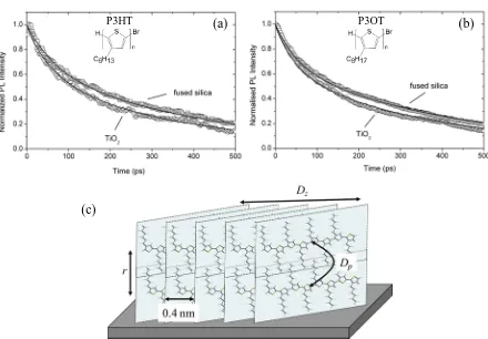
silica substrates with the D values obtained by fitting the decays. We obtained D=2×10⁻⁴ cm2s-1 in P3HT films of medium molecular weight Mn=13800 gmol-1 and D=1×10⁻⁴ cm2s-1 in P3OT films with Mn=13500 gmol-1. We also performed a similar surface quenching experiment using tethered C60 as a quencher[17] and obtained D values which were two times bigger. These results are given in Supporting Information (SI) in Figure S1. There is a non-negligible spectral overlap between the fluorescence of P3HT and absorption of tethered C60 which results in a longer quenching distance by the Förster resonance energy transfer and this slightly overestimates values of D measured using C60 as a quencher. The D values obtained from quenching on TiO2 substrates and from quenching on tethered C60 give the lower and upper limits for the diffusion coefficient respectively.
Submitted to
5
with larger inter-stack distance. In contrast, recent reports showed that films spun from high
Mn P3HT show both, edge-on and face-on orientations of π-π stacks in films thicker than a few nm.[18, 19] In that case fluorescence quenching by TiO2 returns a D value which is between
Dp and a faster diffusion along the stack Dz driven by strong dipole-dipole interactions between π-stacked P3HT chromophores. The fraction of different orientations is unknown, therefore such measurements give only a lower limit for Dz.
2.2. Exciton-exciton annihilation
In order to investigate exciton diffusion and how it is affected by molecular weight, we studied singlet-singlet exciton annihilation. This process occurs at high excitation densities when two excitons can meet on the same chromophore and annihilate to form a higher energy exciton which can then either relax to the lowest energy excited state or dissociate into an electron-hole pair. As a result, one or both excitons are lost per annihilation event. The exciton concentration N is described by a rate equation
(2)
where k is the exciton decay rate in the absence of annihilation and γ is the annihilation rate. We then deduce the time-dependence of γ by measuring at two excitation densities:
(3)
Submitted to
6
Gulbinas et al.[20] except that we use the fluorescence decay at a very low excitation density as a reference. Figure 3 shows exciton population decays measured using different excitation pulse energies which result in different initial exciton concentration and γ(t) calculated using Eq (3). In order to increase the time resolution and to reduce the noise associated with differentiation of the experimental data, we first fitted the experimental decays with a three-exponential decay function convolved with the instrument response and used the fits to calculate γ(t) . We observe a decrease of the γ value in the first 20 ps after the excitation pulse and use an equation derived for diffusion-limited annihilation in the case of preferentially 1D diffusion with a spherical annihilation volume with a radius ra to fit it[21]
(4)
The data shown in Figure 3(d) obtained from transient absorption measurements with 100 fs time resolution fit much better with the anisotropic exciton diffusion model than with an isotropic model. The fit gives Dp=2×10⁻⁴ cm2s-1 and Dz=2×10⁻² cm2s-1 for P3HT film with
Mn=76000 gmol-1. The time resolution of fluorescence measurements (~2 ps), however, was not sufficient to obtain Dp and Dz values independently for polymers of other Mn. Instead, we determine an effective diffusion coefficient Deff = (Dz Dp)1/2 from the time-independent term in Equation 4.
Submitted to
7
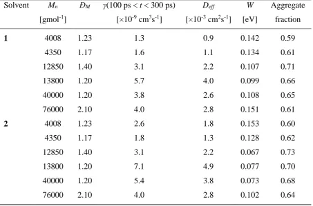
increase if exciton-charge annihilation is active. The γ values however were independent of photon energy, so we rule out a contribution from exciton-charge annihilation at the excitation densities used in our experiments (1018–1019 cm-3). It is worth noting that we indeed observe an increase of γ values at the highest excitation densities at long times t >300 ps (see Figure 4) which is likely to include a contribution from exciton-charge annihilation as more charges can be generated from a higher energy exciton state populated by exciton-exciton annihilation. In order to avoid this charge generation effect and also to avoid the strong time dependence at early times, we take the average of γ from 100 ps to 300 ps giving γ=7.1×10⁻⁹ cm3s-1 for data in Figure 4(c) and γ=5.4×10⁻⁹ cm3s-1 for data in Figure 4(d). Using the previously reported value ra=1.8 nm,[12] we obtain Deff=4.9×10⁻³ cm2s-1 and Deff=3.8×10⁻³ cm2s-1 for a P3HT film with Mn=13800 gmol-1 and Mn=40000 gmol-1 respectively.
2.3. Correlation of exciton diffusion with aggregation and exciton bandwidth
Submitted to
8
affords more time for the polymer chains to self-organize and align themselves to form π -stacked aggregates.
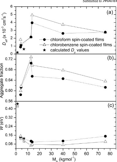
Following previous work of Clark et al.[11] we determine the fraction of the aggregated P3HT chromophores by subtracting the contribution of the amorphous phase in the absorption spectra which is assumed to show a solution-like absorption spectrum. The oscillator strength of planar chains in the aggregates was assumed to be 39% higher than that of non-planar chains in the amorphous phase.[11] The aggregate fraction obtained for different molecular weights in two solvents is displayed in Figure 5(b) and it shows a clear correlation with the effective diffusion coefficient.
The analysis of the absorption spectra shown in Figure 6 also allows us to extract the free exciton bandwidth W in the aggregate using a weakly-interacting H-aggregate model developed by Spano and coworkers.[26] The exciton bandwidth is estimated from the ratio of the 0-0 and 0-1 absorbance peaks using the following expression[7, 9, 26]:
(5)
Submitted to
9
extent of exciton delocalisation i.e. conjugation length. It has been shown by quantum-chemical calculations that increasing the exciton delocalisation along the polymer chain reduces W.[27, 28] Figure 5(c) shows that films spun from chloroform generally display higher
W and hence shorter conjugation lengths while films spun from chlorobenzene exhibit lower
W suggesting longer conjugation lengths. The dependence on Mn suggests that the longest conjugation length is attained in the medium Mn P3HT films.
From the results in Fig. 5 we can conclude that the increased chain aggregation and exciton delocalisation enhances exciton diffusion. This can partially be explained by the observation that excitons diffuse faster along the aggregated stack. The effect of exciton delocalisation is discussed in the next section.
2.4. Theoretical modelling
To explain the experimentally measured effect of molecular weight on exciton diffusion, we have developed a theoretical model for exciton diffusion in P3HT lamellae. The relatively large electronic couplings between the conjugated polymer chains within the stacks yield two-dimensional (2D) excitons delocalised over multiple polymer chains (the actual exciton size depends on the interplay between intermolecular excitonic interactions and the amount of
Submitted to
10
molecules containing 8 and 40 repeated monomers (1360 and 6800 gmol-1, respectively), as in Figure 1(c).
The exciton energy band was built by calculating the eigenvalues of the electronic Hamiltonian, H, where off-diagonal elements Hij representing the electronic couplings
between molecules i and j in a given stack are given by:
m l lm j m i lij Q Q F r
H , 0 ) ( 4 1
, i, j = 1,
…, NP. The Mataga-Nishimoto potential was used to depict the electron interaction potential
F, the atomic transition charge densities Q have been computed through correlated coupled cluster calculations with single and double excitations (CCSD) using the INDO Hamiltonian, and 0 is the vacuum permittivity.[30, 31] The on-site energies (diagonal elements) Hii were sampled from a Gaussian distribution with standard deviation σ = 0.1eV to account for
intrastack static energetic (conformational) disorder. Assuming a simple Gaussian-like shape for the exciton absorption and emission spectra[32] the Förster rate for exciton hopping between stacks is:
2
,
2
,
F k kk kk
k k
k P J F
[6]
22
1
exp ,
2 2
k k ss
kk h h E E F
[7]
where brackets denote averaging over disorder (typically a few thousand realizations), Jkk’ is the electronic coupling between the k-th exciton state of the donor stack and k´-th exciton
state of the acceptor stack ( ki i
k
i with ki the i-th component of the k-theigenfunction obtained by diagonalizing the Hamiltonian H over the isolated stacks and
' '
kk
Submitted to
11
absorption, ss is a fixed Stokes shift (set to 0.2eV[33]), Pk the Boltzmann weighted population
of k-states in the donor stack, and h 2kTss .
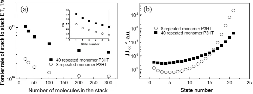
In Figure 7(a), we show the stack-to-stack energy transfer Förster-like rates for two different pairs of identical stacks (1360 and 6800 gmol-1, or 8 and 40 repeated monomers, correspondingly) as a function of the stack size (i.e., the number of polymer chains in the stack). Independently on the stack size, the Förster hopping rate kF is approximately 3.5 times larger for stacks with the higher molecular weight. This trend results from the conjunction of the fact that P3HT chains organize into H-aggregates featuring low-energy weakly dipole allowed electronic excitations and the presence of a rather large (compared to the excitonic bandwidth) conformational energetic disorder. The consequence of the latter is that only a few of the lowest energy states have significant Boltzmann weight at room temperature. The consequence of the former is that these states are optically dark in the absence of disorder and should therefore be inefficient for singlet hopping in the Förster limit (though this is somewhat relaxed in our calculations that go beyond the point dipole approximation). However, the presence of disorder translates into a symmetry breaking effect heading towards (Anderson) localization of the exciton wavefunctions (in the limit of large disorder, the excitations are localized on a single chain). Such localization effects are more pronounced for longer conjugated segments because they have smaller free electronic bandwidth. This can be
better appreciated from the insert to Figure 7(a) where the participation ratios ( ki 4 i
PR
)for the lowest 5 exciton states are portrayed (the PR tends to 1 for complete localization and to 0 for complete delocalisation). It is clear from Figure 7(a) that the PR increases from
Submitted to
12
words, the detrimental effect of the formation of non-emissive H-aggregate-like low-lying excited states is largely suppressed in going to large molecular weights.
The diffusion coefficients for stack-to-stack energy transport Dp can be inferred from the rates above by using a simple variable-range hopping model andis given by the ratio of the squared displacement of exciton with respect to time. The calculated values of Dp=2.0×10⁻⁴ cm2s-1 for
Mn=1360 gmol-1 and Dp=6.2×10⁻⁴ cm2s-1 for Mn=6800 gmol-1 are in good agreement with the lower and upper values of Dp=2x10⁻⁴ cm2s-1 and Dp=4x10⁻⁴ cm2s-1obtained by surface quenching. The result shown in Figure 7(a) suggests that faster inter-stack diffusion Dp is achieved for smaller numbers of molecules in the stack. This means that films with smaller aggregate fraction may display higher Dp which thus can explain why Deff shows just a weak dependence on Mn.
3. Conclusions
Submitted to
13
beneficial effect on inter-stack diffusion. Interestingly, despite smaller electronic interactions between neighboring polymer chains in the stack at large Mn, larger molecular weights also maximize the intra-stack exciton diffusion process owing to a long-range transfer mechanism.[34] The model further illustrates the subtle interplay of interchain coupling, aggregate size and Boltzmann statistics in the energy transfer process. These results suggest that by developing materials with increased crystallinity and conjugation length, exciton diffusion length can be increased.
4. Experimental Section
Materials: The P3HTs studied here were obtained as described in Ref.35. High Mn P3HT (76000 gmol-1) was obtained from Merck Chemicals. All materials had regioregularity of 98% or higher and comparable dispersity values (ĐM) except for the high molecular weight material (see Table 1).
Film preparation: Solutions were prepared in chloroform and chlorobenzene and stirred overnight in a N2-filled glove box at 50ºC and 90ºC respectively. Films were spin-coated onto fused silica substrates. All substrates were first cleaned via ultrasonication for 15 minutes with acetone and propan-2-ol. The films were spin-coated in the glove box using spin speeds of around 1500-2000 rpm. No heat treatment was performed on these films. Films made for transient absorption measurements were sandwiched and sealed between two fused silica substrates in the glove box. For measurements of fluorescence quenching by TiO2, polymer films were spin-coated from p-xylene solution on TiO2 substrates prepared by vacuum deposition.[12] Substrates with tethered C60 used for other fluorescence quenching experiments were prepared as described previously.[17]
Submitted to
14
thicknesses ranged from 12-14 nm. Film thicknesses for fluorescence measurements varied between 23 to 74 nm. Fluorescence decays at low excitation density were measured using 100 fs pulses at 3.1 eV excitation with 80 MHz repetition rate and a Hamamatsu C6860 synchroscan streak camera for detection. For exciton-exciton annihilation in fluorescence and transient absorption measurements the 100 fs laser pulses at a repetition rate of 5 kHz were used. All fluorescence measurements were performed under a vacuum of 10-4 mbar. The energy of the laser pulses was controlled with neutral density filters. The excitation spot was measured with a LaserCam 3D beam profiler and found to be an ellipse with the major and minor diameters of 290 µm and 215 µm in fluorescence measurements. In transient absorption measurements, the ellipse size of the excitation spot was 2.22 mm by 1.64 mm. A film thickness of 170 nm and a probe of 1.0 eV were used for transient absorption measurements.
Supporting Information
Supporting Information is available online from the Wiley Online Library or from the author.
Acknowledgements
Submitted to
15 REFERENCES
[1] R. R. Lunt, J. B. Benziger, S. R. Forrest, Adv Mater, 2010, 22, 1233.
[2] J. Kirkpatrick, P. E. Keivanidis, A. Bruno, F. Ma, S. A. Haque, A. Yarstev, V. Sundstrom, J. Nelson, J Phys Chem B, 2011, 115, 15174.
[3] A. Ruseckas, P. E. Shaw, I. D. W. Samuel, Dalton T, 2009, 10040. [4] M. T. Dang, L. Hirsch, G. Wantz, Adv Mater, 2011, 23, 3597.
[5] A. Zen, J. Pflaum, S. Hirschmann, W. Zhuang, F. Jaiser, U. Asawapirom, J. P. Rabe, U. Scherf, D. Neher, Adv Funct Mater, 2004, 14, 757.
[6] J. F. Chang, J. Clark, N. Zhao, H. Sirringhaus, D. W. Breiby, J. W. Andreasen, M. M. Nielsen, M. Giles, M. Heeney, I. McCulloch, Phys Rev B, 2006, 74, 115318.
[7] J. Clark, C. Silva, R. H. Friend, F. C. Spano, Phys Rev Lett, 2007, 98.
[8] O. G. Reid, J. A. N. Malik, G. Latini, S. Dayal, N. Kopidakis, C. Silva, N. Stingelin, G. Rumbles, J. Polym. Sci. Pt. B-Polym. Phys., 2011, 50, 27.
[9] C. Scharsich, R. H. Lohwasser, M. Sommer, U. Asawapirom, U. Scherf, M. Thelakkat, D. Neher, A. Kohler, J. Polym. Sci. Pt. B-Polym. Phys., 2012, 50, 442.
[10] H. Sirringhaus, P. J. Brown, R. H. Friend, M. M. Nielsen, K. Bechgaard, B. M. W. Langeveld-Voss, A. J. H. Spiering, R. A. J. Janssen, E. W. Meijer, P. Herwig, D. M. de Leeuw, Nature, 1999, 401, 685.
[11] J. Clark, J.-F. Chang, F. C. Spano, R. H. Friend, C. Silva, Appl Phys Lett, 2009, 94, 163306.
[12] P. E. Shaw, A. Ruseckas, I. D. W. Samuel, Adv Mater, 2008, 20, 3516.
[13] L. Luer, H. J. Egelhaaf, D. Oelkrug, G. Cerullo, G. Lanzani, B. H. Huisman, D. de Leeuw, Org Electron, 2004, 5, 83.
[14] O. V. Mikhnenko, H. Azimi, M. Scharber, M. Morana, P. W. M. Blom, M. A. Loi,
Submitted to
16
[15] H. Wang, H.-Y. Wang, B.-R. Gao, L. Wang, Z.-Y. Yang, X.-B. Du, Q.-D. Chen, J.-F. Song, H.-B. Sun, Nanoscale, 2011, 3, 2280.
[16] S. Cook, L. Y. Han, A. Furube, R. Katoh, J Phys Chem C, 2010, 114, 10962. [17] A. J. Ward, A. Ruseckas, I. D. W. Samuel, J Phys Chem C, 2012, 116, 23931. [18] R. J. Kline, M. D. McGehee, M. F. Toney, Nat Mater, 2006, 5, 222.
[19] D. T. Duong, M. F. Toney, A. Salleo, Phys Rev B, 2012, 86, 205205.
[20] V. Gulbinas, I. Mineviciute, D. Hertel, R. Wellander, A. Yartsev, V. Sundstrom, J Chem Phys, 2007, 127, 144907.
[21] U. Gösele, A. Seeger, Philosophical Magazine, 1976, 34, 177.
[22] A. Ruseckas, M. Theander, M. R. Andersson, M. Svensson, M. Prato, O. Inganas, V. Sundstrom, Chem Phys Lett, 2000, 322, 136.
[23] C. X. Sheng, M. Tong, S. Singh, Z. V. Vardeny, Phys Rev B, 2007, 75.
[24] J. Piris, T. E. Dykstra, A. A. Bakulin, P. H. M. van Loosdrecht, W. Knulst, M. T. Trinh, J. M. Schins, L. D. A. Siebbeles, J Phys Chem C, 2009, 113, 14500.
[25] F. Paquin, G. Latini, M. Sakowicz, P.-L. Karsenti, L. Wang, D. Beljonne, N. Stingelin, C. Silva, Phys Rev Lett, 2011, 106.
[26] F. C. Spano, J Chem Phys, 2005, 122, 234701.
[27] E. S. Manas, F. C. Spano, J Chem Phys, 1998, 109, 8087.
[28] D. Beljonne, J. Cornil, R. Silbey, P. Millie, J. L. Bredas, J Chem Phys, 2000, 112, 4749. [29] E. V. Emelianova, S. Athanasopoulos, R. J. Silbey, D. Beljonne, Phys Rev Lett, 2010,
104, 206405.
[30] J. Gierschner, Y. S. Huang, B. Van Averbeke, J. Cornil, R. H. Friend, D. Beljonne, J Chem Phys, 2009, 130, 044105.
[31] Z. Shuai, J. L. Bredas, Phys Rev B, 2000, 62, 15452.
Submitted to
17
[33] F. C. Spano, J. Clark, C. Silva, R. H. Friend, J Chem Phys, 2009, 130.
[34] E. Emelianova, L. J. Wang, D. Beljonne, Z. Masri, I. D. W. Samuel, in preparation. [35] M. Surin, O. Coulembier, K. Tran, J. D. Winter, P. Leclère, P. Gerbaux, R. Lazzaroni, P.
Submitted to
18
Figure 1. Fluorescence decays of (a) P3HT and (b) P3OT film on TiO2 and fused silica substrates. Solid lines are the fits using Equation 1 which give D=2×10⁻⁴ cm2s-1 for P3HT and
D=1×10⁻⁴ cm2s-1 for P3OT. Insets show the chemical structures of P3HT and P3OT. Lamellae of two π-π stacks of regioregular P3HT (c) with nearest neighbour cofacial separation of 0.4 nm and inter-stack distance r. Diffusion coefficient between stacks is Dp whereas diffusion coefficient along a stack is Dz.
(c)
(a) (b)
[image:18.595.77.517.77.383.2]Submitted to
19
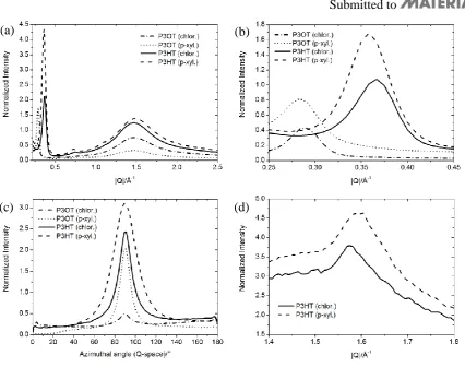
Figure 2. (a) Radial diffraction profiles in out-of-plane direction from different films of P3OT and P3HT. (b) Zoom into first order reflections. (The peaks positions as fitted by Gaussians are given in Table S1 in Supporting Information (SI)). (c) Azimuthal profiles of the first order reflections shown in (b) transformed into Q-space. Fitted values for the full width at half maximum are listed in Table S1 in SI. (d) Radial diffraction profiles in in-plane direction for P3HT films.
(a) (b)
[image:19.595.74.501.63.399.2]Submitted to
[image:20.595.67.517.84.414.2]20
Figure 3. Time dependence of exciton population N(t) in P3HT film (Mn=76000 gmol-1) spin-coated from chlorobenzene solution measured for different excitation densities by (a) time-resolved fluorescence after excitation at 2.2 eV and (b) transient absorption after excitation at 2.4 eV; (c) annihilation rate obtained from time-resolved fluorescence data, (d) annihilation rate obtained from transient absorption data, solid line and dotted lines are simulated rates using Eq. 4 with Dp and Dz values given in the legend.
(a)
(c)
(b)
Submitted to
[image:21.595.77.522.84.405.2]21
Figure 4. Fluorescence decays measured at different excitation densities for P3HT films spin-coated from chlorobenzene solution after excitation at 3.1 eV for (a) Mn=13800 gmol-1 and (b)
Mn=40000 gmol-1. IRF is the instrument function. (c) and (d) show γ(t) extracted from the PL decays shown in (a) and (b) respectively. From the γ values between 100 ps and 300 ps we obtain Deff=4.9×10⁻³ cm2s-1 for Mn=13800 gmol-1 and Deff=3.8×10⁻³ cm2s-1 for Mn=40000 gmol-1. A control experiment using 2.2 eV and 2.45 eV excitation gave very similar results.
(a)
(c)
(b)
Submitted to
22
[image:22.595.106.488.64.594.2]Submitted to
23
Submitted to
24
[image:24.595.76.519.108.273.2]Submitted to
[image:25.595.72.526.130.429.2]25
Table 1. Molecular weights and dispersity values (ĐM=Mw/Mn) of all P3HT samples used along with measured results. P3HT films were spun from chloroform (1) and chlorobenzene (2) solution.
Solvent Mn [gmol-1]
ĐM γ(100 ps < t < 300 ps) [×10-9 cm3s-1]
Deff
[×10-3 cm2s-1]
W
[eV]
Aggregate fraction
1 4008 1.23 1.3 0.9 0.142 0.59
4350 1.17 1.6 1.1 0.134 0.61
12850 1.40 3.1 2.2 0.107 0.71
13800 1.20 5.7 4.0 0.099 0.66
40000 1.20 3.8 2.6 0.108 0.65
76000 2.10 4.0 2.8 0.151 0.61
2 4008 1.23 2.6 1.8 0.153 0.60
4350 1.17 1.8 1.3 0.128 0.62
12850 1.40 3.1 2.2 0.067 0.73
13800 1.20 7.1 4.9 0.077 0.70
40000 1.20 5.4 3.8 0.073 0.68
Submitted to
26
Singlet exciton diffusion in P3HT is studied by a combination of experimental and theoretical methods. We show that exciton diffusion is faster along the co-facial π-π aggregates than between them. The fastest exciton diffusion is observed for intermediate molecular weight polymers and it correlates with the degree of chromophore aggregation and with exciton delocalisation along the polymer chain.
Keyword Exciton diffusion
Zarifi Masri, Arvydas Ruseckas, Evguenia V. Emelianova, Linjun Wang, Ashu K. Bansal, Andrew Matheson, Henrik T. Lemke, Martin M. Nielsen, Ha Nguyen, Olivier Coulembier, Philippe Dubois, David Beljonne, Ifor D. W. Samuel*
Submitted to
27
Copyright WILEY-VCH Verlag GmbH & Co. KGaA, 69469 Weinheim, Germany, 2012.
Supporting Information
for Adv. Energy Mater.,DOI: 10.1002/aenm.((please add manuscript number)) Molecular weight dependence of exciton diffusion in poly(3-hexylthiophene)
Zarifi Masri, Arvydas Ruseckas, Evguenia V. Emelianova, Linjun Wang, Ashu K. Bansal, Andrew Matheson, Henrik T. Lemke, Martin M. Nielsen, Ha Nguyen, Olivier Coulembier, Philippe Dubois, David Beljonne, Ifor D. W. Samuel*
Surface quenching measurements on tethered C60
Time-resolved fluorescence measurements were performed using the time-correlated single photon counting (TCSPC) technique. An excitation of 393 nm with a pulse width at fwhm of about 200 ps from a pulsed Picoquant GaN was used. Measurements were performed under a nitrogen atmosphere.
Figure S1: Fluorescence decays of (a) P3HT and (b) P3OT film on tethered C60 and fused silica substrates with fits obtained using Equation 1 which give D=4×10⁻⁴ for P3HT and
D=2×10⁻⁴ cm2s-1 for P3OT. IRF is the instrument response function.
[image:27.595.76.517.412.577.2]Submitted to
28 X-ray diffraction (XRD) experiments
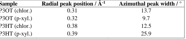
X-ray diffraction experiments were performed at the X-ray centre at Risø DTU on 80 nm thick films of P3HT Mn=13800 gmol-1 and P3OT Mn=13500 gmol-1. The out of plane stacking distances as determined from the first order reflection are 20.3 Å, 19.6 Å, 16.5 Å, 16.1 Å for P3OT (chloroform), P3OT (p-xylene), P3HT (chloroform) and P3HT (p-xylene), respectively. The grazing incidence geometry was employed to suppress background scattering from the substrate by selecting an incidence angle below the angle of total external reflection from the substrate but above the angle of total external reflection from the film, thus maximizing the signal from the film. By nature of the measurements, the out of plane direction is measured with a slight offset. This is also seen as the lack of data points around the 90 azimuth angle in Figure 2 (c).
[image:28.595.77.457.455.535.2]The peak radial positions as fitted by Gaussians are given in Table S1. The fitted values for the full width at half maximum for the first order reflections (azimuthal) are also listed in the table. Using fitted values eliminates possible offsets introduced by measuring only part of the out of plane peak profiles, as discussed above.
Table S1: Radial positions and azimuthal widths of the first order out-of-plane reflections Sample Radial peak position / Å-1 Azimuthal peak width / °
P3OT (chlor.) 0.31 13.7
P3OT (p-xyl.) 0.32 9.7
P3HT (chlor.) 0.38 12.5
Submitted to
29 Calculation of aggregate fraction
[image:29.595.72.314.212.392.2]The aggregate fraction or fraction of film made of planarized and π-stacked polymer chains is estimated by subtracting the dilute solution absorption spectra from the film absorption spectra. As the amorphous phase is made of disordered polymer chains, the absorbance due to these unaggregated polymers can simply be approximated by the absorption spectra from the dilute solution.
Figure S2: Representative absorbance spectra showing calculation of aggregate fraction. The aggregate fraction is thus calculated by taking the ratio of the area under the aggregate and film absorbance curves (Figure S2):
(S1)