CHANGES IN SOME BIOCHEMICAL PARAMETERS AND BODY
WEIGHT OF CHICKEN EXPOSED TO CADMIUM
Khaled Saeed Ali*, Mohammed Abdulla Lakal and Khaldoon Obad
Biology Department, Education Faculty/Sabir, University of Aden.
ABSTRACT
This study was conducted with 6 week old domestic chicken to
determine the effect of supplementation of cadmium to dietary. 10
mg/kg Cadmium chloride added to maize- sesame cake meal diet for 4
weeks. The additional cadmium to the diet induced a decreasing body
weight and changes in biochemical parameters of chicken. Chicken
were divided into two groups. The first group was given a diet
containing the concentration of 10 mg cadmium /kg daily for a period
of 4 weeks and the second group was given diet without cadmium and
used as a control group. The result revealed decrease in the body
weight of treated chicken by 12.7% compared to control group, whose
body weight increased. The plasma glucose concentration, creatinine, aspartate
aminotranseferase (AST), and alanine aminotransferase (ALT) were increased significantly
(P< 0.05) in Cd treated chicken in comparison to the control group. Cadmium accumulation
was observed in the kidney, liver, intestine and muscle. The accumulation of cadmium was
markedly higher (3-4 times) in cadmium-treated animals compared to the control.
KEYWORDS: Cadmium, chicken, Body weight and biochemical parameters.
INTRODUCTION
Cadmium (Cd) belongs to the group of highly toxic heavy metals[1], it is found widely in
nature and present in air, all soils and aquatic systems. Considerable amounts of Cd get
continuously into the environment as a consequence of human activities. The steel industry,
waste disposal, volcanic action, zinc refinery, fossil fuels, and traffic are accounted for the
largest part of emissions of atmospheric Cd. Raised concentrations of Cd in soil may be
found as a result of industrial activities (e.g. mining) or agricultural activities (e.g. sewage
sludge, phosphate fertilizers, and pesticides) containing high concentrations of Cd.[2]
Volume 5, Issue 3, 227-234. Research Article ISSN 2277– 7105
*Correspondence for Author
Khaled Saeed Ali Biology Department,
Education Faculty/Sabir,
University of Aden. Article Received on 04 Jan 2016.
Meat, fish, and fruits generally contain up to 50 μg Cd.kg-1 on fresh weight basis, whereas
vegetables, potatoes, and grain products may contain up to 150 μg Cd.kg-1 fresh weight.
Higher concentrations are found in the kidneys of animals slaughtered for food, in wild
mushrooms, and in seafood such as mussels and oysters.[3]
Consumption of contaminated water is the major way by which humans are exposed to Cd[4]
and the maximum allowable level in drinking-water is 0.005 mg/dL.[5] Animals can be
exposed to Cd pollution by: inhalation of polluted air, ingestion of polluted food and drinking
of polluted water.[6,2,7,8] It is absorbed from gastrointestinal tract to blood, and cadmium is
taken up from the blood into the tissues. The Cd accumulates in human and animals tissues,
especially the liver and kidney, causing their damage.[2] Cadmium induced injury to liver and
kidney organs has been attributed to its ability to enhance free radical formation in vivo.[9,10]
Cadmium in high doses induces structural and function alterations in various vital organs
including liver, kidney, gill and intestine of fishes. Cadmium accumulates in liver of fishes in
high concentrations.[11,12] It also induces various pathological changes in liver tissues
including engorgement of blood vessels, congestion, vacuolar degeneration of hepatocytes,
necrosis of pancreatic cells and fatty changes in the peripancreatic hepatocytes.[13,14] Some
biochemical characteristic features are associated with Cd-induced kidney and liver damage.
Others are marked increase of plasma creatinine or blood urea nitrogen levels[15] as well as
altered plasma glucose level[16], and changes in antioxidant enzymes (GOT and GPT) level,
which produced in damaged liver tissues. The objective of this work is to study the effect of
cadmium on the changes of weight, and some haematological and biochemical parameters in
chicken.
MATERIALS AND METHODS
Six weeks old chickens (n= 16) are collected from their habitat and were housed in cages to
acclimatization for one week in laboratory conditions at the room temperature between 28 –
300C. After the acclimatization period, chicken were divided into two groups (n=8) randomly
and each group was kept in separate cages. The first group was given a maize- sesame cake
meal diet containing the concentration of 10 mg cadmium (Cdcl2) /kg and the second group
was given maize- sesame cake meal diet without cadmium and used as a control group. The
samples were collected with heparinized syringe from the wing vein and was used to obtain
the plasma by centrifugation (5000 rpm) for 5 min. The plasma glucose, craetinine, ALT and
AST were determined using Randox kits.
The animals were sacrificed by cervical dislocation and the kidneys, liver, intestine and
muscle were removed, rinsed in ice-cold saline. Tissues were dried and digested with
HNO3/H2O2. The Cd content of the homogenates were determined with Atomic absorption
spectrophotometer. Cd contents are reported in µg/g dry weight.
Statistical analysis
The data is presented as means ± SD. The mean values of the control and test groups were
compared using Student’s t-test. The significant level was set at P < 0.05.
RESULTS
Body weight gain
The result in table [2] shows the body weight of the control group increased by 37 %,
whereas the body weight of Cd treated group decreased by 12.7 % after 4 weeks. The gain in
body weight of the treated group were lowered in relation to the control which was reduced to
46% after 4 weeks of treatment.
Biochemical parameters
Table (3) shows the changes of some biochemical parameters in Cd treated chickens
compared to control chickens one month of 10mg/kg Cd exposure to diet.
The plasma glucose concentration showed higher significant values (P< 0.05) (196 ±24mg/dl)
in chickens exposed to Cd for 4 weeks than the control chicken group value (127.3 ±15).
The plasma creatinine increased significantly (P< 0.05) in chickens exposed to Cd 4 weeks
(0.75 ± 0.16) when compared to the control group (0.42 ± 0.11). The changes in AST and
ALT activities are used to check liver function in the Cd treated chicken relative to control
group. The Results also revealed that cadmium causes significant (P< 0.05) increasing of
activity of ALT and AST, as seen in table (3). The plasma ALT and AST level reached (7.7 ±
1.3and 141± 16) in treated chickens comparison to control group (5.6 ± 1and 97 ± 14)
As can be seen in Table (4), the accumulation of Cd in the kidneys, liver, intestine and
muscle, where concentrations of cadmium increased in all tissues.The Cd content reached
high level in kidney (186 ± 25 µg/g) and the low level in muscle (18 ± 2.2 µg/g).
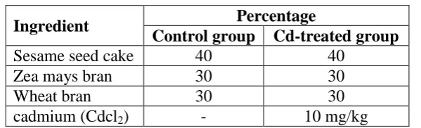
Table (1): Composition of the diet
Ingredient Percentage
Control group Cd-treated group
Sesame seed cake 40 40
Zea mays bran 30 30
Wheat bran 30 30
cadmium (Cdcl2) - 10 mg/kg
Table (2): Growth performance of chickens exposed to Cd in diet (mean ± SD) Items Initial weight Weight after 4 weeks Weight gain
Control 276 ± 20.7 390 ± 25.5 114 ± 11.4
Cadmium treated 284 ± 11.9 222 ± 13.5* 2 ± 14.8
*: P<0.05 comparing with control
Table (3): Cadmium effect on blood glucose, AST, ALT and Creatinine in plasma of the chickens ( mean ± SD)
4 weeks control
Parameter
196 ±24 127.3 ±15
Glucose
141± 16 97 ± 14
AST
7.7 ± 1.3 5.6 ± 1
ALT
0.75 ± 0.16 0.42 ± 0.11
Creatinine
*: P<0.05 comparing with control
Table (4) Cadmium accumulation (µg/g dry weight) in chicken tissues
Items Liver Kidney intestine Muscle
Control 2.8 ± 0.3 5.4 ± 0.45 2.5 ± 0.25 0.8 ± 1.6
Cadmium treated 90 ± 5 186 ± 25 49 ± 6.5 18 ± 2.2
DISCUSSION
The results showed that the chicken exposed to 10 mg/kg Cd in diet showed a significant
lower chicken growth. The final body weight of Cd treated chicken was significantly lower
than that of the control group. In young animals [e.g. calf, swine, rabbit, Japanese quail,
chicken, rat], the different Cd salts can reduce feed intake and growth rate.[17,18,19]
[image:4.595.150.450.165.256.2]Heteropneustes fossilis.[20,21],[22] suggested that hyperglycemia occurred in Atlantic salmon
(Salmo salar) after toxicity with cadmium may be due to changes in liver carbohydrate
metabolism (activation of liver glycogenolysis and glycolysis) as well as increased levels of
plasma glucose.
The activity of AST and ALT enzymes in blood may also be used as a stress indicator. The
significant changes in activities of these enzymes in blood plasma indicates tissue impairment
caused by stress.[23,24] In the present study, there were significant changes in AST and ALT
activities in plasma of chicken exposed to cadmium compared to the control group. The
increase in concentration of AST and ALT in blood plasma indicates impairment of liver. In
addition, the increase of plasma AST and ALT may be attributed to the hepatocellular
damage or cellular degradation by these heavy metal, perhaps in liver, heart or muscle[25]
These results are in agreement with those of[26] who found that sublethal concentration of Cd
caused significant increases in AST and ALT of common carp after 7 and 15 days. Also,[27]
found that Increased activity of serum enzymes ALT and AST in guinea pigs. Similar result
demonstrated by[28] who found that the cadmium exposure induced serum AST, ALT
increasing of rat.
In the present study, kidney function tests showed elevation in creatinine in the treated group.
The results are in agreement with that reported by[28,29] in guinea pigs and rats.[27], mentioned
that the toxic effect of cadmium on the renal tissues was clear as the level of creatinine in
serum increased. The elevation in the creatinine was due to nephrotoxic effect of cadmium on
renal tubules and glomeruli. Also,[30] found significant elevation in serum creatinine
concentration in Cd intoxicated rats. They added that creatinine metabolism was thought to
reflect the amount of glomerular filtration.[26], described a significantly increased creatinine
level in Cd-treated chickens.
The results obtained in this study show that concentration of Cd in liver and kidneys were
significantly increased in animals exposed to Cd (p<0.05).[31], found that increased
accumulation of Cd in the kidneys, liver and intestine in mice and fish liver.[32,33] found that
the diets contained cadium caused accumulation of Cd in liver and kidney of lambs.
REFERENCES
1. NRC (National Research Council, Cadmium. In: (Ed. NRC) Mineral tolerance of
2. KOSTIAL, K., Cadmium. In: (Ed. Mertz, W.) Trace Elements in Human and Animal
Nutrition. Academic Press, Inc., San Diego, Calif., 1986; 2: 319-337.
3. FRIED, K. W. – ROZMAN, K. K. Toxicology and risk assessment: A comprehensive
introduction. Chichester. John Wiley & Sons, Ltd., 2008; 513-655.
4. WHO. cadmium.: World Health Organization. Geneva, 1992; 111–2.
5. WHO report. Guidelines for drinking water quality. Health criteria and other supporting
information. World Health Organisation. Geneva, 1984; 84-90.
6. Van BRUWAENE, R., KIRCHMANN, R., IMPENS, R. Cadmium contamination in
agriculture and zootechnology. Experientia, 1984; 40: 43-52.
7. Bokori, J.,Occurrence of cadmium in the nature and its more important physiological
characteristics (in Hungarian). Magy. Állatorv. Lapja, 1994; 49(2): 69-73.
8. OSHA, Cadmium health effects. Occupational Safety and Health Administration US
Department of Labor, 1999.
9. Bagchi, D., M. Bagchi, E.A. Hassoun and S.J. Stohs. Cadmium-induced excretion of
urinary lipid metabolites, DNA damage, glutathione depletion and hepatic lipid
peroxidation in Sprague-dawley rats. Biol. Trace Elements Res., 1996, 52: 143-154.
10. Vinoth kumar P, Amala bricey A., Veera thamari selvi V, Sudheer kumar C and Ramesh
A., Advances in Applied Science Research, 2010; 1(2): 9-13.
11.Sumet, H.D. and R. Blust, Stress responses and changes in protein metabolism in carp
Cyprinus carpio during cadmium exposure. Ecotoxicol. Environm. Saf, 2001; 48(30):
255-262.
12.Rangsayatorn, N., M. Kruatrachue, P. Pokethitiyook, E.S. Upatham, G.R. Lanza and S.
Singhakaew, 2004.
13.Rani, U.A. and R. Ramamurthi, Histopathological alteration in the liver of freshwater
teleost Tilapia mossambica in response to cadmium toxicity. Ecotoxicol. Environ. Saf,
1989; 17(2): 221-216.
14.Dangre, A.J., S. Manning and M. Brouwer, Effectsof cadmium on hypoxia-induced
expression of hemoglobin and erythropoietin in larval sheepshead minnow, Cyprinodon
variegates.Aquatic Toxicol, 2010; 99(2): 168-175.
15.Horiguchi H., Sato M., Konno N., Fukushima M., Long-term cadmium exposure induces
anaemia in rats through hypoinduction of erytrhopoietin in the kidney. Arch. Toxicol,
17.PRITZL, M. C., LIE, Y. H., KIENHOLZ, E. W., WHITEMAN, C. E. The effect of
dietary cadmium on development of young chickens. Poultry Science, 1974; 53:
2026-2029.
18.Bokori I, J., Fekete, S., Kadar, I., Koncz, J., Vetesi, F., Albert, M., Complex study of the
physiological role of cadmium. III. Cadmium loading trials on broiler chickens. Acta Vet.
Hung, 1994; 43(2-3): 195-228.
19.Turecki, T., Cibulka, J., Slamova, A., Barcalikova, R. Effect of organic and inorganic
forms of dietary cadmium on cadmium, zinc, copper, iron and manganese availability to
rats. J. Anim. Physiol. A. Anim. Nutr, 1997; 78: 119-128.
20.Sastry, K. V and Subhadra, K, In vivo effects of cadmium on some enzyme activities in
tissues of the freshwater catfish, Heteropneustes fossilis. Environ. Res., 1985; 36(1): 32-
45.
21.Khaled S. and Huda A. Effet of cadmium in drinking water on growth, some
haematological and biochemical parameters of chicken, European Journal of
Experimental Biology, 2013; 3(5): 287-291.
22.Soengas, J. L.; Agra-Lago, M. J.; Carballo, B.; Andres, M. D and Veira, J. A. R Effect of
an acute exposure to sublethal concentration of cadmium on liver carbohydrate
metabolism of Atlantic salmon (Salmo salar). Bull. Environ . Contam. Toxicol, 1996; 57:
625- 631.
23.James, R.; Sampath, K.; Jancy Pattu, V and Devakiamma, G., Utilization of Eichhorina
crassipes for the reduction mercury toxicity on food transformation in Heteropneustes
fossilis. J. Aqua. Trop, 1992; 7: 189- 196.
24.Svoboda, M, Stress in fish – review. Bul. VURH Vodnany, 2001; 37: 69- 191.
25.Yamawaki, K.; Hashimoto, W.; Fujii, K.; Koyama, J.; Ikeda, Y. and Ozaki, H.
Hematological changes in carp exposed to low cadmium concentration. Bull of the
Japanese. Soc. Sci. Fish., 1986; 59(3): 459- 466.
26.halaby, A. M., Biochemical and physiological studies on metal contamination in the
common carp (Cyprinus carpio L). Zagazig University, Faculty of Science. (Benha
branch), 1997; 268. [Ph. D. Thesis]
27.Hassan, A. Randa; Dawlat M. Amin; Nariman A. Rahmy; Hatem, M.E. and Dessouky, M.
Clinicopathological, histopathological and immunolog1ical studies on animals exposed to
lead and cadmium under experimental conditions. New York Science Journal, 2012;
28.Deepti gaurav, Shabad Preet and K. K. Dua., Advances in Applied Science Research,
2011; 2 (2): 69-78.
29.Nagyova, A.; Galba, V.S. and Genter, A.C., Cadmium hepatotoxicity in guinea pigs with
different ascorbic acid intake: Histomorphological and biochemical changes." Biologica
Bratislova, 1994; 49(3): 399-404.
30.Kuda, T.; Handa, R. and Tsuritani, I., Progress of renal dysfunction in inhabitants
environmentally exposed to Cd. Arch Evi. Health, 1988; 43: 213- 217.
31.Chwelatiuk et al. Journal of Trace Elements in Medicine and Biology, 2006; 19: 259–265.
32.DOYLE, PFANDER, GREBING AND PIERCE. The Journal of Nutrition, 1974; 104:
160-166.
33.K.S. Ali et al. Biochemical and Biophysical Research communication, 2003; 307: