© 2019 by the Serbian Biological Society How to cite this article: Aydogan Turkoglu S, Okuyan D, Kockar K. TGF-β downregulates CAIII expression via MAPK and PI3K signaling pathways in colon carcinoma and osteosarcoma cells. Arch Biol Sci. 2019;71(3):393-401.
TGF-β downregulates CAIII expression via MAPK and PI3K signaling pathways in colon
carcinoma and osteosarcoma cells
Sumeyye Aydogan Turkoglu1,*, Derya Okuyan2 and Feray Kockar1
1University of Balikesir, Faculty of Science and Literature, Department of Molecular Biology and Genetics, 10145 Balikesir, Turkey
2University of Balikesir, Susurluk Vocational Training Schools, Laboratory and Veterinary Health Program, Balikesir, Turkey
*Corresponding author: [email protected]; [email protected]
Received: October 8, 2018; Revised: February 26, 2019; Accepted: February 27, 2019; Published online: March 25, 2019
Abstract: Identification of cancer-associated genes is critical for developing effective treatments of colorectal cancer (CRC). A limited number of studies have examined the mechanisms and genes underlying CRC. Abnormal transforming growth factor beta (TGF-β) expression was observed at different stages of carcinoma. We examined the effect of cancer-related cytokine TGF-β on carbonic anhydrase (CA) III gene expression in colon cancer HT-29 cells. TGF-β (500 U/mL) down-regulated CAIII gene expression at both the mRNA and protein levels. Transient transfection experiments indicated that different CAIII promoter constructs were active in HT-29 cells. TGF-β reduced transcriptional activity of all promoter constructs, indicating that the potential response element for TGF-β-directed transcription lies within the -108/+86 region of the CAIII promoter. According to the non-Smad pathway inhibitory assay, TGF-β downregulated the CAIII gene through mitogen-activated protein kinase/extracellular signal-regulated kinase (MAPK/ERK) and phosphoinositide-3-kinase (PI3K) pathways. The same decreasing effect was determined in the Saos-2, osteosarcoma cell line, indicating that the effect of TGF-β on CAIII was not tissue-specific. However, examination of PI3K and MAPK/ERK signaling pathways with suitable inhibitors revealed that the PI3K but not the MAPK/ERK pathway was responsible for TGF-β downregulation.
Keywords: CAIII; TGF-β; transcriptional regulation; colon carcinoma; osteosarcoma
393
INTRODUCTION
Colorectal cancer (CRC) is a heterogeneous disease with distinct genetic and epigenetic alterations. The mechanisms leading to CRC development, progression and recurrence are complex. CRC is caused by the ac-cumulation of multiple genetic and epigenetic changes. Somatic mutations in genes encoding for adenomatous polyposis coli (APC), B-Raf proto-oncogene, serine/ threonine kinase (BRAF), Kirsten rat sarcoma 2 viral oncogene homolog (KRAS), phosphoinositide-3-kinase, catalytic, alpha polypeptide (PIK3CA) and tumor protein p53 (TP53) have been frequently observed in CRCs [1-4]. Apart from mutations in these genes, chromosome instability (CIN) such as aneuploidy, am-plification and deletion of subchromosomal genomic regions and loss of heterozygosity (LOH) have been observed in the majority of sporadic CRCs (65-70%). Recent studies [1,5] have also shown that DNA
methyla-tion and covalent histone modificamethyla-tions are common in CRC. Downregulation of the tumor suppressor genes caused by hypermethylation of CpG islands in promoter regions is one of the crucial mechanisms in CRC development and progression.
detection of the genes affected by TGF-β and detailed analysis of these genes are very important.
Carbonic anhydrase (CA) enzymes are very impor-tant for maintenance of pH balance. Some members, CAIX and CAXII, have been proposed as a cancer biomarker in a number of studies. CAIX was shown to be a potential hypoxic CRC biomarker. It has also been suggested that serum CAIX can serve as a potential tool in CRC clinical practice. CAIV is a novel tumor suppressor in CRC, acting through inhibition of the Wnt signaling pathway by targeting the WTAP-WT1-TBL1 axis [10]. CAIII that has lower hydratase activity than other members of the CA family is involved in acute myeloid leukemia and liver carcinoma. It has also been shown that CAIII in hepatocellular carcinoma provides cells with an invasive character via the focal adhesion kinase (FAK) signaling pathway and/or intracellular and extracellular acidification. Overexpression of CAIII increases the resistance to anticancer drugs such as paclitaxel [11] in rat fibroblast cells [11-13]. These activities suggest a potential therapeutic role of CAIII inhibitors in combination therapy with anticancer medication [13,14]. Moreover, there is nothing known about the link between TGF-β and CAIII in CRC. The underlying mechanisms are not well understood, and requires more in-depth characterizations of CAIII regulation in CRC.
When considering the different processes in which TGF-β is involved, the contribution to the regulation of key genes in different cancer types requires exami-nation. Different studies have shown that several CA family members are important in different types of cancer. In particular, CAIX has been proposed as a biomarker in colon carcinoma and many other cancers [15-20]. CAIV has been shown to be hypermethylated in colon carcinoma [21]. Reduction of CAI, CAII, and CAIII in in HCC (hepatocellular carcinoma) has been demonstrated, and this reduction has been shown to increase tumor cell motility and to contribute to tumor growth and metastasis [22]. However, there is no information on the status of CAIII in colon cancer and its regulation by TGF-β. For this purpose, we investigated the effect of TGF-β on human CAIII expression in human colon and sarcoma cell lines. Our data show that TGF-β reduces the expression of CAIII at mRNA and protein levels and also causes transcriptional repression of the promoter.
MATERIALS AND METHODS
Strategies for cloning human CAIII promoter constructs
Genomic DNA was amplified using primers designed for the 939 bp upstream region of the translation start site of the CAIII gene (Access. No. MF374499.). CAIII promoter primer sequences were as follows: 5'-CTC GAG TTG CAA TCT CTC ATT GTA TCT T-'3, 5'-TTC GAA CAT GGT CGC CTT CCT CCG-'3. The 1025 bp CAIII promoter fragment was cloned into a pGEM-T Easy vector (Promega,USA) with a T:A cloning system. Truncated-deletion mutants of the human CAIII promoter region were prepared by a PCR-based technique. HindIII and XhoI restriction sites as possible cloning sites were inserted into the primers used for PCR amplification; 1025 bp, 785 bp, 322 bp, and 194 bp promoter fragments were designated as P1 (-939/+86), P2 (-699/+86), P3 (-236/+86), P4 (-108/+86), constructs, respectively. All PCR-amplified CAIII promoter fragments were confirmed by Sanger sequencing in Refgen, Ankara, Turkey.
Cell culture, transient transfection assays, and luciferase/SEAP assays
Human hepatoma cell line (Hep3B), human osteogenic sarcoma cell lines (MG-63 and Saos-2), human prostate cancer cell lines (PC3 and DU145), human breast car-cinoma cell line (MCF-7), human colon carcar-cinoma cell line (HT-29), human endometrial adenocarcinoma cell line (Ishikawa) and human umbilical vein endothelial cells (HUVEC) were used. Cells were grown in Dul-becco’s modified Eagle’s medium (DMEM; Invitrogen, USA) containing 2 mM L-glutamine and 10% heat-inactivated fetal calf serum (56°C for 1 h). Cells were grown in the incubator containing 5% CO2 at 37°C. Cell viability was followed using trypan blue [23].
activities were measured using the luciferase reporter systems (Clontech, USA) in Luminometer (Thermo, USA). Luciferase activity was normalized to the SEAP values. pMetLuc-control vector was also transfected in HT-29, and Saos-2 cells as a positive control. pMetLuc-Reporter and SEAP reporter vectors served in the transfections as a negative control.
To determine the effects of TGF-β on the CAIII transcriptional activity, cells were grown with bovine serum albumin (BSA; 0.1%) after transfection. TGF-β (500 U/mL) cytokine was administered to the cells at different time intervals. The medium was collected, and luciferase and SEAP activity were measured at each time point (48 and 72 h). Luciferase activity was normalized to the SEAP values. Each transfection experiment was performed in triplicate and indepen-dently at least three times.
MTT Assay
The MTT assay was used for the determination of cytotoxicity of TGF-β [25] as follows: 15000 cells/well were plated in 96-well plates after trypan blue applica-tion. Five hundred U/mL TGF-β were applied to wells at different time intervals (24 to 72 h) in DMEM with BSA. At each time point, 0.5 mg/mL MTT was added into each well and the cells were further incubated for 4 h at 37°C in a medium containing 5% CO2. After 4 h, the medium was removed and the formazan crystals were dissolved in isopropanol containing 0.004 HCl. Absorbance was measured at 550 nm. The results were compared with the absorbance values obtained from the non-treated group. MTT experiments were independently performed at least two times.
RNA isolation and verification of cDNAs
Cell pellets were used for RNA isolation. Total RNA was obtained from pellets using an RNA isolation kit (Thermo, USA). RNAs were stored at -80° C. RT-PCR was used in two separate steps. In the first step, reverse transcriptase (RT) was used for cDNA synthesis. In the second step, amplification of the gene region and polymerase chain reaction (PCR) were carried out using the cDNA obtained from the first step with gene-specific primers. The primer sequences for CAIII were as follows: 5'-TGG GAA GAC CTG CCG AGT TGT
ATT TGA TG-'3 and 5'-TTG ATA GGC TGT GAG GTC GCC AGT TGC-3' were used for RT-PCR; 5'-ACC ACT GGC ATG AAC TTT TCC CAA A-'3 and 5'-TCA GAG CCA TGA TCA TCC GAA GAG C-3' primers were used for RT-PCR for CAIII amplifications. Internal controls primers were: 5'-TCC CTG GAG AAG AGC TAC GA-'3 and 5'-AAG AAA GGG TGT AAC GCA AC-'3 for β-actin; 5'-TTT CTG GCC TGG AGG CTA TC-'3 and 5'-CAT GTC TCC ATC CCA CTT AAC T-'3 for the β-2 microglobulin (B2M) gene. The PCR products were run on a 1% (w/v) agarose gel and analyzed in the gel-doc system (UVP, DigiCam 130). Densitometric analyses were performed with the Image J image processing program, and the expression of CAIII was normalized relative to β2M expression.
Quantitative real time-PCR (qRT PCR)
qRT PCR was used for the expression of CAIII mRNA under cytokine and cytokine-free conditions. In this study, 96-well plates were loaded in a final volume of 10 µL by adding 1 µL cDNA, 5 µL SYBR Green PCR master mix, 0.5 µL forward and reverse primers (50 ng/μL) and 3 µL dH2O. Three replicates of CAIII were performed for each cDNA sample and three replications were performed with β2M primers as an internal control. The results were analyzed according to the LIVAC method [26]. Statistical analyses were performed using the Minitab 14 program.
Immunoblotting
Treatment of cells with inhibitors
Wortmannin, a PI3K inhibitor (99515), and PD98059, mitogen-activated protein kinase 1 (MEK-1) inhibitor (9900S), were obtained from Cell Signaling, USA and were used for inhibition of different cellular signal-ing pathways. Cells were grown in 6-well plates and were then treated with 1 μM wortmannin and 10 μM PD98059 for 30 min. RNA was isolated from the cells, and the changes in CAIII levels were evaluated by RT-PCR [28]. Statistical analyses were performed using the Minitab 14 program.
Statistical analysis
Statistical analysis was performed using one-way analy-sis of variance (ANOVA). A probability (P) of 0.05 or less was deemed as statistically significant.
RESULTS
Analysis of the CAIII mRNA levels in different cell lines and the effect of TGF-β on HT-29 cell proliferation
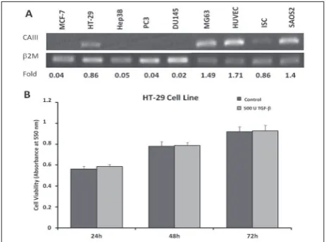
There is limited knowledge of the expression of CAIII in cancer cells. Therefore, our initial aim was to determine the expression profiles of CAIII in different cancer cells, as follows: the human breast cancer cell line (MCF-7), human colon carcinoma cell line (HT-29), human hepatoma cell line (Hep3B), human prostate cancer cell lines (PC3 and DU145), human osteogenic sarcoma cell line (MG-63), human umbilical endothelial cell line (HUVEC), human endometrial adenocarcinoma cell line (Ishikawa) and human osteogenic sarcoma cell line (Saos-2). RT-PCR was performed using 1 µg of RNA after optimizing for non-saturated cycle conditions; β2M was used as an internal control. The resulting bands were analyzed densitometrically. Maximum CAIII mRNA levels were determined, revealing an increase in HUVEC (~1.71-fold), Saos-2 (~1.4-fold) and MG-63 (~1.49-fold) cells, regardless of their ori-gin., HT-29 (~0.86) and ISC (~0.83-fold) indicated the high expression level compared to other cells (Fig. 1A).
Most tumor cells in the neoplastic stage are resist-ant to TGF-β proliferation inhibitory signals. How-ever, TGF-β might exhibit a growth inhibitory effect
through TGF-β receptor I and TGF-β receptor II at the beginning of tumorigenesis. It has been shown that the inhibitory effect of TGF-β on cells leads to loss of TGF-β receptors I and II) [29]. In our study showing TGF-β-related CAIII tissue expression, human colon carcinoma (HT-29) cells were used as model cells. Therefore, we first determined the effect of 500 U/mL of TGF-β in HT-29 cells by the MTT assay. We did not observe any statistically significant growth inhibitory or proliferative effect of TGF-β in HT-29 cells (Fig. 1B).
TGF-β downregulates the expression of CAIII gene in HT-29 cells
In our study, four different CAIII promotor constructs were used as follows: P1: 1025 bp (-939/+86), P2: 785 bp (-699/+86), P3: 322 bp (-236/+86), P4: 194 bp (-108/+86) (Fig. 2A). The basal activities of CAIII promotor
structs in HT-29 cells were higher than in Saos-2 cells (Fig. 2A). This result shows that CAIII is more transcriptionally active in colon carcinoma than in osteosarcoma cells. However, the smallest promoter construct, P4 (194 bp (-108/+86)), had a higher basal activity in Saos-2 cells than in HT-29 cells. This showed that cell type-specific regula-tory elements regulated CAIIIpromoter activity in HT-29 and Saos-2 cells (Fig. 2A).
To the best of our knowledge, nothing is known about cytokine-mediated CAIII expression in CRC. To investigate the role of TGF-β in CAIII gene expression, CAIII promoter fragments were transiently trans-fected to observe transcriptional regulation of CAIII by TGF-β in HT-29 cells. Lucif-erase and SEAP activities were measured 48 h after transfection. We observed that TGF-β decreased the transcriptional activity of all CAIII promoter constructs (Fig 2B). Cells were treated with serum-free media containing 0.5% BSA for 24 h and then with 500 U/mL TGF-β for 48 h. Total RNA was isolated from these cells, and RT-PCR stud-ies were performed with CAIII and β2M as an internal control. As shown in Fig. 2C, the concentration of 500 U/mL TGF-β lowered the level of CAIII mRNA 100-fold compared to the control at all time points.
Immunoblotting was used to observe the effect of TGF-β at the protein level. Cell lysates obtained from cytokine-treated and untreated cell pellets were subjected to Western blotting with rabbit anti-CAIII antibody or β-actin antibody. The level of CAIII protein was decreased, and densito-metric analysis showed that application of 500 U/mL TGF-β at 24 h lowered the level of CAIII protein 1.3-fold (Fig. 2D).
MAPK and PI3K pathways are involved in TGF-β repression of CAIII
TGF-β represses CAIII expression at the mRNA and protein levels. The pathway responsible for this
reduc-tion was examined using specific inhibitors. TGF-β binds to membrane-bound TGF-β receptors I and II, resulting in the activation of the downstream effectors, STAT3, PI3K [30] and MEK pathways. Five hundred U/mL TGF-β was decreased by CAIII mRNA at 6 h. PD98059 and wortmannin partially increased the
reduction of TGF-β-directed CAIII mRNA expression (Fig. 2E). We concluded that TGF-β reduced CAIII mRNA expression via the MEK-1 and PI3K pathways in colon cancer cells.
TGF-β also suppressed CAIII expression in different cancer cells
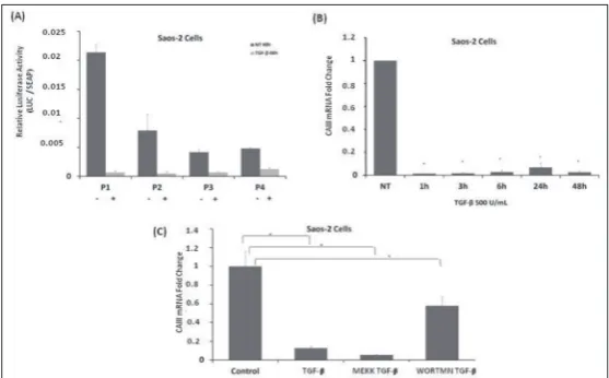
Expression analysis of CAIII mRNA showed that the osteosarcoma Saos-2 cell line expressed a high level of CAIII. Saos-2 cells were used in order to determine whether the contribution of TGF-β to CAIII expres-sion in colon cancer cells was tissue-specific. Studies in this cell line examined transcriptional activity and the level of mRNA. CAIII promoter fragments were transfected into Saos-2 cells, which were then treated with 500 U/mL TGF-β. As shown in Fig. 3A, the ac-tivity of all promoter fragments was reduced in this cell line. To determine the effect of TGF-β on the level of mRNA, total RNA was isolated in transfected and control Saos-2 cells. CAIII mRNA levels displayed a 100-fold reduction after 48 h (Fig. 3B).
As for the pathway inhibition assay, we determined that the TGF-β-directed reduc-tion of CAIII expression uses a different pathway in Saos-2 cells. Treatment with 500 U/mL TGF-β decreased the expression of CAIII mRNA after 6 h. Wortmannin partially recovered the level of CAIII ex-pression, while the MEK inhibitor did not affect the CAIII expression (Fig. 3C). These results indicate that TGF-β represses CAIII mRNA expression via the PI3K pathway in Saos-2 cells.
DISCUSSION
CA enzymes are Zn ion-containing metallo-enzymes that catalyze the reversible reaction of CO2 and HCO3-, which is very important
for physiological and pathophysiological processes. Recent studies have suggested that CAIX and CAXII contribute to cancer progression and can be used as biomarkers in many cancer types. Aberrant expres-sion of CAI, II, CAIV, IX, XII, and XIII has been reported in CRC [15-21,31,32], and CAIX is used as a biomarker for co-lon cancer. CAIV has been identified as a tumor-suppressor gene in colon cancer. In another study, the expression of CA I and II was shown to be important for aggressive distant metastasis in the colon and rectum [33]. However, there is a need to clarify the regulation of each enzyme when different members of the family are thought to play different roles in the cancer process. CAIII is involved in certain cancers such as acute myeloid leukemia and liver carcinoma. Overexpression of CAIII increases the resistance of anticancer medication such as paclitaxel [12]. It was shown in this study that overexpression of CAIII in hepatocellular carcinomas provides the cells with an invasive character through the FAK signaling pathway. However, the role of CAIII in CRC was not elucidated.
In the present study, the role of TGF-β in the regu-lation of the CAIII gene was investigated. Studies have emphasized the heterogeneity of colon cancer cell lines in response to TGF-β due to the presence of different TGF-β receptors [33]. Herein we revealed the effect of TGF-β on colon cancer cells. This protein exerts its
functions by binding to TGF-β type I, type II and type III receptors. It was shown that the inhibitory effect of TGF-β on cells leads to the loss of these cytokine recep-tors (receptor I and receptor II) at the onset of CRC. The TGF-β signal in CRC has not been adequately char-acterized due to the high mutation rates in the TGF-β signaling pathway in human colon cancer cell lines [30]. In our study, TGF-β did not show any statistically significant cytotoxic effects in the related colon cancer model of HT-29 cells. However, TGF-β treatment led to a dramatic decrease in CAIII mRNA and protein expression in HT-29 cells. This decrease was regulated at the transcriptional level, and it could be related to the defense function of CAIII, which was recently re-ported [34]. Results of work on the liver and skeletal muscle suggest that CAIII can function as a scavenger of reactive oxygen species (ROS) [12,35,36]. In fact, CAIII protein has lower hydratase activity than other members of the CA family. In addition, this protein is more resistant to sulfonamides, which are specific CA inhibitors. Therefore, it is thought that CAIII is also involved in other functions in the cell [37-42]. CAIII carries two reactive sulfhydryl groups that are linked to glutathione by disulfide bonds [43,44]. This S-glutathionylation reaction is probably an important component of some sort of cellular defense mechanism that prevents irreversible oxidation of proteins [43,44]. It has also been shown that CAIII plays an antioxidant role in skeletal injury and protects cells from hydrogen peroxide-induced apoptosis [36]. In this respect, TGF-β might downregulate CAIII gene expression to prevent the cell from protecting itself. Thus, CAIII can be a candidate for a tumor-suppressor gene, like the CAIV gene. The downregulation of CAIII by TGF-β is regulated at the transcriptional level, indicating that it is effected via intracellular pathways. Pathway inhibition studies showed that TGF-β uses the MEK and PI3K signaling pathways. TGF-β regulates gene expression via Smad-dependent and Smad-inSmad-dependent routes [29]. TGF-β uses MEK and PI3K signaling pathways through the non-Smad-dependent mechanism in CRC model cells.
To test whether downregulation of CAIII by TGF-β is tissue-specific, osteosarcoma Saos-2 cells were used for TGF-β treatment. The TGF-β superfamily is involved in virtually every aspect of osteoblast cellular activity. Osteoblast differentiation is regulated by the TGF-β signaling pathway [45]. Studies have demonstrated that
members of the CA family of enzymes have also been reported in osteoblasts. CAIX expression was shown to be associated with higher grade tumors, metastasis and poor prognosis of osteosarcoma patients [46]. Ad-ditionally, CAVIII, which does not possess enzymatic activity due to lack of zinc-binding histidine residues, was overexpressed in more aggressive types of human OS cells [47]. Similarly, the same decreasing effect was obtained in the osteosarcoma model, Saos-2 cells. Un-like HT-29 cells, TGF-β used differential pathways in Saos-2 cells. TGF-β repressed CAIII mRNA expression only via the PI3K pathway in Saos-2 cells.
Redox imbalance or oxidative stress results from increased production of reactive oxygen or nitrogen species and a reduced antioxidant capacity [48]. Results obtained in the liver, skeletal muscle and NP (nucleus pulposus) cells suggest that CAIII can function as an oxyradical scavenger [12,34-36]. CAIII protects NP cells from oxidative stress-mediated cell death, medi-ated by Cas3 [36]. On the other hand, TGF-β has been shown to increase ROS production and suppress the antioxidant system, thereby inducing oxidative stress or redox imbalance [48].
To conclude, in both colon cancer and osteosarcoma cells, TGF-β that increased ROS in the cells caused a decrease in CAIII expression that played a role in the mechanism of cell protection from ROS, contributing to the process of carcinogenesis.
Funding: This work was supported by the Turkish Research Council (TUBITAK) project (TBAG 112T669).
Acknowledgments: The authors would like to thanks Dr. Ken-neth Brown and Dr. Dipak Ramji University of Cardiff, UK for the human osteogenic sarcoma cell lines (MG-63 and Saos-2) and Human hepatoma cell line (Hep3B) The authors would like to thank Dr. Kemal Sami Korkmaz, Ege University (EBILTEM) for the prostate cancer cell lines (PC3 and DU145) and human breast carcinoma cell line (MCF-7). The authors would like to thank the Culture Collection of Animal Cells, Foot and Mouth Disease Insti-tute, Ankara (HUKUK) for the human colon carcinoma cell line, HT-29. The authors also wish to thank Dr. Ayhan Bilir, Istanbul University for the Human endometrial adenocarcinoma cell line (Ishikawa) and human umbilical endothelial cell line (HUVEC).
Author contributions: SAT and FK designed the research; DOA and SAT performed the experiments; SAT and FK checked the results; SAT wrote the paper. FK checked the language of the paper.
REFERENCES
1. Balch C, Ramapuram JB, Tiwari AK. The Epigenomics of Embryonic Pathway Signaling in Colorectal Cancer. Front Pharmacol. 2017;8:267.
2. Markowitz SD, Bertagnolli MM. Molecular origins of cancer:molecular basis of colorectal cancer. N Engl J Med. 2009;361(25):2449-60.
3. Vogelstein B, Kinzler KW. The path to cancer–three strikes and you’re out. N Engl J Med. 2015;373(20):1895-8. 4. Wang TL, Rago C, Silliman N, Ptak J, Markowitz S, Willson
JK, Parmigiani G, Kinzler KW, Vogelstein B, Velculescu VE. Prevalence of somatic alterations in the colorectal cancer cell genome. Proc Natl Acad Sci U S A. 2002;99(5):3076-80. 5. Rodriguez-Paredes M. and Esteller M. Cancer epigenetics
reaches mainstream oncology. Nat Med. 2011;17(3):330-9. 6. Jung B, Staudacher JJ, Beauchamp D. Transforming Growth
Factor β Superfamily Signaling in Development of Colorec-tal Cancer. Gastroenterology. 2017;152(1):36-52.
7. Gatza CE, Holtzhausen A, Kirkbride KC, Morton A, Gatza ML, Datto MB, Blobe GC. Type III TGF-β Receptor Enhances Colon Cancer Cell Migration and Anchorage-Independent Growth. Neoplasia. 2011;13(8):758-70 8. Tian M and Schiemann WP.The TGF-β Paradox in Human
Cancer: An Update. Future Oncol. 2009;5(2): 259-71. 9. Li F, Cao Y, Townsend CM Jr, Ko TC. TGF-b Signaling in
Colon Cancer Cells. World J Surg. 2005;29(3):306-11. 10. Grady WM,and Carethers JM. Genomic and epigenetic
instability in colorectal cancer pathogenesis. Gastroenterol-ogy. 2008;135(4):1079-99
11. Dai HY, Hong CC, Liang SC, Yan MD, Lai GM, Cheng AL, Chuang SE. Carbonic Anhydrase III Promotes Transforma-tion and Invasion Capability in Hepatoma Cells Through FAK Signaling Pathway. Mol Carcinog. 2008;47(12):956-63. 12. Roy P, Reavey E, Rayne M, Roy S, Abed El Baky M, Ishii
Y, Bartholomew C. Enhanced sensitivity to hydrogen per-oxide-induced apoptosis in Evi1 transformed Rat1 fibro-blasts due to repression of carbonic anhydrase III. FEBS J. 2010;277(2):441-52.
13. Mohammad HK, Alzweiri MH, Khanfar MA, Al-Hiari YM. 6-Substituted nicotinic acid analogues, potent inhibitors of CAIII, used as therapeutic candidates in hyperlipidemia and cancer, Med Chem Res. 2017;26(7):1397-1404
14. Pinard MA, Mahon B, McKenna R. Probing the Surface of Human Carbonic Anhydrase for Clues towards the Design of Isoform Specific Inhibitors. Biomed Res Int. 2015;2015:453543.
15. Kivela AJ, Saarnio J, Karttunen TJ, Kivelä J, Parkkila AK, Pastorekova S, Pastorek J, Waheed A, Sly WS, Parkkila S, Rajaniemi H. Differential expression of cytoplasmic carbonic anhydrases, CA I and II, and membrane-associated isozymes, CA IX and XII, in normal mucosa of large intestine and in colorectal tumors. Dig Dis Sci. 2001;46(10):2179-86 16. Saarnio J, Parkkila S, Parkkila AK, Haukipuro K,
Pas-toreková S, Pastorek J, Kairaluoma MI, Karttunen TJ. Immu-nohistochemical study of colorectal tumors for expression of a novel transmembrane carbonic anhydrase, MN/CA IX, with potential value as a marker of cell proliferation. Am J Pathol. 1998;153(1):279-85.
17. Niemelä AM, Hynninen P, Mecklin JP, Kuopio T, Kokko A, Aaltonen L, Parkkila AK, Pastorekova S, Pastorek J, Waheed A, Sly WS, Orntoft TF, Kruhøffer M, Haapasalo H, Parkkila S, Kivelä AJ. Carbonic anhydrase IX is highly expressed in hereditary nonpolyposis colorectal cancer. Cancer Epide-miol Biomarkers Prev. 2007;16(9):1760-6.
18. Kivelä A, Parkkila S, Saarnio J, Karttunen TJ, Kivelä J, Park-kila AK, Waheed A, Sly WS, Grubb JH, Shah G, Türeci O, Rajaniemi H. Expression of a novel transmembrane carbonic anhydrase isozyme XII in normal human gut and colorectal tumors. Am J Pathol. 2000;156(2):577-84.
19. Kummola L, Hämäläinen JM, Kivelä J, Kivelä AJ, Saarnio J, Karttunen T, Parkkila S. Expression of a novel carbonic anhydrase, CA XIII, in normal and neoplastic colorectal mucosa. BMC Cancer. 2005;18(5):41.
20. Viikilä P, Kivelä AJ, Mustonen H, Koskensalo S, Waheed A, Sly WS, Pastorek J, Pastorekova S, Parkkila S, Haglund C. Carbonic anhydrase enzymes II, VII, IX and XII in colorec-tal carcinomas. World J Gastroenterol. 2016;22(36):8168-77. 21. Zhang J, Tsoi H, Li X, Wang H, Gao J, Wang K, Go MY, Ng
SC, Chan FK, Sung JJ, Yu J. Carbonic anhydrase IV inhibits colon cancer development by inhibiting the Wnt signalling pathway through targeting the WTAP–WT1–TBL1 axis. Gut. 2016;65(9):1482-93.
22. Kuo WH, Chiang WL, Yang SF, Yeh KT, Yeh CM, Hsieh YS, Chu SC. The differential expression of cytosolic carbonic anhydrase in human hepatocellular carcinoma. Life Sci. 2003;73(17):2211-23.
23. Türkoğlu S, Köçkar F. SP1 and USF differentially regulate ADAMTS1 gene expression under normoxic and hypoxic conditions in hepatoma cells. Gene. 2016;575(1):48-57. 24. Yıldırım H, Kockar F. TGF-b upregulates tumor-associated
carbonic anhydrase IX gene expression in Hep3B cells. Cell Biol Int. 2009;33(9):1002-7
25. Tokay E, Kockar F. Identification of intracellular pathways through which TGF-β1 upregulates URG-4/URGCP gene expression in hepatoma cells. Life Sci. 2016;144:121-8. 26. Livak K.J, Schmittgen TD. Analysis of relative gene
expres-sion data using real-time quantitative PCR and the 2 (−Delta Delta C(T)) Method. Methods. 2001;25(4):402-8.
27. Laemmli UK. Cleavage of structural proteins during the assembly of the head of bacteriophage T4. Nature. 1970;227(5259):680-5.
28. Alper M, Kockar F. IL-6 upregulates a disintegrin and metal-loproteinase with thrombospondin motifs 2 (ADAMTS-2) in human osteosarcoma cells mediated by JNK pathway. Mol Cell Biochem. 2014;393(1-2):165-75
29. Massagué J. TGFβ in Cancer. Cell. 2008;134(2):215-30 30. Zanetti D, Poli G, Vizio B, Zingaro B, Chiarpotto E, Biasi F.
4-Hydroxynonenal and transforming growth factor-b1 expres-sion in colon cancer. Mol Aspects Med. 2003;24(4-5):273-80 31. Yang GZ, Hu L, Cai J, Chen HY, Zhang Y, Feng D, Qi CY,
Zhai YX, Gong H, Fu H, Cai QP, Gao CF. Prognostic value of carbonic anhydrase II expression in colorectal carcinoma. BMC Cancer. 2015;15:209.
33. Bekku S, Mochizuki H, Yamamoto T, Ueno H, Takayama E, Tadakuma T. Expression of carbonic anhydrase I or II and correlation to clinical aspects of colorectal cancer. Hepato-gastroenterology. 2000;47(34):998-1001.
34. Silagi ES, Batista P, Shapiro IM, Risbud MV. Expression of Carbonic Anhydrase III, a Nucleus Pulposus Phenotypic Marker, is Hypoxia-responsive and Confers Protection from Oxidative Stress-induced Cell Death. Scientific Reports. 2018;8:4856.
35. Zimmerman UJ, Wang P, Zhang X, Bogdanovich S, Forster R. Anti-Oxidative Response of Carbonic Anhydrase III in Skeletal Muscle. IUBMB Life. 2004;56(6):343-7.
36. Räisänen SR, Lehenkari P, Tasanen M, Rahkila P, Härkönen PL, Väänänen HK. Carbonic anhydrase III protects cells from hydrogen peroxide-induced apoptosis. Faseb J. 1999:13(3):513-22
37. Feng HZ, Jin JP. Carbonic Anhydrase III Is Expressed in Mouse Skeletal Muscles Independent of Fiber Type-Specific Myofilament Protein Isoforms and Plays a Role in Fatigue Resistance. Front Physiol. 2016;7:597.
38. Cabiscol E, Levine RL. The phosphatase activity of carbonic anhydrase III is reversibly regulated by glutathiolation. Proc Natl Acad Sci USA. 1996;93(9):4170-4
39. Chai YC, Jung CH, Lii CK, Ashraf SS, Hendrich S, Wolf B, Sies H, Thomas JA. Identification of an abundant S-thiolated rat liver protein as carbonic anhydrase III; characterization of S-thiolation and dethiolation reactions. Arch Biochem Biophys. 1991;284(2):270-8
40. Carter ND, Hewett-Emmett D, Jeffrey S, Tashian RE. Testos-terone-induced, sulfonamide-resistant carbonic anhydrase isozyme of rat liver is indistinguishable from skeletal muscle carbonic anhydrase III. FEBS Lett. 1981;128(1):114-8.
41. Wistrand PJ. Carbonic anhydrase III in liver and muscle of male rats purification and properties. Ups J Med Sci. 2002;107(2):77-88.
42. Sanyal G, Swenson ER, Pessah NI, Maren TH. The carbon dioxide hydration activity of skeletal muscle carbonic anhy-drase. Inhibition by sulfonamides and anions. Mol Pharma-col. 1982;22(1):211-20.
43. Lii CK, Chai YC, Zhao W, Thomas JA, Hendrich S. S-thio-lation and irreversible oxidation of sulfhydryls on carbonic anhydrase III during oxidative stress: a method for studying protein modification in intact cells and tissues. Arch Bio-chem Biophys. 1994;308(1):231-9.
44. Thomas JA, Poland B, Honzatko R. Protein sulfhydryls and their role in the antioxidant function of protein S-thiolation. Arch Biochem Biophys. 1995;319(1):1-9.
45. Yang R, Piperdi S, Zhang Y, Zhu Z, Neophytou N, Hoang BH, Mason G, Geller D, Dorfman H, Meyers PA, Healey JH, Phinney DG, Gorlick R. Transcriptional Profiling Iden-tifies the Signaling Axes of IGF and Transforming Growth Factor-b as Involved in the Pathogenesis of Osteosarcoma. Clin Orthop Relat Res. 2016;474(1):178-89.
46. Park HY, Seo J, Bacchini P, Bertoni F, Park YK. Expres-sion of Carbonic Anhydrase IX Correlates with Histologic Grade and Metastasis in Osteosarcoma. Korean J Pathol. 2010;44(4):384-9.
47. Wang TK, Lin YM, Lo CM, Tang CH, Teng CL, Chao WT, Wu MH, Liu CS, Hsieh M. Oncogenic roles of carbonic anhydrase 8 in human osteosarcoma cells. Tumour Biol. 2016;37(6):7989-8005.