CRUSH SYNDROME INFLUENCE ON THE ULTRASTRUCTURE
OF HEPATOCYTES AND NEURONES
K
arapetyan, G. R.
[a], Kukurtchyan, N. S.
[a], Kevorkian, G. A.
[a]Keywords: Crush syndrome, PRP-1, TEM, neurone, hepatocyte, mitochondria (Mch).
The aim of this study was to find the dynamic of ultrastructural changes of liver at CS and the role of PRP-1 (proline rich peptide) as a protector. Our investigation demonstrated that Mch (mitochondria) of brain are more subject to changes at 2 h of compression than liver Mch, but one time administration of the PRP-1 in a dosage of 10 g/100 g of the animal weight before decompression period prevent the development of the destructive changes of the hepatocytes of the liver and neurons of the rat cortex (injection of PRP on the background of CS prevent progress of ultrastructural changes typical at 4 h of decompression, and leads to Mch fission).
Corresponding Authors: Karapetyan G.R. Fax: +374 -10-28 19 51
E-Mail: [email protected]
[a] H. Buniatian Institute of Biochemistry, of National Academy of Sciences of Republic Armenia, Paruir Sevag str. 5/1, 0014, Yerevan, Republic of Armenia.
INTRODUCTION
Earthquake is one of the most dangerous nature cataclysms.1 Sudden buildings destroy as well as the
damages and death of the people caused by the earthquake is more than 90 %. At last 20 years developed the knowledge basis and the tactics chose at such cases.2 In
current study we were chosen the experimental model of traumatic stress - crush syndrome (CS).3
CS is a special type of traumatic injury of the organism with a specific clinical development of the pathogenesis with a high lethality (up to 70 %).4
Today one of the most actual questions is studying the pathology of Crush syndrome (CS) and searching the ways prevent the metabolism disturbances caused by toxins formed in ischemic muscle during decompression. CS follows by acute hemodynamic shock, myoglobinuria, acute renal insufficiency and lethal endotoxicity.5
CS is characterized by serious changes in the most parameters of homeostasis, which lead to improve of the patients’ treatment. It is already developed the general conceptions of the medical help to the patients with CS. However it could lead to complications especially in excretory and detoxification functions of the organism. There are numerous data indicating that the main intoxication of the organism occurs during decompression, in which toxic metabolic products (toxic peptides) as product from proteolisis of myoglobin are released into the blood from damaged tissue and kidney and accumulated at the targeted organ. Liver damages are wide-spread cause of people disease and death.6-8
An assumption may be suggested that the ultrastructural manifestations of the cell pathology of various organs may occur in all the stages of developing crush syndrome. The
manifestations mentioned have to do both with the cells' and non-cellular structures' components of membranous structure (plasmolemma, mitochondria, nuclear shell) and those of non–membranous structure.
In this work as a protector we have chosen the prolin rich peptide (PRP-1) which is an immunmodulator and neuroprotector with a wide profile of action.9 The
following important properties of PRP-1 were identified: inhibition of the proappoptotic capasas 3 and 9, activation of 2 and 6 capasas,10 stimulation of the immuncompetetnt
cells (Т, В and macrofagus),11 involving in the
mechanisms of interleukin expression (TNF, IL-1, IL-6) in fibroblasts, macrophages and astrosites, neuroprotection against many toxic products produced in organism, has an antimicrobial and antivirus influence.12
As at CS we observe the toxic products produced by organism itself (in the ischemic muscle during compression and released into the blood stream during decompression), so in this model, we decided to use PRP-1, taking into consideration all its properties, especially its neuroprotection effect against many toxic products produced in the organism.
Experimentally proved that under compression, white rats that received intraperitoneal PRP-1 before decompression period survived up to 100% (without PRP-1 administration the death of the animals was up to 25-30%), and the damage corrected by PRP-1, in some cases reached to intact level.
It was shown that PRP lead to increase of glucose utilization in the brain during different periods of decompression, which was different in the myocardium and kidneys.
Previous morphological studies were carried out with an hour compression and 1-3 days decompression period, respectively. However, the question of the brain and liver ultrastructure at longer period of compression still remains unknown.
DOI: 10.17628/ECB.2013.2.726
MATERIAL AND METHODS
Reagent: powder paraformaldehyde; OsO4; Sodium
cacodylate trihydrate; 90º ethyl alcohol, acetone, Epon 812, Epon Hardener MNA, Epon Hardener DDSA, Epon accelerator DNP-30, uranil acetate, citrate Na, Nitrate Pb, photo plates.
All reagent used were of analytical grade and purchased from Sigma Chemical Co. (USA).
Animals: All procedures involving animals were
approved by the Institutional Review Board/ Institutional Animal Care and Use Committee (H. Buniatian Institute of Biochemistry, Yerevan, NAS RA) conformed to the European Communities Council directives (86/609/EC). For all experiments, two-month-old male rates weighing 150-200 g obtained from our breeding colony were used. Animals were maintained at normal room temperature with free access to food and tap water. The experimental model of Crush syndrome (CS) was induced by compression of femoral soft tissues using a special press with a compression force 100 kg kg-1 of body weight for 2
h.
Rats were divided randomly into four groups (n=5 in each group). Group I – intact animals; Group II – control (2 h compression); and 2 experimental Groups (III and IV): 4 h decompression; PRP-1 injection and 4 h decompression at the end of using PRP-1 (10 g/100 g of animal weight).
Treatment of material: The bioptates taken
immediately after decapitation put in cold 4 ºC mix of paraformaldehyde on cacodilate buffer and glutaraldehyde for 12 hours with following post fixation in 1 % OsO4
solution during 2 hours; dehydratation in ascending series of spirits; saturation in a mixture of acetone and epon resins of different proportions and pouring in gelatinous capsules into epon.
Obtaining of ultrathin slices and its treatment: The ultrathin slices (up to 500 Å) were made using ultracut LKB (Swedish) and Reichert (Austria). Ultrathin slices were double contrasted with uranil acetate and lead citrate.
Observation under TEM: Obtained ultrathin slices
were observed under the transmission electron microscope (Phillips CM 10) with resolution X 10-20.000.
Statistical Analysis: Data were expresses as the mean ± S.E.M. All data were analyzed using a one-way analysis of variance (ANOVA) (SigmaStat 3.5 for Windows). Differences were considered as significant at P 0.05.
RESULTS
As have shown the results of our study, in control group of animals (2 h compression) the ultrastructural changes of neurons of cortex and hepatocytes of liver (in particular Mch, Nc, GER and SER) are quite different from ultrastructure of intact group. Thus the specific alterations between organelles of neurons and hepatocytes are also observed. So, it was shown that GER of neurons is in
process of fragmentation and vesiculation, while in hepatocytes it is presented as separate tubules fragments. This indicates on the fact of hypersensibility of cells to the influence of catecholamine’s level increasing caused by stress.
Endoplasmatic reticulum is one of the major organelles demonstrate expressed symptoms of a stress and dysfunction at pathology.13 Disfunction of ER can cause to
organ dysfunction at a strong inflammation caused by endotoxins.14
As about Mch, at 2 h compressions of soft tissues, in neurons of a brain the tendency of 2-3 organelles fusion is observed (Fig. 1). Thus the cristae are of varies shapes: tubular or in a process of fragmentation.
Figure 1. The ultrastructure of neurons at 2 h compression. X 20.000.
In hepatocytes of liver Mch are in groups where the organelles are close contacted to each other, thus no fusion is observed. As about inner membrane of Mch, we can indicate that cristae are generally tubular, practically no process of fragmentation is observed.
Мch are also critical metabolic organelles known as «power system of cells».15,16 In addition to their role in
cellular bio-energetic, Mch initiate the general forms of the programmed cellular death (apoptosis) through realizing of proteins, such as cytochrome C from an inner membrane.17
Hepatocytes consist double nuclei, compare to neurons of cortex.
In the first experimental group of animals (2 h compression - 4 h decompression) the ultrastructural changes are developed acutely both in neurons, and in hypatocytes.
As have shown the results of our study in the following group total GER vesiculation process is observed, while SER is presented both in a type of fragmented tubules and by fields consists of vesicules.
Such organelles condition at 4 h decompression caused by the influence of toxins and toxic peptides,18 produced
Such toxic peptides composed of 5-7 amino acids realize into the blood and reach to the target organs lead to the destructive changes and functional disorders.
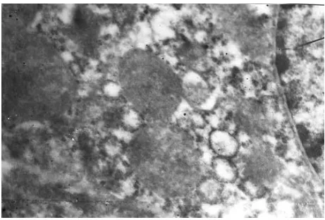
It’s important to mention that Mch of cortex can form conglomerates consist of a great number of fussed Mch. Mch matrix has a swollen character. The inner membrane of Mch also has specific alterations. The number of tubular cristae decreased, as well as the number of fragmented cristae, however the process of vesiculation is presented. The Mch of liver hepatocytes are also significantly altered. At 4 h decompression the Mch fuse and the tendency of megamitochondria formation is observed (Fig. 2).
The matrix of Mch is in the process of swollen. The cristae are presented as in a form of tubuls as well as in fragmentation type; it is shown the increasing process of vesiculation tendency.
Increasing of the Mch fussion and fission processes lead to desorganisation of inner membrane of Mch and formation of the tubular and vesiculated cristae.
The ultrastructural architecture of the Mch mostly depends on the quantity and form of cristae. A great number of criastae is observed in tissues with a high need in energy. Such variety in cristae architecture could be explained by different metabolism of organells.23
As about nuclei we have to mention that as in neurons and in hepatocytes they are swollen with diffused chromatin.
As have shown the results of our study in the second experimental group of animals (2 h compression- PRP-1 injection – 4 h decompression) no ultrastructural changes occur in hepatocytes as well as in neurons. It was shown , that one time PRP-1 administration directly in muscle before decompression neutralize the toxic peptides produced in the ischemic muscle during compression. And inhibit its negative influence on the target organs, in our case on liver and cortex. It was shown that GER in neurons is in a form of tubules and vesicles, while in hepatocytes it’s presented by nets. This indicate on the positive effect of PRP-1 on both organs, however its better occurred in hepatocytes, which is caused by sensitivity of neurons to catecholamine’s influence, while hepatocytes are less sensitive.
Compare with the Mch of the second group the organelles of the first group do not form conglomerates, and in spite of the fact that there are still observed the fussed organelles, but the neurons with single Mch are more common (Fig. 3).
It is important to mention about the influence of the PRP-1 on the inner membrane of Mch. Based on the dates obtained via electron grammas (from tissues and fraction) we indicate increasing of the quantity of tubular and decreasing of vesicular cristae.
As about the Mch of liver hepatocytes we have to say that single injection of PRP-1 lead to the positive changes in the ultrastructure of Mch. It was shown that Mch are closely contact to each other however no process of fusion Figure 2. Megamitochondria formation in hepatocytes at 4 h decompression. X 60.000.
DOI: 10.17628/ECB.2013.2.726 was observed. The cristae of Mch were presented in
tubules.
DISCUSSION
Today one of the most actual questions is the nature of the toxic damages of organs. It could have as direct and indirect effect, be a result of acute and chronic influence.7,24-26
Organs toxic injuries are widespread cause of the diseases and death in populations.6,27-31
The detoxication reactions are performed by the ferments of endoplasmatic reticulum and Mch,32 that’s
why any changes in its ultrastructure could have negative effect on the detoxication function of the liver and on the whole its function and on organism. The role of Mch in detoxication is presented by the use of fluorescent microscopy.32
The influence of hepatotoxic matter lead to damages of parenchyma of liver and destroying of its metabolic fermentative functions. One of the common reasons of death at CS is hyperglycemia at decompression period. At CS develop the immobilization stress activated by toxic peptides form in ischemic muscle.33 At the compression
period the catecholamine level increase which leads to cramp of arterioles and precapillaries lead to falling down the speed of blood flow, endovascular aggregation of erythrocytes and development of thrombus formation. At post compression period toxic products of different etiology produced in ischemic muscles and kidneys realized to the blood and spread to the organism. Such toxic peptides (the toxic products of methabolism) consist of 5-9 amino acids. Which of these toxic peptides is more aggressive from toxical point of view is still unknown.18
As have shown the results of our study at CS take place the process of aggregation of erythrocytes in sinusoids, which become more critical at 4 h decompression period. Progression of microcirculation dysfunction lead to development of acute hypoxia and as result to ischemia of different organs and tissues.19-21 Ischemic damage of
different organs and tissues is still one of the most actual problems in medicine and biology. One of the most important factors in the pathogenesis of CS is cells energy deficit, which lead to disruption of intra and extra cellular transport, as well as damage of protein synthesis apparatus of cells.3,34 Typical for the 24 h decompression after 1 h
compression is damages of the organelles ultrastructure, such as destroying of cristae of Mch. Fragmentation of GER, observation of ribonucleotides masses in cytoplasm as well as partial necrosis foci in hepatocytes.
According to literature dates obtained by different authors, it could be mentioned that alterations ultrastructure of Mch indicate the functional condition of the organelles, depend on the level of energy metabolism.35,36 Mch change their forms dynamically by
fusion and fission processes.37
Structural changes of Mch divided into 2 main categories: simple swelling38 and formation of mega
mitochondria (ММ).39,40 There are different points of view
concerning the ways of MM formation. Some authors consider, that MM form as a result of nearby Mch fusionх,41-43 the others suggest that MM are formed by
growth of single Mch, and not by fusion of small Mch.44
The process of formation of atypical by the structure and size Mch in cells are related to the different diseases or physiological condition of the organism, such as erythroleicosis in bone marrow, in endometric cells before ovulation process, etc.40,45 For each disease Mch have its
unique typical just for current disease form. However such giant Mch could accumulate in post mitotic cells depend on the disease type, as well as on the age dependant alterations. It must be mentioned that such organelles have a low inner membrane potential and have no ability to fuse to the other giant or normal Mch.46 As have shown the
results of our study the morphology of Mch is different depend on CS. So, at 2 h compression there is no viewable alteration in Mch structure however their physiological condition is changed. They are presented in groups where the organelles are closely contacted to each other. As a response to the pain the level of catecholamine in blood increase making Mch more tension, which could be explained by increasing of energy need in hepatocytes. However, increasing of catecholamine level in blood does not influence on the structural safeness of Mch. At 4 h decompression organelles condition become more critical. In spite of the fact that organelles are presented as groups, the size and forms of Mch are very different, and the polymorphism of Mch is observed. Organelles are swollen; in some cases the tendency of MM formation is observed. In literature dates is known that at ischemia, partial hepatoectomia etc, take place the process of Mch swelling.38 It must be mentioned that 24 h after
hepatoectomia not only swelling of Mch but also cristae number reducing tendency is observed, which however recovered 96 hr, after partial hepatoectomia.47 So if at 2h
compression cristae had tubular or vesicle type, then at 4h decompression it is observed almost completely reduction of the cristae, which has its influence on the functional activity of Mch. It’s known that at chronically liver diseases take place MM formation with reduction of cristae.48 However at 4 h decompression at CS the
influence of stress factor has a short time and unlike the dates mentioned above we observe just a tendency of MM formation with reduction of cristae, while the normal size Mch with reduced cristae are often observed, which is suggested as an nonspecific adaptive cellular response to the influence of toxin produced by ischemic muscles. It is well known that alterations in Mch ultrastructure such as remodeling of cristae are included in initiation of cell death.49 Mch lost their cristae normal morphology can’t
realize their function and falling out of cell energy supplying function, lead to disruption of energy dependent processes of metabolism such as liver detoxication function.
presented by whole nets, while in CS group there was a fragmentation and vesiculation process.
The results of present study update the information about the influence of traumatic stress on the ultrastructure of neurones and hepatocytes, as well as give new dates about PRP-1 as a corrector. Obtained dates have an important theoretical significance because of the increasing number of toxically injures in population. Morphological reconstruction observed at different stresses indicate to the necessity of new drugs developments lead to prevent neurons and hepatocytes ultrastructure injures.
CONCLUSION
CS as a specific type of traumatic damages lead to the whole complex of ultrastructural pathology. Our investigation demonstrated that Mch of brain are more subject to changes at 2 h of compression than liver Mch, but one time administration of the PRP-1 in a dosage of 10 g/100g of the animal weight before decompression period prevent the development of the destructive changes of the hepatocytes of the liver and neurons of the rat cortex (injection of PRP on the background of CS prevent progress of ultrastructural changes typical at 4 h of decompression, and leads to Mch fission).
REFERENCES
1Briggs, S. M., Surg. Clin. North Am., 2006, 86(3), 537-44. 2Ashkenazi, I., Isakovich, B., Kluger, Y., Alfici, R., Kessel, B.,
Better, O. S., Prehosp. Disaster Med., 2005, 20(2), 122-133.
3Kevorkian, G. A., Kanayan, A. S., Voskanian, L. H.,
Khachatrian, H. F., J. Neurochem, 1998, 71, 70.
4Wang, L., He, Q., Li, G. S., Zhongua Neike Zazhi, 2008, 47(9),
711-4.
5Kevorkian, G. A., Guevorkian, A. G., Kukurtchyan, N. S.,
Galoyan, A. A., J. Neurochem.,2003, 87(1), 145-149.
6Lewis, J. H., Curr. Pract. Med., 1999, 2, 49-58.
7Mochizuki, M., Shimizu, S., Urasoko, Y., Umeshita, K.,
Kamata, T., Kitazawa, T., Nakamura, D., Nishihata, Y., Ohishi, T., Edamoto, H., J. Toxicol Sci.,2009, 34(2), 175-81.
8Zimmerman, H. J., Maddrey, W. C., Diseases of the liver, 7th ed.
Schiff, L. and Schiff, E. R. eds., 1993, 707-783.
9Galoyan, A. A., Proc. Int. Conf Biochem. Mol. Biol., Aspects of
the Brain Immune System. Sep. 15-19, Yerevan-Tsakhkadzor, Encyclopedia Armenica, Armenia, 2001, 22-34.
10Galoyan, A. A., Terio, T., Berg, M., Marks, N., Neurochemistry
(RAS and NAS RA), 2000, 17, 185-188.
11Galoyan, A. A. and Aprikian, V. S., Neurochem. Res.,2002, 27,
305-312.
12Galoyan, A. A., Neurochem. Res., 2000,25, 1343-1355. 13Margaret G. Gregor and Gokhan S. Hotamisligil., The Journal
of Lipid Research,2007, 48, 1905-1914.
14Kozlov A. V., Duvigneau JC, Miller I., Nurnberger S.,
Gesslbauer B., Kungl A., Ohlinger W., Hartl RT., Gille L., Staniek K., Gregor W., Haindl S., Redl H., Biochim Biophys Acta.,2009, 1792 (6): 521-30.
15Wei, Y., Wang, D., Topczewski, F., and Pagliassotti, M. J., Am
J. Physiol. Endocrinol. Metab., 2006, 291(2), E275-81.
16Wei Y., Rector, R. S., Thyfault, J. P., Ibdah, J. A., World J.
Gastroenterol., 2008, 14(2), 193-9.
17Sun, M. G., Williams, J., Munoz-Pinedo, C., Perkins, G. A.,
Brown, J. M., Ellisman, M. H., Green, D. R., Frey, T. G.,
Nat. Cell Biol., 2007, 9(9), 1057-65.
18Kevorkian, G., Kanayan, A., Hayrapetyan, H., Guevorkian A.,
Melkonyan, L. and Galoyan, A.. FEBS J.,2006, 273, 255.
19Lameire, N. H., De Vrise, A. S., Vanholder, R. Curr. Opin. Crit.
Care, 2003, 9(6), 481-490.
20Malinoski, D. J., Slater, M. S., Mullins, R. J., Crit. Care Clin.,
2004, 20(1), 171-192.
21Ismailov, R. M., Shevchuk, N. A., Khusanov, H., Biomed. Eng.
Online, 2005, 4(1), 24.
22 Gevorkian R., Melkonyan L., Hayrapetyan H., Guevorkian A,
Galoyan A., The FEBS Journal,2006,273, 77-78.
23John, G. B., Shang, Y.-L., Li, L., Renken, C., Mannella, C. A.,
Selker, J. M. L., Rangell, L., Bennett, M. J. and Zha, J.-P.,
Mol. Biol. Cell, 200516(3), 1543-1554.
24Maddrey WC, J. Clin. Gastroenterol., 2005, 39 (4 Suppl 2):
S83-9.
25Doi H., Horie T., Chem. Biol. Interact., 2010, 183 (3),
363-368.
26 Kalender S., Uzun FG., Durak D., Demir F., Kalender Y.,
Food and Chemical Toxicology,2009,48 (2), 633-638.
27Bass, N. M., Ockner, B. A., Hepatology: a textbook of liver
disease, 3rd ed., Philadelphia,1996, 962-1017.
28Holland, E. G., Degruy, F. V., Am. Fam. Physician. 1997, 56(7),
1781-1792.
29Schiodt, F. V., Rochling, F. A., Casey, D. L., Lee, W. M., New
Engl. J. Med., 1997, 337(16), 1112-1117.
30Plevris, J. N., Schina, M., Hayes, P. C., Aliment. Pharmacol.
Ther.,1998, 12(5), 405-418.
31Bond, G. R., Hite, L. K., Acad. Emerg. Med., 1999, 6(11),
1115-1120.
32 Fellous, R., Coulaud, D., El Abed, I., Roques, B. P., Le Pecq,
J. B., Delain, E., Gouvette, A., Cancer Res.,198848(22),
6542-9.
33 Rawlins, M., Gullichsen, E., Kuttila, K., Peltala, O., Niinikoski,
J., Eur. Surg. Res., 1999, 31(5), 9-18.
34 Mikaelian, N. P. Pathol. Physiol. Exp. Ter.,1992, 1, 19-21. 35 Hackenbrock C. R., J. Cell Biology, 1968, 37(2), 345-369. 36 Guenthard, J., Wyler, F., Fowler, B., Baumgartner, R., Arch.
Dis. Childh., 1995, 72(3), 233-236
37Huang P., Yu T., Yoon Y., Eur J Cell Biol.,2007, 86 (6),
289-302.
38Wilasrusmee, C., Siritheptawee, S., Kanchanapanjapon, S.,
Sopon, P., Vanichanon, C., Limpthong, W., Pongchailerks, P., Lertsithichai, P., Wilasrusmee, S., Kittur, D. S., J. Hepatobil. Pancreat. Surg., 2004, 11(4), 266-71.
39Zamora SA., Pinto A., Scott RB., Parsons HG., J Inherit Metab
Dis.,1997,20 (4), 509-16.
40Guastadisegni, C., Balduzzi, M., Mancuso, M. T., Di Consiglio,
E., J. Toxicol. Environ. Health A., 1999,57(6), 415-29
41Tandler, B., Dunlap, M., Hoppel, C. L., Hassan, M., Ultrastruct.
Pathol., 2002, 26(3), 177-83.
42Wakabayashi, T., Horiuchi, M., Sakaguchi, M., Misawa, K.,
Onda, H., Iijima, M., Allmann, D. W., Eur J. Biochem.
1984, 143(2), 455-65.
43Bakeeva, L. E., Manteifel', V. M., Rodichev, E. B., Karu, T. I.,
Mol. Biol. (Moscow), 1993, 27(3), 608-17.
DOI: 10.17628/ECB.2013.2.726
45Asano, M., Wakabayashi, T., Ishikawa, K., Kishimoto, H., Acta
Path. Jap., 1978, 28, 205-213.
46Navratil,M., Terman, A., Arriaga, E. A., Exp. Cell Res., 2008,
314(1), 164-72.
47Ferri, D., Moro, L., Mastrodonato, M., Capuano, F., Marra, E.,
Liquori, G. E., Greco, M., Biol. Cell., 2005, 97(4), 277-88.
48Krähenbühl, S., Pharmacol. Ther., 1993, 60(1), 1-38.
49Cheung, E. C., McBride, H. M., Slack, R. S., Apoptosis,2007,
12(5), 979-92.