University of South Carolina
Scholar Commons
Theses and Dissertations
12-14-2015
The Role of Ovarian Function in the Progression of
Cachexia in the APC
MIN/+
Mouse
Kimbell Louise Hetzler
University of South Carolina - Columbia
Follow this and additional works at:https://scholarcommons.sc.edu/etd Part of theMedicine and Health Sciences Commons
This Open Access Dissertation is brought to you by Scholar Commons. It has been accepted for inclusion in Theses and Dissertations by an authorized administrator of Scholar Commons. For more information, please [email protected].
Recommended Citation
T
HER
OLE OFO
VARIANF
UNCTION IN THEP
ROGRESSION OFC
ACHEXIA IN THEA
PCMIN/+M
OUSEby
Kimbell Louise Hetzler
Bachelor of Science Clemson University, 2010
Submitted in Partial Fulfillment of the Requirements
For the Degree of Doctor of Philosophy in
Exercise Science
The Norman J. Arnold School of Public Health
University of South Carolina
2015
Accepted by:
James A. Carson, Major Professor
J. Larry Durstine, Committee Member
J. Mark Davis, Committee Member
E. Angela Murphy, Committee Member
ii
iii
A
CKNOWLEDGEMENTSThe body of work represented in this dissertation would have been impossible to
achieve without the love and support of my fiancé, Brian Bell, and my parents, Dale and
Susan Hetzler. I am indebted to them for standing by me through the figurative and
literal blood, sweat, and tears over the past five years.
I am also indebted to my advisor, Dr. James Carson, who has guided me through
this journey with patience and understanding; and who has pushed me to do work of a
higher caliber and quality than I believed possible.
I would also like to thank my dissertation committee: Dr. J. Larry Durstine, Dr. E.
Angela Murphy, and Dr. J. Mark Davis. Each has given willingly of their time, effort,
and wisdom in order to help guide me through this process.
Aside from these key people, making it to this point in my doctorate career would
have been impossible without help from the USC Counseling and Human Development
Center, motivation and further counseling from my former swimming coach Pat Murphy,
Hi-Caf Teas, Immaculate Consumption’s “Motivator™,” and the soothing sounds of
Pandora’s Stevie Nicks Radio.
And of course, the hundreds of mice that have sacrificed their lives and tissues for
iv
A
BSTRACTCachexia is a devastating a life-threatening condition that occurs secondary to
underlying disease including cancer, AIDS, COPD and comprises severe loss of muscle
and fat mass. Muscle mass atrophy and wasting with cachexia is especially dire, as
skeletal muscle mass is associated with quality of life, functionality, and ability to
respond to chemotherapeutics. While much investigation has been done in the male
animal to elucidate the inflammatory pathways and muscle signaling underlying
cachexia, very little work has occurred in the female. The overall purpose of this study is
to determine if ovarian function can alter cachexia progression in the female ApcMin/+
mouse through IL-6 signaling and the regulation of skeletal muscle metabolism. Specific
aim #1 sought to determine the relationship between circulating IL-6 and cancer cachexia
progression in the female ApcMin/+mouse. We found that the canonical IL-6 signaling
pathway that is a key point of regulation in the male ApcMin/+is dysregulated in the
female, and that IL-6 levels do not correlate with body weight loss and severity as they
do in the male. Specific aim #2 sought to determine whether ovarian function loss or
dysfunction could influence IL-6 regulation of cancer cachexia progression in the female
ApcMin/+mouse. The loss of ovarian function due to disease did cause an increase in
Il-6-related and other inflammation, while ovariectomy (OVX) alleviated much of the
related inflammation. Specific aim #3 sought to determine whether
cachexia-induced skeletal muscle metabolic dysfunction is regulated by ovarian function in the
v
dysregulation increased with increasing cachexia severity; however, OVX brought these
measures back towards baseline. These findings provide insight into the intricate
regulation of cachectic pathways by ovarian endocrine function, and will provide
vi
T
ABLE OFC
ONTENTSACKNOWLEDGEMENTS ... iii
ABSTRACT ... iv
LIST OF TABLES ... ix
LIST OF FIGURES ...x
LIST OF ABBREVIATIONS ... xii
CHAPTER 1:INTRODUCTION ...1
CHAPTER 2:REVIEW OF LITERATURE ...8
2.1COLON CANCER ...9
2.2CANCER CACHEXIA ...9
2.3APCMIN/+MOUSE MODEL ...11
2.4CACHEXIA AND SKELETAL MUSCLE ...11
2.5IL-6 IN TUMORIGENESIS AND CACHEXIA ...13
2.6SEX DIFFERENCES IN CANCER AND CACHEXIA ...16
2.7OVARIAN FUNCTION ...17
2.8OVARIAN FUNCTION AND IL-6 ...23
2.9OVARIAN FUNCTION AND CANCER ...26
2.10OVARIAN FUNCTION AND SKELETAL MUSCLE ...27
vii
CHAPTER 3:SEX DIFFERENCES IN THE RELATIONSHIP OF IL-6SIGNALING TO CANCER
CACHEXIA PROGRESSION ...30
3.1ABSTRACT ...31
3.2INTRODUCTION ...32
3.3METHODS ...34
3.4RESULTS ...40
3.5DISCUSSION ...46
3.6ACKNOWLEDGEMENTS ...51
3.6FIGURE LEGENDS...55
CHAPTER 4:THE ROLE OF OVARIAN FUNCTION IN IL-6REGULATION OF INFLAMMATION DURING CANCER CACHEXIA PROGRESSION ...62
4.1ABSTRACT ...63
4.2INTRODUCTION ...64
4.3METHODS ...68
4.4RESULTS ...75
4.5DISCUSSION ...83
4.6ACKNOWLEDGEMENTS ...88
4.7FIGURE LEGENDS...93
CHAPTER 5: CACHEXIA-INDUCED MUSCLE METABOLIC DYSREGULATION IN THE FEMALE ...105
5.1ABSTRACT ...106
5.2INTRODUCTION ...107
5.3METHODS ...110
5.4RESULTS ...115
viii
5.6ACKNOWLEDGEMENTS ...123
5.7FIGURE LEGENDS...126
CHAPTER 6:OVERALL DISCUSSION ...136
REFERENCES ...144
APPENDIX A–DETAILED METHODS ...164
APPENDIX B–PROPOSAL ...184
APPENDIX C–RAW DATA ...223
ix
L
IST OFT
ABLESTable 3.1 Characteristics of female ApcMin/+mice ...52
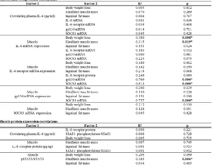
Table 3.2 Relationships between circulating IL-6 and skeletal muscle signaling during cachexia development ...53
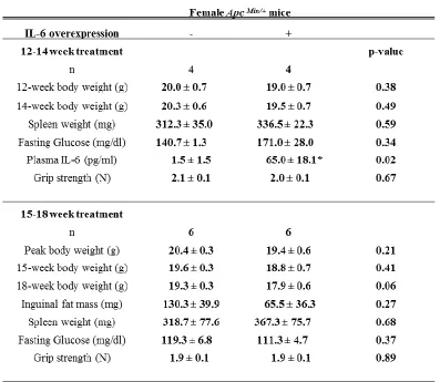
Table 3.3 Characteristics of 14-week and 18-week female ApcMin/+mice with IL-6
overexpression ...54
Table 4.1 Characteristics of female ApcMin/+ mice stratified by ovarian status ...89
Table 4.2 Characteristics of 18-week female C57BL/6 and ApcMin/+mice with and
without ovaries intact ...90
Table 4.3 Circulating cytokine levels in mice with present and absent estrus cycling ...91
Table 4.4 Effects of ovariectomy and IL-6 overexpression in 15-week female ApcMin/+
mice ...92
Table 5.1 Characteristics of 18-week female B6 and ApcMin/+ mice stratified by
cachexia severity ...124
x
L
IST OFF
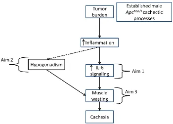
IGURESFigure 1.1 Overall Working Model ...6
Figure 3.1 Characteristics of male and female ApcMin/+mice ...57
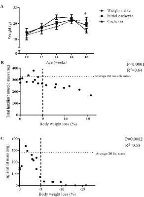
Figure 3.2 Cachexia progression in female ApcMin/+mice ...58
Figure 3.3 Muscle IL-6 signaling-associated mRNA levels ...59
Figure 3.4 Muscle IL-6 signaling-associated protein levels ...60
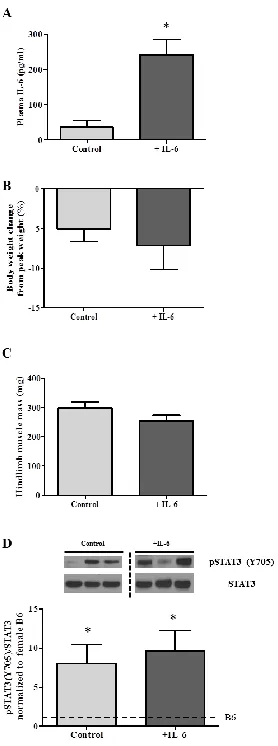
Figure 3.5 Effects of IL-6 overexpression in 18 week-old female ApcMin/+mice ...61
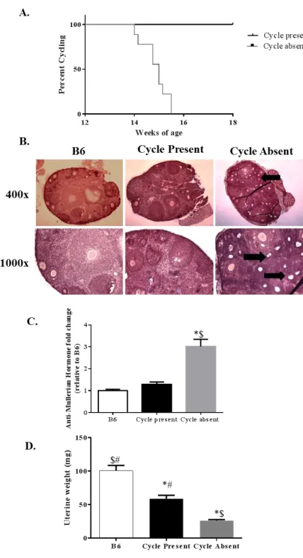
Figure 4.1 Estrus Cycle Cessation with Cachexia ...97
Figure 4.2 Morphological and functional changes with ovarian dysfunction ...98
Figure 4.3 Morphological and functional changes with ovariectomy ...99
Figure 4.4 Morphological changes with ovariectomy and IL-6 overexpression ...100
Figure 4.5 Effect of loss of ovarian function and IL-6 overexpression on tumor burden and circulating IL-6 signaling ...101
Figure 4.6 Effect of loss of ovarian function on circulating inflammatory factors ...102
Figure 4.7 Changes in muscle inflammatory signaling with loss of ovarian function and IL-6 overexpression ...103
Figure 4.8 Changes in muscle inflammatory receptors with ovariectomy and IL-6 overexpression ...104
Figure 5.1 Canonical cachexia-related pathways with loss of ovarian function in female ApcMin/+ mice ...129
Figure 5.2 Canonical cachexia-related pathways with overexpression of IL-6 in ovariectomized female ApcMin/+ mice ...130
xi
Figure 5.4 IL-6-related downstream signaling with loss of ovarian function in female ApcMin/+ mice ...132
Figure 5.5 IL-6 receptor expression and related downstream signaling with
overexpression of IL-6 in ovariectomized female ApcMin/+ mice ...133
Figure 5.6 Mitochondrial biogenesis, dynamics, and function with loss of ovarian
function in female ApcMin/+ mice ...134
xii
L
IST OFA
BBREVIATIONSAIDS ... Acquired Immunodeficiency Syndrome
ApcMin/+ ... Adenomatous polyposis coli heterozygote (Multiple intestinal neoplasms)
B6 ...Black-6 mouse strain
C26 ... Colon-26 Carcinoma
C57BL/6 ...Black-6 mouse strain
cDNA ... Complementary DNA
CMV ... Cytomegalovirus
COPD ... Chronic Obstructive Pulmonary Disease
CRP ...C-reactive protein
DEPC ... Active Cas
DNA ... Deoxyriboonucleic Acid
EGFR ...EGF Receptor
ELISA ... Enzyme-Linked Immunosorbent Assay
ERK... Extracellular Receptor Kinase
FDA... Food and Drug Administration
GAPDH ... Glyceraldehyde-3-phosphate dehydrogenase
Gp130 ... Glycoprotein-130
Gp80 ... Glycoprotein-80
IL-6 ... Interleukin-6
IL-6r ... IL-6 receptor
JAK ...Janus Kinase
xiii
LLC ... Lewis Lung Carcinoma
mIL-6r ... Membrane-bound IL-6 receptor
mRNA ... Messenger RNA
ms ... Millisecond
n...Number (of samples)
NfκB ... Nuclear factor-kappa B
p... Probability
p38... 38-kilodalton protein
PCR ... Polymerase chain reaction
R2... Correlation
RNA ... Ribonucleic Acid
RT-PCR... Real-time PCR
SOCS3... Suppressor of Cytokine Signaling-3
STAT3... Signal transducer and activator of transcription-3
TNFα ... Tumor necrosis factor-alpha
ul ... Microliters
1
CHAPTER
1
2
Cachexia is a devastating condition that occurs secondary to several chronic
diseases, including AIDS, COPD, chronic renal failure, and many forms of cancer [1].
There is no cure or FDA-approved treatment for cachexia, though it occurs in 30-50% of
cancers [1-3] and has an annual mortality rate of 80% [1], underlining the necessity of
investigation into its etiology and progression. The most recent definition of cachexia
includes an unintentional weight loss of 5% or more secondary to a chronic condition in
addition to other symptoms including anemia, fatigue, muscle weakness, increased
inflammatory markers and plasma triglycerides, and insulin resistance [1]. Cachexia
comprises a severe loss of adipose and skeletal muscle mass, the latter of which has
serious implications for quality of life, function, wound healing, and ability to respond to
chemotherapeutic treatments. The loss of both lean and fat mass is largely mediated by
inflammatory cytokines, including IL-1β, IL-6, and TNFα [2]. The ApcMin/+ mouse is an
established model of colon cancer and cachexia [4-6] that has been widely used by our
lab and others. The mouse has a nonsense mutation in the adenomatous polyposis coli
(Apc) gene, a tumor suppressor gene [7] which is also mutated in the majority of human
colon cancer cases [8] as well as familial adenomatous polyposis (FAP), a congenital
disease leading to about 15% of human colorectal cancer cases [9]. Apc encodes the APC
protein, which regulates cell proliferation via a direct interaction with, and leading to
ubiquitin-mediated destruction of, β-catenin, a protein regulating multiple
proto-oncogenes [9]. The ApcMin/+ mouse develops a large number of intestinal neoplasms
beginning at 4 weeks of age and plateauing by 12 weeks, after which cachexia develops
3
IL-6 is a pro-inflammatory cytokines closely involved with cancer cachexia
progression in many animal models, as well as the human condition [11, 12], including
the ApcMin/+mouse. Our laboratory has previously shown that overexpression of IL-6 in a
male ApcMin/+mouse can induce [13] and aggravate [14] cachexia progression; inversely,
global IL-6 knockout (KO) in a male ApcMin/+alleviates tumor burden and severity of
cachexia symptoms [13, 14]. The role of IL-6 during cachexia progression is less clear in
the female, however. While our lab has begun to elucidate the differences in IL-6-related
signaling that occur during cachexia in the female [15], many gaps remain in our
knowledge of the true etiology of cachexia in the female, as well as the exact role of IL-6
in the female. Plasma levels of IL-6 do not correlate with weight loss in the female [15]
as they do in the male [13], and overexpression experiments in the female ([15],
unpublished data) have shown no abrogation of cachexia severity.
Dysregulation of muscle metabolism leading to muscle wasting with cachexia is
regulated by a number of pro-inflammatory and pro-apoptotic factors. Four major
pathways exist that are involved in proteolysis due to various wasting conditions [3]; the
major pathway at work during cancer cachexia is the ubiquitin-proteasome system (UPS),
which is an ATP-dependent mechanism that involves tagging (ubiquitinating) proteins for
breakdown by the 26s proteasome [16]. This process is regulated upstream by
inflammatory factors including IL-6, STAT3, ERK, and p38 MAPK [17]. Many of the
pathways are redundant.
Though cachexia has been well-characterized in the male by our lab and others
[4-6, 10, 18-24], almost no research into the condition has taken place in the female. It
4
leading to cachexia; however, data from our laboratory has shown differences in the
response of the male and female [15]. Though both sexes undergo cachexia, data from
our lab has shown that the female loses less body weight than the male, and seems to
have a differential response to IL-6 from the male [20]. Cachectic male and female
ApcMin/+ mice have similar plasma IL-6 levels [15]; however, induced overexpression of
IL-6 in the male abrogates the cachectic response [5, 6, 20], but has no effect in the
female. There is no increase in STAT3 signaling in the female ApcMin/+ mouse with
overexpression of IL-6 [15], indicating a differential regulation of this pathway.
Importantly, it has been shown that estrogen, specifically 17β-estradiol, can block
transcription of IL-6 [25, 26] and signaling downstream of STAT3 [27]. IL-6 is a major
putative regulatory point in the pathophysiology of cancer cachexia as it is increased in
most cachexia models [11] and activates several pathways leading to eventual muscle
wasting [11, 28].
Ovarian function plays a major role in the health and disease outcomes of
females. The role of estrogen, in particular, in the health of both males and females has
come into focus in the past decade. Body composition is regulated in part by estrogen
signaling, and it has been shown that the ablation of such has a significant deleterious
impact on regulation of muscle and fat mass [29-32]; this dysregulation has potentially
dire consequences when coupled with the innate dysregulation of cachexia. Estrogen has
well-documented benefits against inflammation [33-35], particularly IL-6 [36, 37], that
may provide some benefit to the female during the onset and progression of cancer
cachexia. In addition, the relatively recent discovery of estrogen receptors α and β in
5
and strength [30, 38, 39]. Specifically, estrogen is necessary for recovery from atrophy
[29, 40]. Preliminary data from our lab shows that estrous cycling, which normally
occurs in the C57BL/6 mouse until around 14 months of age [41], ceases during the
progression of cancer cachexia in the female ApcMin/+ mouse, which indicates interplay
between the ovary and cachexia. Apart from estrogen, the other major ovarian hormone
progesterone and its analogs show potential for cachexia therapeutics; progestins
including megestrol acetate (MA) and medroxyprogesterone acetate (MPA) have been
shown to improve caloric intake and and anorexia symptoms in cachexia patients [3].
IL-6 plays a role in the normal cyclic signaling of the ovary [42], but its dysregulation is
also linked to ovarian pathology [42, 43]. This supports the hypothesis that there is
interplay between ovarian function and IL-6. This, together with the gaps in
understanding of the mechanisms underlying cachexia-related muscle wasting in the
female provides the rationale for this study. The novelty of this study arises from it
being, to our knowledge, the first study to specifically look at mechanisms of cachexia in
the female ApcMin/+ mouse, as well as the first study to investigate a potential role of
ovarian endocrine function.
Purpose & Specific Aims
The overall goal of this proposal is to determine if ovarian function can alter cachexia
progression in the female ApcMin/+ mouse through IL-6 signaling and the regulation of
skeletal muscle metabolism. The central hypothesis of the proposed study is that loss of
ovarian function in the female ApcMin/+ mouse causes abrogated progression of cachexia
through altered IL-6 sensitivity, which increases skeletal muscle metabolic dysfunction
6
study include body composition, in vivo functional capacity, abnormal blood
biochemistry, tissue IL-6 signaling, and disrupted skeletal muscle metabolism.
In order to prove the proposed study’s overall hypothesis three specific aims are
proposed.
Specific Aim #1 will determine the relationship between of circulating IL-6 to cancer cachexia progression in the female ApcMin/+mouse (revised 4/23/15)
Specific Aim #2 will determine if IL-6 regulation of cachexia progression is regulated by ovarian function in the female ApcMin/+ mouse.
Specific Aim #3 will determine whether cachexia-induced skeletal muscle metabolic dysfunction is regulated by ovarian function in the female ApcMin/+ mouse.
Figure 1.1 Overall Working Model: Working model of the focus of each of the three specific aims. First, the proposal will examine sex differences in the role of IL-6 signaling during cancer cachexia progression in ApcMin/+ mice (AIM 1). Second, the proposal will examine the role of loss of ovarian function (or ovariectomy) and its effect on inflammatory signaling during cancer cachexia progression in the female ApcMin/+mouse. Hypogonadism has an established role in cachexia progression of the male ApcMin/+
7
8
CHAPTER
2
9
2.1 Colon Cancer
Colorectal cancer is one of the leading forms of cancer in both men and women [7]. The
adenomatous polyposis coli (Apc) gene is a tumor suppressor gene that accounts for one
of the highest rates of loss-of-function in human colon cancer [7]. The Apc gene lies on
5q21 and includes 15 exons; the translated protein is fairly large at 309 kilodaltons [9].
Some studies have shown that obesity affects men’s risk of colon cancer more so than
women’s; other studies have shown no effect [44]. Sporadic colon cancer accounts for
85% of cases, whereas familial adenomatous polyposis accounts for the other 15% [9].
This syndrome causes the development of thousands of intestinal and rectal adenomas in
the second decade of life and is due to a germline mutation of the Apc gene [9]. In fact,
most colon cancers involve loss-of-function mutations in Apc [8]. Cancer cachexia
frequently occurs secondary to colon cancer and accounts for 30-50% of deaths due to
gastrointestinal cancers [45]. In cancer it is unclear whether cytokines are produced by
tumor cells or inflammatory cells in response to tumor burden [3].
2.2 Cancer cachexia
Cachexia develops secondary to many chronic disease, including AIDS, COPD, chronic
renal disease, and cancer [1]. Gastrointestinal cancer patients are particularly susceptible
to cancer cachexia [20]. Estimates of prevalence in cancer patients range from 28-57%
[1, 2], with an annual mortality rate of 80% [1] and contributing to 20% of cancer-related
deaths [2, 4]. This translates to over two million cancer-related cachexia deaths per year
[1]. The most recent definition of cachexia includes an unintentional weight loss,
10
symptoms including anemia, fatigue, anorexia, decreased muscle strength, and increased
inflammatory markers [1]. During experimental cachexia, fat stores are mobilized prior
to muscle catabolism [2]. With some cancers, patients show high levels of TNFα, IL-1β,
and IL-6, which roughly correlate with tumor progression [3]. Antibodies against each of
these factors have been used with some success, but none has completely attenuated
cachexia progression [3]. IL-6 is an important mediator of cachexia [20].
Cachexia is essentially an inflammatory process. Several inflammatory cytokines
are known to play roles in the etiology and progression of cachexia. TNFα leads to
extensive apoptosis of skeletal muscle cells during cachexia [2]. IL-1β and IL-6 promote
TNFα-mediated cachexia, which then leads to an increase in FoxO1 signaling [3] as well
as NfκB signaling through TNFR1[2]. IL-1β and IL-6 also contribute to the loss of lean
body mass during cachexia [46]. Inactive NfκB is suppressed by the IKB family of
proteins; when stimulated by TNFα, the IKB kinase complex (IKK) degrades IKB, which
releases and thereby activates NfκB [2]. NfκB plays a major role in the upregulation of
inflammation during cachexia [3]. Toll-like receptors (TLRs) are pathogen-recognizing
receptors present on immune cells, tumor cells, and muscle cells that play a role not only
in pathogen-related inflammation, but cachexia-related inflammation as well [46]. TLR2
and TLR4 are expressed in skeletal muscle and lead to further increases of cytokine
production (including IL-1β, IL-6, and TNFα) when stimulated [46]. The transforming
growth factor (TGF) family of proteins is yet another inflammatory system capable of
regulating cachexia-induced muscle wasting; TGFβ is the major player, signaling through
11
2.3 ApcMin/+mouse model
The ApcMin/+ mouse has a nonsense mutation in codon 850 of the Adenomatous polyposis
coli gene, (Apc) causing it to spontaneously develop colon polyps [4]. This model is
especially relevant, as the Apc gene is mutated in the majority of human colorectal
cancers [9]. Apc is a tumor-suppressor gene that regulates cell proliferation through
physical interaction with β-catenin, which leads to the ubiquitination and proteasomal
degradation of β-catenin [9]. When Apc heterozygosity and expression is lost, activation
of β-catenin by Wnt signaling is increased, leading to increased transcription of β-catenin
target genes including several proto-oncogenes [9]. Min/+ mice lose heterozygosity of
Apc in some cells, which leads to overexpression of β-catenin and eventually causes
tumors [24]. Min/+ mice generally have tumors in the small intestine and not the colon,
unlike humans; however, the loss of both Apc and estrogen receptor beta (ERβ) in tumor
cells make it a good model for the human disease [24]. Polyp number plateaus by 12
weeks of age, but tumor size continues to increase as cachexia develops, up to 20 weeks
of age [4].
2.4 Cachexia and Skeletal Muscle
Hypermetabolism due to cancer cachexia results in accelerated breakdown of
adipose tissue and skeletal muscle in order to provide energy substrates to the tumor and
host organism [16]. Skeletal muscle atrophy due to cachexia can lead to disability,
weakness, lessened wound healing, and decreased response to chemotherapeutic agents
12
Skeletal muscle is highly affected by cancer cachexia, though all fiber types do not
undergo wasting uniformly [16, 20]. Fast-twitch (type II) muscle fibers are more
susceptible to wasting with cancer cachexia than slow-twitch (type I) fibers, as the
oxidative phenotype is protective against cachexia [2, 20]. Initiation of muscle mass loss
with cachexia occurs as myonuclei undergo caspase-3-mediated apoptosis, decreasing the
size of the myonuclear domain [2]. Additionally, dysregulated protein synthesis and
myofiber regeneration [48], in addition to increased protein degradation, contribute to net
muscle wasting with cachexia.
There are four major proteolytic pathways involved in the loss of skeletal muscle
mass during cancer cachexia: Calcium-dependent (calpain/calpastatin-dependent),
lysosomal, caspase-dependent, and ubiquitin/proteasome [2, 3]. The calpain and
calpastatin proteolytic systems disrupt myofibrillar proteins and lead to eventual
ubiquitination and proteasomal degradation of such [2]. Lysosomal proteases play only a
small role in cachexia [2]. The ubiquitin/proteasome pathway is the major proteolytic
player during cachexia [16], and is regulated by FoxO3 [3]. The other major proteolytic
system involved in cachexia-related muscle wasting is autophagy; this mechanism is also
regulated largely by FoxO3 [17]. FoxO3 is generally phosphorylated, and therefore
inactivated and excluded from the nucleus of the cell, by PI3K/Akt [3]. When activated,
FoxO3 translocates to the nucleus, where it acts as a transcription factors for skeletal
muscle-specific E3 ligases including muscle ring finger (MuRF)1 and atrogin-1/MAFbx
[3], which are highly activated with cachexia [16]. Upstream of PI3K/Akt, the pathway
is regulated by reactive oxygen species (ROS), TNFα, PGC1α, and IGF-1[3].
13
MAPK signaling [17]. The UPS is an ATP-dependent process, and can therefore
contribute to lower efficiency and further dysregulation of mitochondrial energy
synthesis [16].
In recent years, it has become apparent that mitochondrial dysfunction plays a
major role in the etiology of cachexia and directly leads to many of the metabolic
abnormalities and overall atrophy of skeletal muscle due to cachexia. Indeed, it is
thought that mitochondria are the site of energy waste in skeletal muscle that leads to the
degradation of protein and overall wasting [49]. Oxygen consumption in mitochondria
isolated from cachectic muscle is lower than in healthy mitochondria [16]. Initial
cachexia does not affect mitochondrial content, though PGC1α and Mfn1, mitochondrial
proteins, are decreased at this stage [50]. As cachexia progresses, these changes become
more apparent and mitochondrial content decreases [50]. Cytochrome C oxidase (COX)
IV protein levels [14] and activity decrease with cachexia; the latter of which is
correlated with decreased oxygen consumption in mitochondria [16]. Higher levels of
uncoupling protein-3 (UCP3) have been noted in wasting muscle cells [49]. It has been
proposed that this decrease in mitochondrial content leads to an increase in reactive
oxygen species (ROS), which leads to an increase in oxidative stress, energy stress, and
apoptosis susceptibility leading to AMPK and FoxO1 activation [50]. However, Apc min/+
mice show these metabolic changes independent of oxidative stress [50].
2.5 Role of IL-6 in tumorigenesis and cachexia
Interleukin-6 (IL-6) is a pro-inflammatory cytokine secreted by many types of immune
14
elevated IL-6 is associated with many pathophysiological conditions including cachexia
and insulin resistance [51]. IL-6 suppresses insulin signal transduction via SOCS1 [2].
In humans, IL-6 and c-reactive protein (CRP) plasma levels correlate with insulin
resistance [53]. IL-6 has further been shown to inhibit hepatic insulin signaling in vitro
and in vivo, though it does not affect insulin receptor signal transduction in skeletal
muscle [53].
Classic IL-6 signaling involves IL-6 binding to its membrane-bound receptor
comprising IL6Rα, the ligand-binding domain, and gp130, the signal-transducing domain
[54, 55]. The membrane-bound IL-6 receptor has limited tissue expression, including
hepatocytes, immune cells, and skeletal muscle cells [55]. The alternative signaling
paradigm is known as trans IL-6 signaling, and is thought to be responsible for many of
the pro-inflammatory effects of IL-6 during cancer cachexia [56]. Inflammatory
responses are largely mediated by endothelial cells, which lack mIL-6r and therefore rely
on trans signaling [56]. Trans IL-6 signaling involves binding of the soluble IL-6
receptor-IL-6 complex to the ubiquitously-expressed gp130 receptor [55], allowing for
IL-6 signaling in nontraditional tissues. Soluble IL-6 receptor (sIL6r) is found in many
body fluids. Structurally identical to membrane-bound IL-6r and with the same affinity
for IL-6 as the membrane-bound receptor [56], it can be formed by alternative mRNA
splicing of the IL-6r gene (IL6ra) (10% of sIL-6r protein), or by shedding of the
membrane-bound protein by metalloproteinase ADAM17 (90%) [52]. The cleaved
protein can then dimerize and bind IL-6 in circulation, thereby increasing its half-life
[57]. This complex formed can then bind to gp130 which is ubiquitously expressed on
15
would not [42]. Cancer patients have higher levels of plasma sIL-6r compared with
healthy controls [56].
Activation of the membrane-bound IL-6 receptor or gp130 receptor activates
signal transducer and activator of transcription 3 (STAT3), which causes many of IL-6’s
downstream deleterious effects [27]. Further, STAT3 is essential for gp130-mediated
cell survival, cell cycle phase transition, cell movement, and cell differentiation [58].
Suppressor of cytokine signaling-3 (SOCS3) is the major negative regulator of STAT3,
and exerts its effects on STAT3 signaling by binding simultaneously to gp130, JAK1,
and JAK2 on the gp130/6r/JAK/STAT3 complex [59]. SOCS3 can thereby inhibit
IL-6-induced STAT3 signaling and further aggravation of this pro-inflammatory pathway.
However, when the epidermal growth factor (EGF) receptor (EGFR) is active and
present, STAT3 can be re-phosphorylated by a second influx of IL-6 despite the
continued presence of SOCS3 [59]. In muscle, IL-6 can also act as a mitogen, activating
satellite cell and myocyte proliferation [51]. In addition to the STAT3 pathway, IL-6 can
activate AMPK, p38 MAPK, and NfκB signaling [11, 60]. Muscle contraction, functional
overload, and recovery from disuse all increase IL-6 secretion from muscle [51]. When
IL-6 is knocked out in mice, IGF-1 signaling decreases 80% [51].
Cells secrete proteins, a process known as “shedding,” by releasing extracellular
vesicles [61]. Cancer cells shed more proteins, including IL-6, than normal cells, thus
providing a means by which tumors secrete IL-6 [61]. IL-6 causes high levels of
hepatocellular proliferation, eventually to hepatocellular carcinoma (HCC), the most
common form of liver cancer [25]. Experimental ablation of IL-6 in mice abolished sex
16
Phosphorylated (activated) STAT3 is higher in 22-week old male ApcMin/+ mice
than in 13-week old mice, indicating that IL-6 signaling is involved in the development
of cachexia [20]. Fbxo32 (Atrogin-1) mRNA and protein are also increased by 18-22
weeks of age in male mice, and correlate with the decrease seen in gastrocnemius muscle
weight [20]. When IL-6 was overexpressed in male ApcMin/+ mice for two weeks, the
cross-sectional area of type IIB muscle fibers decreased by 11% in the gastrocnemius
muscle, indicating that IL-6 is involved in the skeletal muscle wasting seen with cachexia
in the male [20]. However, overexpression of IL-6 in wildtype mice did not have the
same effect, indicating the IL-6 is not sufficient to induce muscle wasting [20].
Conversely, administration of an IL-6 receptor antibody (IL-6ra) to cachectic ApcMin/+
mice attenuated further cachexia progression as well as loss of mitochondria and changes
in mitochondrial dynamics [50].
2.6 Sex differences with Cancer and Cachexia
Sex differences have been noted with muscle mass and strength in cancer and other forms
of cachexia [62-64]; however, the vast majority of mechanistic work in cancer cachexia
has been performed in the male. In healthy mice, there are no differences in
intermuscular amino acid kinetics. In the postabsorptive state, however, there are
significant differences in amino acid turnover between the sexes. Additionally, men have
a higher overall protein turnover rate than women, which also may play a factor in the
sex differences seen in cachexia. Interestingly, differences between the sexes in cachexia
have been seen on the molecular level as well. After 14 days of synergist ablation in
male and female (Atrogin-1) MAFbx-KO rats, male rats were able to induce muscle
17
classical cachectic pathways are differentially regulated by the sexes. Interestingly, our
lab has shown that hypogonadism play a major role in the etiology of cachexia
progression in the male [66], however, it is unknown if a similar effect happens in
females. Leptin is a molecule that reports the organism’s overall energy status to the
brain, Leptin levels are lower in cachectic cancer patients than in healthy controls [67];
interestingly, women’s leptin levels tend to be higher than men’s when matched for body
fat [68]. Sex differences have long been seen in immune function; men generally have
lower immune responses than women do to a stimulus [69]. Men also have naturally
higher plasma levels of proinflammatory cytokines including TNFα and IL-6, two key
cytokines involved in the progression of cachexia. Hepatocellular carcinoma (HCC) is the
most common liver cancer; however, it occurs three to five times more often in men than
in women [25]. This cancer is caused by high levels of IL-6, which are abolished in
women by circulating estrogen [25]. Interestingly, several single-nucleotide
polymorphisms (SNPs) along the IL-6 gene promoter that affect outcome in IL-6-related
conditions have sex-related differences in frequency [70].
2.7 Ovarian Function
The ovary comprises oocytes in various stages of maturity, surrounded by
granulosa and theca cells, which produce estrogen and progesterone, respectively, among
other secreted hormones and cytokines. These functional units of the ovary are known as
follicles [71].
The hypothalamic-pituitary-gonadal (HPG) axis is similar across all animals, and
18
function in rodents can be monitored outwardly via the estrus cycle [73]. The estrus
cycle lasts 4-5 days and consists of four stages based on relative estrogen levels and cell
population present. Stages include diestrus, in which estrogen levels are lowest;
proestrus, featuring rising estrogen levels; estrus, during which estrogen levels are
highest and ovulation occurs, and metestrus, during which estrogen levels fall [73]. The
cycle is very regular and is not disrupted by stresses commonly found in animal facility
settings [73]. Similar to humans, the estrogen spike is caused by a surge in LH and FSH.
At the antral (early) stage of follicle development, the majority of follicles undergo
atresia and die, unless they are stimulated to be ovulated [74]. Antral follicles produce
Anti-Müllerian hormone (AMH), which is used as a marker of ovarian reserve [71]. As
woman approach menopuase, FSH levels rise as inhibin, a negative feedback molecule,
levels decrease [72]. At the time of menopause, Inhibins A, B, and C each decrease
before there is a noticeable decrease in estrogen levels. Eventually, there are very low
levels of estrogen, progesterone, and AMH, and ovulation stops [72].
Ovariectomized mice have increased body fat, which can be reversed by estrogen
replacement [32]. Along with higher body fat, ovariectomized mice have higher blood
glucose, higher insulin, a higher insulin resistance index, higher cholesterol, and higher
TNFα levels [32]. Before menopause, the incidence of type II diabetes is lower in
women than in men. After menopause, insulin sensitivity decreases, coinciding with a
rise in inflammatory factors and a decrease in proper glucose metabolism [75]. Kidneys
of ovariectomized mice show decreases in estrogens receptors α and β, as well as
adiponectin; as the kidney is estrogen-sensitive, this indicates a shift in proper kidney
19
OVX mice also have lower levels of energy expenditure and higher levels of
energy intake [76]. These animals also have a chronically activated inflammatory
response in adipose tissue [76]; this suggests that ovarian hormones ate involved in the
modulation of inflammation.
Estrogen is an ovarian steroid hormone that typically works through a genomic
pathway; that is, it binds to estrogen-response elements (EREs) on promoter sequences of
target genes to activate transcription [77]. However, the estrogen/receptor complex can
also work nongenomically by directly activating targets such as
phosphatidylinositol-3-kinase (PI3K) and mitogen-activated protein phosphatidylinositol-3-kinase (MAPK), which have roles in cell
proliferation, protection against oxidative stress, and survival [77-80]. This is the
mechanism by which estrogen conveys its osteoporosis-preventing effects in osteocytes,
degeneration-preventing effects in neurons, and protective effects in many other cell
types [77, 79-81]. Estrogen prevents apoptosis in many cells types by upregulating
Bcl-2, an anti-apoptotic mitochondrial protein, and by activating PI3K [81, 82]. Women
have improved outcomes after ischemia/reperfusion injury to cardiomyocytes, largely due
to estrogen’s effects in the cardiovascular system; specifically, estrogen activates the
PI3K/Akt pathway in cardiomyocytes, leading to less inflammation, oxidative stress and
apoptosis than men [33, 83].
Estrogen has significant effects on metabolism. In a healthy animal muscle cell,
insulin signals for upregulation of GLUT4 at the cell membrane through the PI3K
pathway [38]. Estrogen receptor α-knockout mice have very reduced GLUT4 signaling,
indicating that estrogen is an important modulator of insulin sensitivity [38]. The kidney
20
by the kidney, leading to metabolic imbalances [32]. In fact, estrogen receptor
α-knockout mice show many of the symptoms of the metabolic syndrome, including
glucose intolerance, insulin resistance, and adiposity [32]. It has been shown that
overexpression of estrogen sulfotransferase (EST), an enzyme responsible for the
deactivation of estrogens, causes inhibitions of insulin signaling, indicating that a certain
level of active estrogen is necessary to maintain proper insulin signaling [31]. Estrogen
receptor β activation has been shown to increase PGC-1, thereby modulating lipid
metabolism [31]. Estrogen receptors α and β have further been implicated in the
maintenance of body composition. Experimentally, estrogen receptor α has been shown
to increase hormone-sensitive lipase (HSL) in adipose tissue, which causes conversion of
triglycerides to free fatty acids, and increases AMPK expression in skeletal muscle [31].
Estrogen receptor β has been shown to keep body weight stable in high fat diet
(HFD)-fed mice [31]. Because it plays such a major role in glucose and lipid metabolism,
estrogen receptor β has become a target of great potential for metabolic disease [31].
Estrogen’s effects in skeletal muscle are not well-studied, though estrogen
receptors α and β are both present [84]. It is thought that many of estrogen’s beneficial
effects in skeletal muscle are due to its antioxidant capacity and anti-inflammatory
properties. It has been hypothesized that estrogen stabilizes the cell membrane,
protecting muscle cells from damage [84]. Though more study is needed, it has been
shown in various animal muscle atrophy models that estrogen is necessary for recovery
of muscle size and fiber cross-sectional area [40, 84]. Estrogen receptor α-knockout mice
have lower contractile strength than wildtype mice, indicating that estrogen has important
21
perimenopause in women is associated with a decrease in muscle mass and strength,
though hormone replacement therapy (HRT) has been shown to increase both variables
[85].
Exposing skeletal muscle cells to estrogen before damaging stimuli protects them
from apoptosis, as has been seen in many other cell types [86]. Estrogen regulates
myoblast proliferation and differentiation [86]. In skeletal muscle, estrogen increases
PPARδ and FoxO1, both of which are involved in the genesis of type I muscle fibers
[87]. Ovariectomized mice showed significant decreases in genes associated with type I
muscle fibers including PPARδ, FoxO1, myogenin, myosin light chain, and troponin C,
and significant increases in genes associated with type II muscle fibers including MyoD
[87]. However, ovariectomized mice also showed decreases in atrogin-1 and MuRF1
expression, which may indicate less muscle wasting [87].
The rate of colon cancer in men is 35% higher than in women [88, 89], and some
part of this is due to the beneficial effects of estrogen. In fact, hormone replacement
therapy (HRT) is associated with a 30-40% reduction of risk of colorectal cancer [24].
Population studies have shown menopause is associated with obesity and colon cancer
[44]. The effects of estrogen on colon epithelium are numerous and lead to overall lower
rates of colon cancer. Hormone replacement therapy is associated with a 30-40%
reduction in colon cancer risk Estrogen reduces plasma IGF-1, a risk factor for tumor
development [90], and increases insulin sensitivity [91]. Insulin and IGF-1 are mitogenic
to colon cancer cells, and epidemiologic studies have shown a correlation between
colorectal cancer and insulin levels [91]. Interestingly, however, the loss of estrogen in
22
colonocytes from transforming into malignancies. [24, 88, 91]. Estrogen reduces growth
in colonocytes via ERβ-mediated effects, including higher rates of apoptosis in
pre-malignant cells [44, 88]. The first sign of colon tumors are often aberrant crypt foci
(ACFs); mice treated with estrogen had fewer ACFs than untreated mice, though this
effect is ablated in ERβKO mice [88].
The effects of ERβ on colon epithelium have been well-established. Estrogen can
reduce the incidence of chemically-induced polyps in ERαKO mice, but not ERβKO
mice, indicating that this receptor mediates the effects [88]. ERβ controls cell cycling in
colon epithelium [89-91], and expression is frequently lost in colon tumors [24, 89, 91].
In fact, ERβ expression is inversely related to tumor differentiation in colon epithelium
[91]This effect occurs via estrogen responsive element (ERE)-binding dependent
transcription, as well as through activator protein 1 (AP1) and SP1 [90]. ERαKO and
ERβKO mice have increased colon tumor numbers and abnormal colon histology [24].
It has been found that injecting mice with SW480 (colon cancer) cells as well as an ERβ
construct decreases size of tumors compared to mice injected with only SW480 cells
[90]. Contrary to the effects of ERβ in colon epithelium, ERα expression has been
associated with increased incidence of cancer cell proliferation [44]. This follows with
the general functions of the ERs: ERα induces proliferation and ERβ suppresses
proliferation [44].
Conversion of androgen into estrogens is favored in pro-inflammatory
environments [92]. Bacterial lipopolysaccharide (LPS; endotoxin) induces multiple
pro-inflammatory factors including TNFα and NfκB, leading to apoptosis; these are inhibited
23
(IL-1β; IL-6) and tumor necrosis factor alpha (TNFα), all pro-inflammatory inflammatory
factors, are able to activate the hypothalamic-pituitary-adrenal (HPA) axis. Estrogen
modulates the HPA axis response, but has different modulatory effects on HPA responses
to endotoxin and IL-1 [37]. HPA responses to estrogen are attenuated by ovariectomy
[37]. Estrogen inhibits NfκB, which activates monocyte chemoattractant protein-1
(MCP1) [26], and along with other co-factors is involved in the transcription of IL-6,
thereby blocking pro-inflammatory responses [26]. In fact, estrogen and NfκB have a
mutually inhibitory relationship [33].
Many of estrogen’s anti-inflammatory properties have been most thoroughly
investigated in the context of ischemia-reperfusion (I-R) injury of myocardial cells.
Estrogen has been shown to decrease p38 MAPK-mediated inflammation following I-R
[33]; this protection is ablated in ovariectomized female rodents. Indeed, OVX was
shown to increase phosphorylated p38, IL-1β, IL-6, TNFα, and caspase-3 cleavage in the
myocardium [33].
Menopause is associated with an increase in inflammatory factors, including
TNFα, IL-6, and plasminogen activator inhibitor-1 (PAI-1) [75], indicating that estrogen
regulates levels of these factors in the pre-menopausal state.
2.8 Ovarian Function and IL-6
Granulosa cells of the ovary secrete IL-6 and other cytokines throughout the
menstrual or estrous cycle. Granulosa cells present both mIL-6r and gp130 [93].
Increasing amounts of IL-6 inhibit estrogen production by these cells, regardless of the
24
of IL-6 [58, 94]. TGFβ and IL-6 are involved in the post-ovulation healing process of the
ovary [95]. In humans, during the follicular phase of the menstrual cycle (during which
estrogen is high), IL-6 levels do not change but TNFα levels increase [57]. TNFα levels
as well as sIL-6r levels increase during the luteal phase, during which there are low
estrogen levels [57]. Across the whole menstrual cycle, women have higher TNFα and
sIL-6r plasma levels than males [57]. Levels of IL-6 mRNA are increased in ovarian
epithelial cells and cancer cells that have been stimulated with luteinizing hormone (LH)
and follicle-stimulating hormone (FSH) [58]. Gonadotropins including LH and FSH have
been shown to be more potent than sex steroids at stimulating IL-6 production [58].
Circulating IL-6 increases after menopause [92]. IL-6 levels have been shown to
increase in younger women who undergo oophorectomy, but this is abolished by
hormone replacement therapy [92]. Additionally, the hypogonadism seen with
menopause is associated with increased plasma levels of IL-6, and more negative
outcomes associated with IL-6 [85]. Hormone replacement therapy after menopause is
associated with increases in ovarian cancer rates [58]. Interestingly, postmenopausal
women have increased plasma levels of soluble gp130 and soluble 6 receptor [85].
IL-6 has been implicated in the pathogenesis of polycystic ovarian syndrome (PCOS) [42].
Women with PCOS had higher serum gp130 concentrations than women without PCOS;
women with PCOS have significantly lower sIL6r concentrations than women without,
indicating that IL-6 signaling is occurring via a different mechanism in this syndrome
[42]. Serum gp130 is related to SHBG levels [42].
Elite female gymnasts who had not reached menarche have increased IL-6 and TNFα
25
had estrogen levels that were 200% of those athletes who had not yet reached menarche
[96].
In intact mice, it has been shown that both androgens and estrogens are able to
inhibit IL-6 production [37]. Estrogen directly inhibits IL-6 synthesis and release from
bone marrow cells through ERα-mediated gene transcription [37, 58]. Estrogen has been
reported to increase IL-6 expression in circulating mononuclear cells, but to inhibit IL-6
synthesis in other cell types including osteoblasts [58]. Estrogen prevents necessary
transcription factors, including NfκB, C/EBPβ [25], and p65 [26] from binding to the
IL-6 promoter, thereby inhibiting transcription [97]. The estrogen receptor α (ERα) also
blocks NfκB from binding to the MCP-1 promoter and blocks TNFα from inducing high
levels of MCP-1[26]. Ovariectomized mice show higher plasma IL-6 levels than intact
mice, but estrogen replacement attenuates this increase [37]. Further, estrogen
upregulates SOCS3 in hepatocytes [25]. Administration of estrogen has been shown to
decrease plasma levels of IL-6, IL-1β, and TNFα [85]. In mice given diethylnitrosamine
(DEN) to induce hepatocellular carcinoma (HCC), a form of liver cancer, estrogen
abolishes IL-6 secretion from hepatic Kupffer cells by inhibiting IKKβ and Myd88,
protecting the female from IL-6-induced tumorigenesis [25]. Estrogen can block
downstream signaling of STAT3 [27]; further, estrogen can upregulate SOCS3 (a
downstream negative inhibition molecule which inhibits STAT3 signaling) in
hepatocytes [98]. Administration of estrogen to males and ovariectomized females
protected them as well as intact females [25]. It was found that STAT3 was being
activated by IL-6 in the males, but not the females, leading to an important finding about
26
IL-6 inhibits cilia activity in fallopian tubes; gp130 and IL-6r are also present in
fallopian tubes [54]. In the uterus, estrogen can both positively and negatively regulate
IL-6 [54]. Interestingly, estrogen induces a time-dependent decrease in IL-6r expression;
this can be blocked by inhibiting ERα [54].
In multiple myeloma cells, 17β-estradiol completely abolishes IL-6-inducible cell
proliferation and STAT3 DNA binding [27]. Importantly, estrogen does not inhibit
STAT3 activation by IL-6; it blocks downstream activity of STAT3 [27].
In breast cancer cells, IL-6 is a key factor in the secretion of estrogen. It strongly
synergizes with estrogen to activate aromatase and steroid sulphatase in order to further
stimulate production [99]. Further, IL-6 activates transcriptional activity of ERα via
estrogen response elements (EREs) [99].
2.9 Ovarian Function and Cancer
Hypogonadism is apparent in 40-90% of male cancer patients [66]. Though this has not
yet been proven in females, the hypogonadism seen with menopause negatively affects
the metabolism of multiple organ systems including skeletal muscle and the immune
system [85]. Sex hormone binding globulin (SHBG) is increased in cancer patients,
leaving less unbound (biologically active) hormone available [66]. Progestins, including
megestrol acetate (MA) and medroxyprogesterone acetate (MPA), have been shown to
decrease anorexia seen with cachexia, and possibly inhibiting IL-6 and TNFα [3].
Unfortunately, it is very difficult to study the role of ovarian function with cancer, as
most chemotherapeutic agents ablate ovarian function, making most women who undergo
27
adulthood; however, women tend to have fewer primordial and antral (growing, healthy)
follicles in their ovaries than age-matched controls, generally due to the ionizing effects
of radiation or chemotherapy [101].
2.10 Ovarian Function and Skeletal Muscle
OVX mice have significant decreases in gene expression of PPARδ, FoxO1,
PDK4, and UCP2 in skeletal muscle [87]. PPARδ and FoxO1 affect muscle fiber type;
both tend to shift towards type I [87]. In addition, OVX mice have decreased myosin
light chain and troponin C proteins, both of which are prevalent in type I fibers [87].
Interestingly, these mice also have more MyoD than sham mice, which drives
development of type II fibers [87]. OVX mice also have decreased atrogin-1 and MuRF1
[87], two essential skeletal muscle-specific E3 ligases involved in the degradation of
skeletal muscle protein with cancer cachexia. Metabolically, OVX is associated with
increased adiposity due to upregulation of genes involved with lipid storage including
lipoprotein lipase (LPL), acetyl-CoA carboxylase-1 (ACC-1), fatty acid synthase (FAS),
and others; supplementation of estrogen downregulated these genes [102].
Estrogen has multiple mechanisms by which it can enhance skeletal muscle
function and quality, including antioxidant properties, stabilizing myofiber membranes
by intercalation, and downstream gene expression [103]. After muscle damage or
unaccustomed exercise, estrogen plays a role in stimulating muscle repair and
regeneration, in part by activating satellite cells [103]. Due to membrane rupture,
damaged skeletal muscle cells release creatine kinase (CK) and myoglobin after damage
28
CK, women do tend to have lower levels in circulation which is directly attributable to
estrogen [103].
In C2C12 mouse myoblast culture, exposing cells to 17β-estradiol before
exposure to apoptotic stimuli including hydrogen peroxide (H2O2) protects them from
damage [86]. One of the protective mechanisms of estrogen on skeletal muscle is
increased expression of heat-shock proteins (HSPs), which control cell homeostasis by
acting as protein-folding chaperones and protecting protein structure from physiological
stresses [86]. Expression of HSPs 25, 27, 70, and 90 is directly modulated by estrogen
[86].
Estrogen causes lower skeletal muscle mass in ovaries-intact mice or
estrogen-replete mice compared to OVX mice; this is at least in part attributable to the effects of
ubiquitin-specific peptidase 19 (USP19), which is upregulated by estrogen receptor alpha
(ERα) and during muscle atrophy [104]. The promoter of USP19 contains estrogen
response elements (EREs), to which ERα binds in female mice and male mice given
estrogen; this does not occur in OVX female mice or control male mice [104].
2.11 Conclusion
Though much work has been done in the areas of ovarian function, largely including
estrogen’s roles, in cancer and skeletal muscle, it is clear that many gaps remain in our
knowledge. Very little is known of ovarian function’s effects on muscle outside of what
we have learned from menopausal women or from animal studies of estrogen’s direct
effects on muscle. Additionally, beyond estrogen’s effects in cancer, there is a paucity of
29
progression. As far as cachexia, there is an embarrassing lack of research regarding the
roles of either ovarian function or estrogen in the treatment or role in the etiology of
cachexia. Going forward, it is intended that the present studies will fill some of these
gaps and begin to fill in the picture of how ovarian function contributes to the etiology
30
CHAPTER
3
S
EXD
IFFERENCES IN THER
ELATIONSHIP OFIL-6
S
IGNALING TOC
ANCERC
ACHEXIAP
ROGRESSION11
Hetzler KL, Hardee JP, Puppa MJ, Narsale AA, Sato S, Davis JM, Carson
JA. 2015.
Biochimica et Biophysica Acta
1825: 816-25.
31
3.1
AbstractA devastating aspect of cancer cachexia is severe loss of muscle and fat mass. Though
cachexia occurs in both sexes, it is not well-defined in the female. The Apc Min/+ mouse is
genetically predisposed to develop intestinal tumors; circulating IL-6 is a critical
regulator of cancer cachexia in the male Apc Min/+ mouse. The purpose of this study was
to examine the relationship between IL-6 signaling and cachexia progression in the
female Apc Min/+ mouse. Male and female Apc Min/+ mice were examined during the
initiation and progression of cachexia. Another group of females had IL-6 overexpressed
between 12-14 weeks or 15-18 weeks of age to determine whether IL-6 could induce
cachexia. Cachectic female Apc Min/+ mice lost body weight, muscle mass, and fat mass;
increased muscle IL-6 mRNA expression was associated with these changes, but
circulating IL-6 levels were not. Circulating IL-6 levels did not correlate with
downstream signaling in muscle in the female. Muscle IL-6r mRNA expression and
SOCS3 mRNA expression as well as muscle IL-6r protein and STAT3 phosphorylation
increased with severe cachexia in both sexes. Muscle SOCS3 protein increased in
cachectic females but decreased in cachectic males. IL-6 overexpression did not affect
cachexia progression in female Apc Min/+ mice. Our results indicate that female Apc Min/+
mice undergo cachexia progression that is at least initially IL-6-independent. Future
studies in the female will need to determine mechanisms underlying regulation of IL-6
response and cachexia induction.
32
3.2 Introduction
Cachexia is a devastating condition that occurs secondary to several chronic diseases,
including AIDS, COPD, chronic renal failure, and many forms of cancer [1]. There is no
FDA-approved treatment for cachexia, though it occurs in 30-50% of cancers [1-3] and
has an annual mortality rate of 80% [1]; the investigation into its etiology and progression
is therefore essential. The most recent definition of cachexia includes an unintentional
5% weight loss over twelve months, comprising the loss of muscle and fat [1]. Other
abnormalities associated with cachexia include anemia, fatigue, muscle weakness,
increased plasma triglycerides and inflammatory markers, and insulin resistance [1].
There has been considerable improvement in our understanding of the regulation of
cancer cachexia progression due to research employing male mouse models [5, 14, 21,
105, 106]. However, there is evidence that sex differences exist in the development of
cachexia in rodents [107]. Additionally, sex differences have been observed in the loss of
muscle strength associated with cachexia severity in humans [108]. Although there is
clear evidence that cachexia occurs in both males and females, the fundamental
differences in the pathophysiology due to sex and underlying mechanisms are unknown.
Systemic inflammation related to cancer is thought to be a mediator of cachexia, as
several pro-inflammatory pathways are enhanced during the progression of cachexia
[109, 110]. Sex hormones can modulate the inflammatory response to a variety of stimuli
[33, 37, 111]. Specifically, estrogen is known to inhibit NfB and tumor necrosis factor
(TNF) signaling [35, 111], C-reactive protein (CRP)-induced interleukin-6 (IL-6)
33
downstream of IL-6 [27]. In several rodent models of cachexia and some human cancers,
IL-6 is associated with the development of muscle wasting and body weight loss [5, 13,
14, 112, 113]. Classical IL-6 signaling involves binding of the cytokine to the
membrane-bound IL-6 receptor (mIL-6r) on target tissues, which include hepatocytes, immune cells,
and skeletal muscle [42, 55, 114]. mIL-6r is a heterodimer comprising the ligand-specific
gp80 unit and the signal-transducing gp130 unit [54]. Activation of mIL-6r induces
downstream activation of many signaling pathways, including JAK/STAT, p38, and ERK
[11, 55, 115, 116]. Several of these pathways have been implicated in the regulation of
muscle mass loss during cancer cachexia [11, 28].
The ApcMin/+ mouse is genetically predisposed to develop intestinal tumors and
becomes cachectic secondary to the tumor burden [20, 105]. Our lab initially
characterized cachexia in retired female ApcMin/+ breeders [18]; however, the progression
and etiology of cachexia in the female ApcMin/+ mouse has not been described.
Development of cachexia in the male ApcMin/+mouse has an established IL-6 dependence
[4-6, 14, 20, 22, 106]. Administration of an IL-6 receptor antibody to cachectic male
ApcMin/+ mice can attenuate further cachexia progression [106]. When IL-6 is
systemically overexpressed in the male, more body weight is lost and cachexia is more
severe, indicating a causative role [5, 11, 14]. However, the response of female ApcMin/+
mice to IL-6 overexpression has not been examined. Sex differences have been noted in
the inflammatory milieu of humans [68, 69] and mice [25, 33] under multiple
pathological circumstances. Additionally, estrogen has been shown to inhibit IL-6
transcription and signaling in several tissues [25, 27, 36, 92, 97], which may also lead to
34
purpose of the present study was to examine the relationship of circulating IL-6 to cancer
cachexia progression in the female ApcMin/+ mouse. Our hypothesis is that cachexia
progression in the female ApcMin/+ mouse would not be associated with increased
circulating IL-6 levels as has been reported in the male. This hypothesis was tested
through three experiments. The first experiment followed a cohort of female and male
ApcMin/+ mice to 18 weeks of age, at which point the association between cachexia
severity and circulating IL-6 level was determined. In the second experiment, 12-week
old female ApcMin/+ mice had IL-6 systemically overexpressed for two weeks (until 14
weeks of age) to determine if supraphysiological IL-6 levels could induce cachexia as we
have previously shown in the male [5, 6, 60]. In the third experiment, 15-week old female
ApcMin/+ mice had IL-6 systemically overexpressed for three weeks (until 18 weeks of
age) to determine if supraphysiological IL-6 levels could accelerate cachexia progression
as we have previously shown in the male [20].
3.3Methods
Animals
Female ApcMin/+mice (N=32), male ApcMin/+ mice (N=12), female C57BL/6 mice (N=6),
and male C57BL/6 mice (N=6) were bred and maintained at the University of South
Carolina Animal Resource Facility. ApcMin/+ mice used were offspring from breeders
originally purchased from Jackson Labs (Bar Harbor, ME, USA). Male and female mice
were taken during the three-month period from a standing inbred ApcMin/+ breeding
colony. ApcMin/+ mice used were on a C57BL/6 background. Mice were kept on a 12:12h
35
rodent chow (Harlan Teklad Rodent Diet, #8604). Mice were weighed weekly. Blood
was collected by retro-orbital eye bleed at 12, 14, 16, 18, and 20 weeks for IL-6 analysis.
All experiments were approved by the University of South Carolina’s Institutional
Animal Care and Use Committee.
Procedures
Three experiments were performed. Experiment 1 examined the progression of cachexia.
Female ApcMin/+mice (n=18) were sacrificed at 18 weeks of age. Male ApcMin/+ mice
were sacrificed at 18-20 weeks of age. Female (n=6) and male (n=6) C57BL/6 (B6) were
sacrificed at 18 weeks of age as non-cancer controls. Prior to sacrifice, blood was taken
for analysis of IL-6 levels. Experiment 2 examined 2 weeks of IL-6 over-expression in 12
week-old, weight stable female ApcMin/+ mice as we have previously completed in the
male [60]. Female ApcMin/+ mice were electroporated with a control plasmid or with an
IL-6 overexpression plasmid (n=4 per group) at 12 weeks and were sacrificed at 14
weeks. Blood was taken at sacrifice (post-treatment) to determine plasma IL-6 levels.
Mice from this experiment were not used for any other analysis. Experiment 3 examined
IL-6 over-expression in 15-week-old female ApcMin/+ mice corresponding with the
initiation of cachexia, a time course we have previously examined in the male [20]. At 15
weeks of age mice were randomly separated into IL-6 over-expression (n=6), control
plasmid (N=3), or non-electroporated control (n=3) treatment groups. All
female ApcMin/+ mice were sacrificed at 18 weeks of age. Blood was taken at sacrifice
(post-treatment) for determination of plasma IL-6 levels. No differences were found in
body weight (p=0.78) or muscle mass (p=0.36) between control plasmid and