CELLULAR & MOLECULAR BIOLOGY LETTERS http://www.cmbl.org.pl
Received: 30 June 2011 Volume 17 (2012) pp 171-181 Final form accepted: 12 January 2012 DOI:10.2478/s11658-012-0001-z Published online: 24 January 2012 © 2012 by the University of Wrocław, Poland
# Paper authored by participant of the international conference: 18th Meeting, European
Association for Red Cell Research, Wrocław – Piechowice, Poland, May 12-15th, 2011.
Publication cost was covered by the organizers of this meeting.
* Author for correspondence. e-mail: [email protected], tel: +386 1 5437602, fax: +386 1 5437601
Mini review
RED BLOOD CELL SHAPE AND DEFORMABILITY IN THE CONTEXT OF THE FUNCTIONAL EVOLUTION
OF ITS MEMBRANE STRUCTURE #
SAŠA SVETINA*
Institute of Biophysics, Faculty of Medicine, University of Ljubljana and Jožef Stefan Institute, 1000 Ljubljana, Slovenia
Abstract: It is proposed that it is possible to identify some of the problems that had to be solved in the course of evolution for the red blood cell (RBC) to achieve its present day effectiveness, by studying the behavior of systems featuring different, partial characteristics of its membrane. The appropriateness of the RBC volume to membrane area ratio for its circulation in the blood is interpreted on the basis of an analysis of the shape behavior of phospholipid vesicles. The role of the membrane skeleton is associated with preventing an RBC from transforming into a budded shape, which could form in its absence due to curvature-dependent transmembrane protein-membrane interaction. It is shown that, by causing the formation of echinocytes, the skeleton also acts protectively when, in vesicles with a bilayer membrane, the budded shapes would form due to increasing difference between the areas of their outer and inner layers.
Key words: Red blood cell, Vesicle shapes, Deformability, Membrane skeleton, Functional evolution
INTRODUCTION
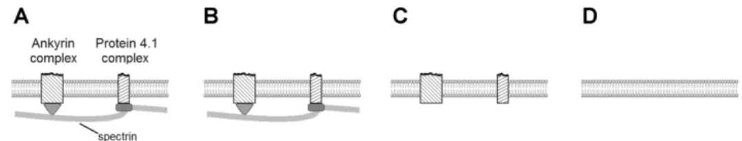
membrane properties that guarantee its stability. The RBC membrane is a composite of a lipid bilayer densely occupied by transmembrane proteins and, linked with it, spectrin-based membrane skeleton (Fig. 1A) [1]. The effects of RBC structural elements on its behavior can be fully appreciated only in the context of the purpose for which they emerged and were developing. The evolutionary origin of spectrin and its accompanying proteins ankyrin and 4.1 has been revealed, with a remark that it still remains to be answered what further adaptations occurred in the evolution of enucleated mammalian erythrocytes [2]. In view of this it seems sensible to complement the studies of the evolution of the RBC membrane structure by focusing on the development of its function. It is plausible to assume that in general an improvement of a given RBC function required the membrane structure to increase its complexity. A possible approach to study the evolutionary development of the RBC membrane from the perspective of the development of its function is therefore to study the differences between the behavior of the normal RBC membrane and analogous systems that lack some of its structural features.
Fig. 1. Schematic representation of membranes of different complexity that exhibit different partial characteristics of the RBC membrane. A – Symbolic representation of the normal RBC membrane. Most of the RBC transmembrane proteins participate in the portrayed ankyrin and actin–protein 4.1 based complexes. B – The RBC membrane as a trilayer structure. C – The RBC membrane deprived of its skeleton. D – A lipid bilayer membrane.
The aim of this minireview is to identify some problems that had to be solved in the course of evolution for the RBC to achieve its present day effectiveness, by studying the shape behavior and deformability of systems featuring different partial characteristics of the RBC membrane (Figs. 1B, C, D). This contribution will also bring together and strengthen different concepts related to the evolution of the RBC function, which we were developing and are scattered around in several previous publications [3-6]. The treatment will be restricted to red blood cells without nuclei, such as the human red blood cell.
skeleton by causing the formation of echinocytes prevents the formation of budded shapes that would in a bilayer occur due to the increased difference between the areas of its outer and inner layers. In order to properly express the accompanying arguments we will first provide the necessary theoretical background.
THEORETICAL BACKGROUND
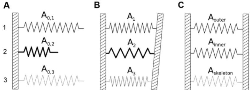
At the macroscopic level the RBC membrane can be viewed as a trilayer with the property that its three layers keep the distances between their neutral surfaces fixed while, in their lateral direction, they are free to slide, one over the other. The elastic behavior of such a system can be visualized by a model of three springs exhibiting different lengths and spring constants (Fig. 2A) [3]. If these springs are constrained on both sides by solid bars, some of them are compressed (springs 1 and 3) and some extended (spring 2) so that their lengths differ from their equilibrium lengths (Fig. 2B). RBC membrane layers are also elastic, albeit in two dimensions. They are constrained because they form a closed surface, so that in general, their areas (Ai , with i = 1, 2, 3) differ from their equilibrium areas (A0,i).
Fig. 2. Spring representation of the equilibrium state of three coupled layers. A – Three unconstrained springs with different equilibrium lengths (denoted as A0,i with i = 1, 2, 3)
and different spring constants (with their strengths depicted by the thickness of the lines). B – The same three springs constrained on both of their sides by solid bars. Their lengths are denoted by Ai. C – Representation of the elastic properties of the RBC membrane
layers. Layers 1 and 2 are the outer and inner layers of the bilayer, respectively, and layer 3 represents the skeleton. In each case the notation for spring lengths symbolizes the areas of the membrane layers.
The corresponding constraints can be expressed as the relationships between the areas Ai. The difference between the areas Ai and Aj (i < j = 1, 2, 3) depends on the cell shape and the distance between their neutral surfaces hi,j. It is equal to
M
h
A
A
A
i,j
i
j
i,j
, (1)
c
c
dA
M
(
1 2)
(2)Of the three constraints (Eq. 1) only two are independent [3, 7]. It is therefore possible to express the elastic energy of the whole trilayer membrane with a closed surface as the sum of the in-plane elasticity (including area compressibility and shear) and the local and nonlocal bending energies.
The local bending energy is
k c c dA
Wblocal c 2
2 1 , ( ) 2 1 (3)
where the local bending constant kc is the sum of the bending constants of the constituent layers. Membrane spontaneous curvature [8] is neglected because it is not, in the present discussion, essential.
The nonlocal bending energy is proportional to (M – M0)2, where M0 is the material constant effectively representing the equilibrium value of the integral of the sum of the two principal curvatures over the membrane area. The latter depends on the equilibrium areas A0,i and interlayer distances hi,j [7]. Here we shall only refer to the corresponding expression for the bilayer which can be presented as 2 0 0 2 2 , 1 ,
(
)
2
1
A
A
A
h
k
W
r nonlocalb
, (4)where kr is the nonlocal bending constant. From Eq. 1 we have A = h1,2M, while M0 in this case is A0/h1,2, with A0 = A0,1 – A0,2 [9]. Such a nonlocal bending term can relax by the transfer of constituent lipids from the denser into the less dense layer [10, 11].
THE RBC VOLUME TO MEMBRANE AREA RATIO IS OPTIMAL
The appropriateness of the physiological RBC volume to membrane area ratio for its circulation in the blood will be revealed on the basis of the analysis of the shape behavior of phospholipid vesicles (Fig. 1D). To a first approximation, the shape behavior of vesicular structures is governed by the membrane bending energy so that, because of the negligible bending energy of the skeleton, the shape behavior of an RBC and phospholipid vesicles could be similar.
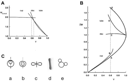
A convenient measure of the volume to membrane area ratio is the reduced volume, v, which is the ratio of the cell volume, V, to the volume of a sphere with the same membrane area, A: v = 6π1/2V/A3/2 [14]. At each value of v there is a vesicle shape that has the lowest possible local bending energy (Eq. 3). Within different ranges of v these shapes belong to different shape classes (Fig. 3A): at higher reduced volumes (0.63 < v < 1), to the class of tube shapes; within a short, intermediate interval (0.58 < v < 0.63), to the class of discoid shapes; and at lower reduced volumes (0.00 < v < 0.58), to the class of cup shapes [14]. These shapes differ in their values of the integral M (Eq. 3), which is, in the case of the vesicle with the bilayer membrane, conveniently expressed in terms of the
Fig. 3.The dependence of vesicle properties on reduced volume. A – Reduced local bending energy of the vesicle (wb,local = Wb,local/8πkc, where 8πkc is the local bending energy of the
reduced difference between the areas of the two membrane layers, Δa, defined relative to the equivalent difference for the sphere with the same membrane area,
Δa = ΔA/4h1,2(πA)1/2 (Fig. 3B). Fig. 3B also shows the a(v) dependence of the two types of limiting shapes. Examples of the corresponding shapes, for v = 0.6, are presented in Fig. 3C.
In fact, there is only partial correspondence between the shape behavior of RBC and that of phospholipid vesicles [6]. The stress-free shape of the RBC is a discocyte and thus identical to the shape of a vesicle with the same reduced volume. The shape correspondence also holds for lower a values where the minimum energy shape is the cup shape. In contrast, the two systems behave differently at higher a values, which, as will be discussed, can be ascribed to the effects of the RBC skeleton. It is however possible, despite this inconsistency, to speculate, on the basis of vesicle shapes, about the functional significance of the choice for the RBC stress-free shape.
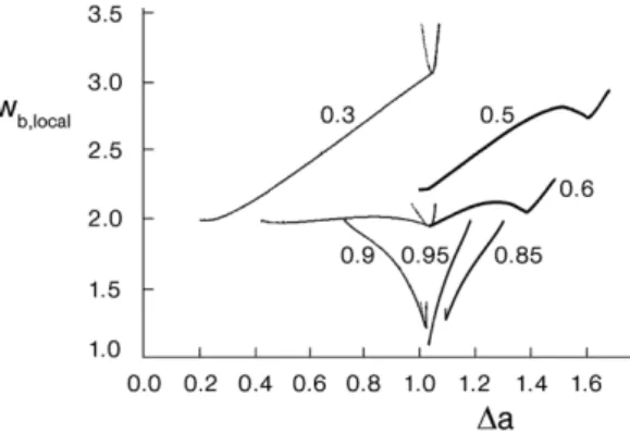
In order to squeeze easily through narrow capillaries it is, for geometrical reasons, convenient for an RBC to have as small a reduced volume as possible. What is then the advantage for the RBC of having a reduced volume of around 0.6? It is evident from Fig. 3A that, in the region of reduced volumes around 0.6, the very different tube, disc and cup shapes (Fig. 3C) exhibit rather similar local bending energies. This property of vesicles becomes even more apparent when comparing vesicle local bending energies at different vesicle reduced volumes. Some earlier calculated dependences of local bending energy on the reduced area difference Δa are collected in Fig. 4. It is clear that, in the region of reduced volumes in which the shape of the lowest energy is a discocyte, the variation of the local bending energy is least. In either larger or lower reduced volumes any change of vesicle shape – characterized by the variation of the Δa value – would require much stronger increases or decreases of local bending energy.
Any deformation of an RBC causes its nonlocal bending energy (Eq. 4) to change. With regard to this energy it can be assumed that the RBC stress-free condition includes the relaxed nonlocal bending energy, with the consequence that Δa0 = Δa. When changing shape from a discocyte to the extended cylindrical shape attained in the capillary, the Δa value of the RBC must therefore increase to a value similar to that of the tube-like shape “d” of Fig. 3C, thereby, according to Eq. 4, correspondingly increasing its nonlocal bending energy. At a slightly larger reduced volume, the stress-free shape would, by itself, be tube-like (Fig. 3A). However, it appears that it is of more advantage for the RBC to maintain a lower reduced volume and to pay for the corresponding increase of the nonlocal bending energy. Moreover, as will be discussed below, the discocyte shape is optimal also because it is safer for an RBC to have a Δa0 value as different as possible from that of the budded shape “e” (Fig. 3C). Why then is the reduced volume not even smaller than 0.6? In the case of cup shapes, the required increase of the difference Δa – Δa0 would be at least twice as large as in the case of the discocyte (Fig. 3B), which means that the necessary increase of the nonlocal bending energy would now be appreciably larger than in the case of the discocyte. It can be concluded that, from the point of view of bending energy changes and the requirement to have as small a volume as possible, RBC volumes around v = 0.6 are optimal.
THE RBC SKELETON IMMOBILIZES TRANSMEMBRANE PROTEINS
The RBC bilayer is crowded with the transmembrane proteins needed for its performance, e.g., for sustaining its osmotic balance and for exchanging anions. Most of these proteins or their complexes are linked with the skeleton (Fig. 1A). A possible approach to understanding the role of the bilayer-skeleton linkages is to envisage the consequences of the skeleton being absent (Fig. 1C).
proportion of membrane with favorable curvatures increases and the fraction of unfavorable curvatures decreases [19, 21].
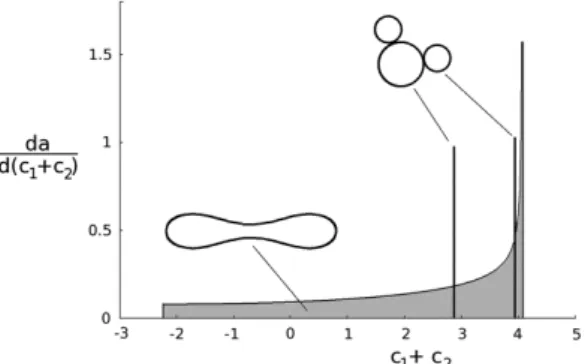
Fig. 5, which shows how much area belongs to different curvatures for the discocyte and the budded shape, illustrates the relevance of the described behavior for an RBC. The budded shape differs completely from that of the discocyte in involving only the curvatures of the spherical mother part and of the two daughter spheres. In the absence of inclusions, the local bending energy of the shape “e” is about three times as large as that of the discocyte [14]. However, if the intrinsic curvature of an inclusion is close to the curvature of one of the buds of the budded shape, this shape would, at a sufficiently large number of inclusions, become the shape with the lowest free energy of the system [4]. In the RBC, the free mobility of its transmembrane proteins and their complexes is diminished because of their attachment to the skeleton. The skeleton thus maintains membrane stability by immobilizing them. In this manner it prevents the RBC from transforming into a budded shape composed of a large mother sphere and spherical buds connected to it by narrow necks, such as in shape “e” in Fig. 3C. Such shapes are hazardous for the RBC because their buds could easily be pinched off from the mother sphere by shear forces in the blood stream, causing a deleterious increase of the RBC volume to membrane area ratio.
Fig. 5. Distribution of the membrane area over the sum of principal curvatures for the RBC disc shape (shape “c” in Fig. 3C) and for the budded shape (shape “e” in Fig. 3C). Curvatures are given in units of the reciprocal value of the radius of a sphere with the area of the RBC membrane. The distribution for the discocyte is continuous while that for the budded shape is discrete. In the latter case the heights of the two bars indicate the relative proportions of the areas corresponding to the two different curvatures of the budded shape. BUDDED SHAPES AND THE FORMATION OF AN ECHINOCYTE
membrane layer [10, 11] – which can for instance occur because of too long a stay of an RBC in a capillary where a > a0. As already indicated, a possible protection against budding in this case is for the a0 of the stress-free shape to be sufficiently different from the Δa of the budded shape.
Protection against budding resulting from an increase of a0 can also be based on having a low initial value of a0 and a high value of the a of the budded shape. This is achieved in the case of the RBC by the formation of echinocytes. Shapes of echinocytes are characterized by a large number of spiky evaginations, and the budding of such shapes can only occur at values of a much larger than that of shape “e” in Fig. 3C. The occurrence of echinocytes can thus be identified as an additional protective feature of the skeleton for maintaining RBC mechanical stability.
The explanation for the formation of echinocytes can be based on the elastic properties of the RBC membrane as a trilayer (Fig. 1B). Any change of the geometry of the skeleton causes an increase in its elastic energy. The RBC membrane is thus assumed to attain the shape that requires the smallest increase of the sum of the bending energy of its bilayer and the elastic energy of its skeleton. Analyses based on a minimal model of the RBC membrane elasticity, that describes its main functional requirements and emphasizes the complementary roles of the bilayer and the skeleton, predicted the echinocyte as a possible minimum energy shape [12, 13].
IMPLICATIONS
The RBC membrane comprises a large number of components (Fig. 1A) which may all play one role or another in its function. As has been deduced from the behavior of various RBC mutants, many of these components are involved in protecting the RBC against its mechanical malfunction [23]. However, their functional role is not yet fully understood. Here I have attempted to show that the reasons for the evolutionary acquisition of at least some of the acquired properties of RBC can be understood by comparing the mechanical behavior of model membranes exhibiting different partial characteristics of the RBC membrane. On this basis we have interpreted the attainment of the proper cell volume to membrane area ratio and the protection of RBC against budding. Of particular interest is the role of the RBC skeleton.
transmembrane proteins in different membrane regions, whereas in the RBC its function appears to be rather to distribute them uniformly over the whole membrane area. Its function in RBCs is thus of a different nature, but is basically still the same in the sense that it controls the membrane position of transmembrane proteins. Considering the evolution of the spectrin skeleton function, it can be assumed that the protective roles of the skeleton developed consecutively. In this sense it can be speculated that in the RBC the specialization of its skeleton regarding the formation of the echinocyte appeared as a later modification and improvement.
Acknowledgement. The work was supported by the Slovenian Research Agency through grants P1-0055 and J3-2268.
REFERENCES
1. Mohandas, N. and Gallagher, P.G. Red cell membrane: past, present, and future. Blood 112 (2008) 3939-3948.
2. Baines, A.J. Red cell skeleton. Evolution of the spectrin-based membrane skeleton. Transfus. Clin. Biol. 17 (2010) 95-103.
3. Svetina, S., Brumen, M. and Žekš, B. Mechanical behavior of closed layered membranes. in: Biophysics of Membrane Transport (Kuczera, J. and Przestalski, S., Eds.), Proceedings of the Ninth School on Biophysics of Membrane Transport, Polanica Zdroj, Poland, 1988, 151-169.
4. Svetina, S., Iglič A., Kralj-Iglič, V. and Žekš, B. Cytoskeleton and red cell shape. Cell. Mol. Biol. Lett. 1 (1996) 67-78.
5. Svetina, S. and Žekš, B. Shape behavior of lipid vesicles as the basis of some cellular processes. Anat. Rec. 268 (2002) 215-225.
6. Svetina, S., Kuzman, D., Waugh, R.E., Ziherl, P. and Žekš, B. The cooperative role of membrane skeleton and the bilayer in the mechanical behaviour of red blood cells. Bioelectrochemistry 62 (2004) 107-113. 7. Svetina, S. and Žekš, B. The elastic deformability of closed multilayered
membranes is the same as that of a bilayer membrane. Eur. Biophys. J. 21 (1992) 251-255.
8. Helfrich, W. Elastic properties of lipid bilayers: theory and possible experiments. Z. Naturforsch. 28c (1973) 693-703.
9. Božič, B., Svetina, S., Žekš, B. and Waugh, R.E. Role of lamellar membrane structure in tether formation from bilayer vesicles. Biophys. J. 61 (1992) 963-973.
10.Raphael, R.M., and Waugh R.E. Accelerated interleaflet transport of phosphatidylcholine molecules in membranes under deformation. Biophys. J. 71 (1996) 1374-1388.
12.Mukhopadhyay, R., Lim, G. and Wortis, M. Echinocyte shapes: bending, stretching, and shear determine bump shape and spacing. Biophys. J. 82 (2002) 1756-1772.
13.Kuzman, D., Svetina, S., Waugh, R.E. and Žekš, B. Elastic properties of the red blood cell membrane that determine echinocyte deformability. Eur. Biophys. J. 33 (2004) 1-15.
14.Svetina, S. and Žekš, B. Membrane bending energy and shape determination of phospholipid vesicles and red blood cells. Eur. Biophys. J. 17 (1989) 101-111.
15. Svetina, S. and Žekš, B. The mechanical behaviour of cell membranes as a possible physical origin of cell polarity. J. Theor. Biol. 146 (1990) 115-122. 16.Svetina, S., Božič, B., Majhenc, J. and Žekš, B. Mechanical properties of
closed lamellar membranes and cellular processes. Nonlin. Anal. 47 (2001) 269-280.
17.Käs, J., Sackmann, E., Podgornik, R., Svetina, S. and Žekš, B. Thermally induced budding of phospholipid vesicles - a discontinuous process. J. Phys. II France 3 (1993) 631-645.
18.Ziherl, P. and Svetina, S. Nonaxisymmetric phospholipid vesicles: Rackets, boomerangs and starfish. Europhys. Lett. 70 (2005) 690-696.
19.Kralj-Iglič, V., Svetina, S. and Žekš, B. Shapes of bilayer vesicles with membrane embedded molecules. Eur. Biophys. J. 24 (1996) 311-321. 20.Kralj-Iglič, V., Heinrich, V., Svetina, S. and Žekš, B. Free energy of closed
membrane with anisotropic inclusions. Eur. Phys. J. B 10 (1999) 5-8. 21.Božič, B., Kralj-Iglič, V. and Svetina, S. Coupling between vesicle shape
and lateral distribution of mobile membrane inclusions. Phys. Rev. E 73 (2006) 041915(11).
22.Sheetz, M.P. and Singer, S.J. Biological-membranes as bilayer couples - molecular mechanism of drug-erythrocyte interactions. Proc. Nat. Acad. Sci. USA 71 (1974) 4457-4461.