ORIGINAL ARTICLE
Linear and exponential TAIL-PCR: a
method for efficient and quick amplification
of flanking sequences adjacent to Tn5
transposon insertion sites
Xianbo Jia
1,2†, Xinjian Lin
1†and Jichen Chen
1*Abstract
Current genome walking methods are very time consuming, and many produce non-specific amplification products. To amplify the flanking sequences that are adjacent to Tn5 transposon insertion sites in Serratia marcescens FZSF02, we developed a genome walking method based on TAIL-PCR. This PCR method added a 20-cycle linear amplifica-tion step before the exponential amplificaamplifica-tion step to increase the concentraamplifica-tion of the target sequences. Products of the linear amplification and the exponential amplification were diluted 100-fold to decrease the concentration of the templates that cause non-specific amplification. Fast DNA polymerase with a high extension speed was used in this method, and an amplification program was used to rapidly amplify long specific sequences. With this linear and exponential TAIL-PCR (LETAIL-PCR), we successfully obtained products larger than 2 kb from Tn5 transposon insertion mutant strains within 3 h. This method can be widely used in genome walking studies to amplify unknown sequences that are adjacent to known sequences.
Keywords: LETAIL-PCR, Genome walking, Linear amplification, Tn5 transposon
© The Author(s) 2017. This article is distributed under the terms of the Creative Commons Attribution 4.0 International License (http://creativecommons.org/licenses/by/4.0/), which permits unrestricted use, distribution, and reproduction in any medium, provided you give appropriate credit to the original author(s) and the source, provide a link to the Creative Commons license, and indicate if changes were made.
Introduction
Tn transposons are widely used to study functional genes in bacteria by insertion knockouts (Reznikoff 2008). Amplification of flanking regions adjacent to the inser-tion sites is necessary for the detecinser-tion of mutant genes generated by random Tn transposon insertion. However, the flanking sequences adjacent to the known sequences cannot be amplified by conventional PCR because the sequences are unknown; therefore, specific primers pairs cannot be designed to amplify such regions. To solve this issue, currently, the random primed method and the ligation-mediated method are the most commonly used methods for flanking sequence amplification (Uchiyama
and Watanabe 2006; Yan et al. 2003). The random primed method is based on primer design and the optimization of the PCR protocol. For example, Liu and Whittier (1995) developed a version of random primed PCR termed thermal asymmetric interlaced PCR (TAIL-PCR), which employs three rounds of PCR with three specific (SP) primers that have a high Tm from the known sequences
and four arbitrary degenerate (AD) primers that have a low Tm for binding to the unknown sequences. This
method utilizes the specificity of PCR through increas-ing the bindincreas-ing efficiency of SP primers and decreasincreas-ing the binding efficiency of AD primers at higher anneal-ing temperatures. Subsequently, Liu and Chen (2007) improved the efficiency of TAIL-PCR and developed a new procedure termed high-efficiency thermal asym-metric interlaced PCR (hiTAIL-PCR). This procedure combines the advantages of TAIL-cycling and suppres-sion-PCR, thus significantly improving the amplifica-tion efficiency of the target flanking sequences. However,
Open Access
*Correspondence: [email protected]
†Xianbo Jia and Xinjian Lin contributed equally to this work 1 Institute of Soil and Fertilizer, Fujian Academy of Agricultural
and Sciences, Wusi Road, Fuzhou 350001, Fujian, People’s Republic of China
non-specific products are also often amplified when using the random primed methods. Completing three or more rounds of PCR is also time consuming. Ligation-mediated methods are based on digesting sequences with restriction enzymes, ligating sequences into a plasmid, transforming bacteria with plasmids and screening for strains containing the flanking regions. For example, to identify the flanking sequences of T-DNA insertions in plants, (Cottage et al. 2001) digested plant genomic DNA with blunt-cutting restriction enzymes and annealed an adapter sequence to the blunt-ended unknown region. Then, specific primers for the T-DNA and specific primers for the adaptor sequence were used to amplify the unknown flanking sequences using a conventional PCR procedure. A genome walking procedure termed loop-linker PCR was developed by adding a loop-linker adapter to the unknown region digested with restric-tion enzymes, which enhances amplificarestric-tion efficiency by suppressing the generation of nonspecific PCR prod-ucts (Trinh et al. 2012). Several other ligation-mediated methods have also been reported (Yin and Largaespad 2007; Rosenthal and Jones 1990; Huang et al. 2000; Tsaf-taris et al. 2010), these Ligation-mediated methods are effective and highly specific, but they require restriction enzyme digestion, ligation, DNA purification and clon-ing, which are laborious and time-consuming and are not suitable for high-throughput use, such as screening a mutant library with hundreds of clones. Random primed methods rely on PCR protocols, which are faster and simpler than ligation-mediated methods. However, these random primed methods produce non-specific amplifi-cation. Additionally, to amplify long flanking sequences, the extension time of the PCR procedure in these meth-ods is usually more than 3 min/cycle, resulting in these methods taking more than 1 day to complete, even for products < 1.5 kb (Liu and Chen 2007). So, a method that is fast, efficient and specific has yet to be considered.
On the basis of comparing the advantages and disad-vantages of the methods mentioned above, here, we pre-sent a more effective method termed LETAIL-PCR to obtain the flanking sequences adjacent to Tn5 transpo-son insertion sites. On the basis of hiTAIL-PCR, linear
pre-amplification combined with high-fold dilution of the pre-amplification product was used to decrease non-specific amplification. A version of high speed DNA polymerase was used to shorten the amplification time. Using this method, long PCR products can be obtained with high specificity in a short time.
Materials and methods
Strains and DNA template preparation
Serratia marcescens FZSF02 was isolated from soil in Fuzhou, China and was deposited in China Gen-eral Microbiological Culture Collection Center (CGMCC) with the strain number CGMCC 1.16177. Tn5 mutant strains of Serratia marcescens FZSF02 were obtained using the EZ-Tn5™ <KAN-2>Tnp Transpo-some™ Kit (Epicentre, USA). DNA templates were pre-pared with a Bacteria Genomic DNA Kit (TIANGEN, China) by extracting the genomes of wild-type FZSF02 and mutant FZSF02 strains. Three DNA samples from three FZSF02 mutant strains were used in this study. Flanking sequences adjacent to Tn5 transposon insertion sites of these three samples were submitted to GenBank with the accession numbers of MF034039 (sample 1), MF034040 (sample 2) and MF034041 (sample 3).
Primers and reagents
The non-specific primers LAD1 and LAD3 and the spe-cific primer AC1 were designed by Liu and Chen (2007). The primers F389, F536 and F772 were designed from the known region of the insertion site (Additional file 1). All primers are shown in Table 1.
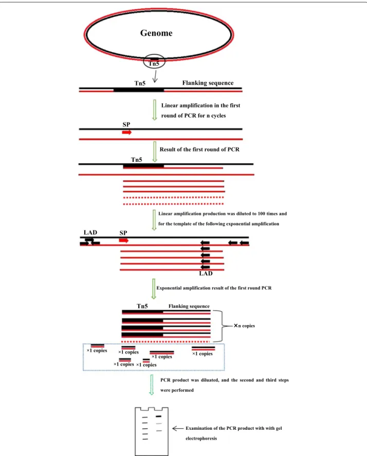
Overview of the method
This method is divided into three steps. The first step includes two reactions:a linear reaction and an exponen-tial reaction. For the linear reaction, a total PCR reac-tion volume of 20 μL was prepared containing 10 μL of prime STAR Max DNA polymerase (TAKARA Japan), 5 μL of primer F389 (100 pmol), 1 μL of genomic DNA (about 100 ng) and 4 μL of ddH2O. The thermal
condi-tions of the linear reaction are shown in Table 2. The product was diluted 100-fold and used as the template
Table 1 Primers used in this method
Primer name Sequence Tm (°C) Description
F389 5′-TCAAGCATTTTATCCGTACTCCTG-3′ 55.39 Designed by our lab
F536 5′-CGGTTGCATTCGATTCCTGTTTGTA-3′ 58.73 Designed by our lab
F772 5′-TAGGTTGTATTGATGTTGGACGAG-3′ 55.09 Designed by our lab
LAD1 5′-ACGATGGACTCCAGAG(G/C/A)N(G/C/A)NNNGGAA-3′ (Liu and Chen 2007) LAD3 5′-ACGATGGACTCCAGAG(T/A/C)N(A/G/C)NNNCCAC-3′ (Liu and Chen 2007)
for the subsequent exponential reaction. For the expo-nential reaction, a total PCR reaction volume of 20 μL was prepared containing 10 μL of prime STAR Max DNA polymerase (TAKARA Japan), 1 μL of primer F389 (20 pmol), 0.5 μL of primer LAD1 (10 pmol), 0.5 μL of LAD3 (10 pmol), 1 μL of template and 7 μL of ddH2O.
The reaction conditions for this exponential reaction are shown in Table 2. The PCR product was diluted 100-fold and used as the template for the second step. The second step also included two reactions: a linear reaction and an exponential reaction. For the linear reaction, a total PCR reaction volume of 20 μL was prepared containing 10 μL of prime STAR Max DNA polymerase (TAKARA Japan), 5 μL of primer F536 (100 pmol), 1 μL of the diluted PCR product from the first step as the template and 4 μL of ddH2O. The conditions of this linear reaction are shown
in Table 2. The product was diluted tenfold and used as the template for the following exponential reaction. For the exponential reaction, a total PCR reaction volume of 20 μL was prepared containing 10 μL of prime STAR Max DNA polymerase (TAKARA Japan), 2 μL of primer F536 (40 pmol), 2 μL of primer LAC1 (40 pmol), 1 μL of the diluted PCR product from the linear reaction as the template and 7 μL of ddH2O. The reaction conditions of
this exponential reaction are shown in Table 2. The PCR product was diluted ~ 10- to 50-fold for use as the tem-plate of the next third step. The third step contains one exponential reaction. For this reaction, a total PCR reac-tion volume of 20 μL was prepared containing 10 μL of prime STAR Max DNA polymerase (TAKARA Japan), 1 μL of primer F772 (20 pmol), 1 μL of primer LAC1 (20 pmol), 1 μL of the diluted PCR product from the sec-ond step as the template and 7 μL of ddH2O. The
reac-tion condireac-tions of this exponential reacreac-tion are shown in Table 2.
Sequence analysis
PCR products from the three exponential amplifications were electrophoresed on 1% agarose gels and visualized after ethidium bromide staining. The DNA bands on the gels were purified with a Gel Extraction Kit (OMEGA USA) and were sequenced directly with the Sanger
method. Chromas 2.22 was used to evaluate the sequence quality by examining the chromatograms.
Results
Amplification of the flanking unknown sequence
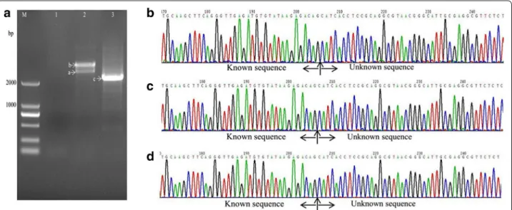
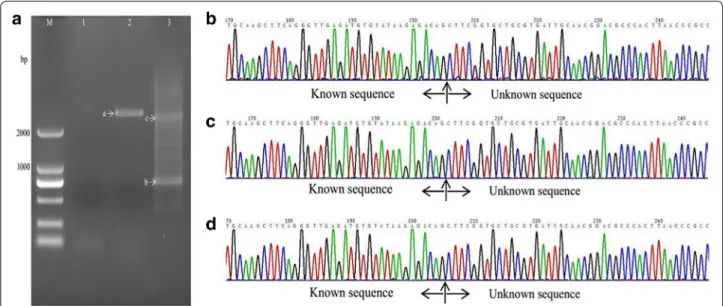
With the method described in this study (Fig. 1, Table 2), samples from all three mutant strains showed one or two visible bands larger than 2 kb (Figs. 2a, 3, 4a) after the second exponential amplification step. After the third step, the results varied significantly between the three samples. A distinct band that was slightly larger than 2 kb appeared in sample 1 (lane 3 of Fig. 2a), a weak band of approximately 1.5 kb appeared in sample 2 (lane 3 of Fig. 3a), and a weak band larger than 2 kb and a weak band of approximately 750 bp appeared in sample 3 (lane 3 of Fig. 4a). There were no visible bands in the three lanes of the gels for the DNA sample of the wild-type strain without Tn5 transposon (Fig. 5a). A non-linear amplification control was carried out with sample 1, and the result shows only lane 3 and lane 6 (Fig. 5b), which correspond to the third amplification step, have disper-sion zones.
Sequencing of the amplified DNA bands
The visible DNA bands on the gels of sample 1, sample 2 and sample 3 were sequenced, and the chromatograms showed that these band all contained target sequences adjacent to the inserted sequences (Figs. 2b–d, 3b–d, 4b–d). Moreover, the chromatograms showed almost no miscellaneous peaks, demonstrating that the DNA bands were very pure. Detailed DNA sequences of these DNA bands are shown in Additional file 1.
Discussion
The principle basis of LETAIL-PCR is outlined in Fig. 1. In detail, linear amplifications with a specific primer were carried out before the exponential amplifications to increase the relative quantity of the target sequences. For the first step, the linear amplification product was diluted 100-fold to decrease the relative genomic DNA concentration. Therefore, after the linear ampli-fication and dilution, non-specific ampliampli-fication in the
Table 2 Thermal conditions used in this method
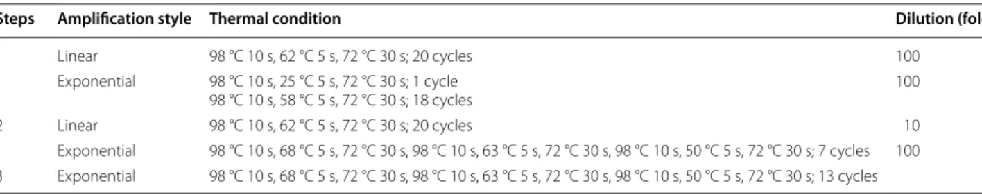
Steps Amplification style Thermal condition Dilution (fold)
1 Linear 98 °C 10 s, 62 °C 5 s, 72 °C 30 s; 20 cycles 100
Exponential 98 °C 10 s, 25 °C 5 s, 72 °C 30 s; 1 cycle
98 °C 10 s, 58 °C 5 s, 72 °C 30 s; 18 cycles 100
2 Linear 98 °C 10 s, 62 °C 5 s, 72 °C 30 s; 20 cycles 10
Exponential amplification result of the first round PCR
Examination of the PCR product with with gel electrophoresis
Tn5
Tn5 Flanking sequence
SP
LAD SP
LAD
Tn5
Tn5 Flanking sequence
×1 copies
×1 copies
×1 copies ×1 copies
×1 copies ×1 copies
Genome
Linear amplification in the first round of PCR for n cycles
PCR product was diluated, and the second and third steps were performed
Result of the first round of PCR
Linear amplification production was diluted to 100 times and for the template of the following exponential amplification
n copies
exponential amplification with the specific primer F389 and degenerate LAD primers is significantly decreased. For the second step, the product of the first step was diluted 100-fold for use as the template, linear ampli-fication with only the specific primer F536 was carried out, and the amplification product was diluted tenfold for use as the template for the following exponential amplification. After the two linear amplifications and
the two exponential amplifications described above were complete, the specific amplification was increased. There are several important minor points for the lin-ear amplification. First, the primer concentration was relatively high (100 pmol/20 μL reaction mixture) to enhance the specific annealing of the primer with the target sequence. Second, the annealing temperature of the linear amplification was set at a higher temperature
Fig. 2 Amplification and sequencing results of the sequence flanking the Tn5 insertion site of sample 1. a Amplification products shown on an agarose gel. Lanes 1–3 show the results of steps 1–3, respectively. b Chromatograms of DNA in band a. c Chromatograms of DNA in band b. d
Chromatograms of DNA in band c
Fig. 3 Amplification and sequencing results of the sequence flanking the Tn5 insertion site of sample 2. a Amplification product shown on an agarose gel. Lanes 1–3 show the results of steps 1–3, respectively. b Chromatograms of DNA in band a. c Chromatograms of DNA in band b. d
of 62 °C to reduce the possibility of non-specific ampli-fication. Third, the exponential amplifications of the first and second steps were both set at ~ 18–21 cycles, rather than the normal ~ 30 cycles, to decrease the generation of non-specific products. Fourth, the 100-fold dilution after the linear amplification in the first step decreases the genomic DNA concentration to avoid non-specific
amplification of the LAD1 and LAD3 primers with genomic DNA. The 100-fold dilution of the first expo-nential amplification product before the linear amplifi-cation of the second step decreases the concentration of LAD1 and LAD3 primers to avoid non-specific amplifi-cation in this linear amplifiamplifi-cation. These minor points all ensured the specificity of the amplification.
Fig. 4 Amplification and sequencing results of the sequence flanking the Tn5 insertion site of sample 3. a Amplification product showed on agarose gel. Lanes 1–3 show the results of steps 1–3, respectively. b Chromatograms of DNA in band a. c Chromatograms of DNA in band b. d
Chromatograms of DNA in band c
This LETAIL-PCR method also adopted the reported TAIL-PCR method (Liu and Chen 2007). TAIL-PCR uses specific primers from the known sequence (F389, F536 and F772 in this study) with Tm values higher than that of
the primer for the unknown sequence (LAC1). Therefore, in the PCR process, the target sequence can be amplified with higher annealing temperatures. Compared with the previous reports, linear amplification before exponential amplification further increased the specificity of TAIL-PCR. In our study, PCR products showed more than one band on the agarose gels (Figs. 2a, 3a, 4a), but all bands contained target sequences adjacent to the Tn5 transpo-son insertion sites. Specific products were found in the second step, which is accordance with the description of a similar protocol in a previous report (Luo et al. 2011).
In traditional random primed based methods, DNA polymerases have a theoretical extension speed of approxi-mately 1 kb/min. The extension time required for every cycle in these PCR procedures is more than 3 min to obtain long PCR products, which is very time consuming. In this study, to obtain longer unknown sequences, amplifying long enough single stranded DNA by linear amplification is necessary. So those faster DNA polymerases which can amplify longer DNA sequences in much shorter time are more suitable than the common Taq DNA polymerases. In this presented method, a type of fast DNA polymer-ase, PrimeSTAR Max DNA Polymerase (Takara, Japan), with a high extension speed of 5 s/kb was used. The PCR program for the method with this kind of DNA polymer-ase decrepolymer-ased the time requirement to 3 h compared with approximately 7 h for other genome walking methods (Liu and Chen 2007; Li et al. 2015). Though significantly less time was spent using this DNA polymerase and PCR program, the obtained PCR products were more than 2 kb, which are much longer than those in many previous reports (Singer and Burk 2003; Schmidt et al. 2007; Zhang et al. 2011; Ma et al. 2014; Reddy et al. 2008; Xu et al. 2013).
In conclusion, a modified LETAIL-PCR method was developed to amplify the flanking sequence of the Tn5 transposon insertion sites. Two linear amplifica-tions combined with high-fold dilution can significantly enhance the specificity of the amplification. The DNA polymerase and the corresponding amplification pro-gram allowed larger PCR products to be obtained in a short amount of time using this method. The high effi-ciency and short time required make this method very appealing for genome walking studies, including Tn transposon insertion sequence in microbes and flanking sequence identification for T-DNA insertion in plants.
Abbreviation
TAIL-PCR: thermal asymmetric interlaced polymerase chain reaction.
Authors’ contributions
JC designed and conducted the experiments, evaluated the results and drafted the manuscript. XJ and XL designed and conducted the study and drafted the manuscript. All authors read and approved the final manuscript.
Author details
1 Institute of Soil and Fertilizer, Fujian Academy of Agricultural and Sciences,
Wusi Road, Fuzhou 350001, Fujian, People’s Republic of China. 2 Institute
of Applied Ecology, Fujian Agriculture and Forestry University, Fuzhou, People’s Republic of China.
Acknowledgements
This work was supported by the Special Fund for Agro-scientific Research in the Public Interest of China, Social Research in the Public Interest of Fujian Finance and the Special Fund for Research institutes in the Public Interest of Fujian China.
Competing interests
The authors declare that they have no competing interests.
Availability of data and materials
All of the data are presented in the main paper or Additional file 1.
Consent for publication
All authors gave their consent for publication.
Ethical approval and consent to participate
This article does not contain any studies with human participants or animals performed by any of the authors.
Funding
This work was supported by the Special Fund for Agro-scientific Research in the Public Interest of China (201303094-05), Social Research in the Public Inter-est of Fujian Finance (2060302) and the Special Fund for Research institutes in the Public Interest of Fujian China (2014R1022-3).
Publisher’s Note
Springer Nature remains neutral with regard to jurisdictional claims in pub-lished maps and institutional affiliations.
Received: 23 April 2017 Accepted: 26 October 2017
References
Cottage A, Yang A, Maunders H, de Lacy RC, Ramsay NA (2001) Identification of DNA sequences flanking T-DNA insertions by PCR-walking. Plant Mol Biol Rep 19(4):321–327
Huang G, Zhang L, Birch RG (2000) Rapid amplification and cloning of Tn5 flanking fragments by inverse PCR. Lett Appl Microbiol 31(2):149–153 Li H, Ding D, Cao Y, Yu B, Guo L, Liu X (2015) Partially overlapping primer-based
PCR for genome walking. PLoS ONE 10(3):e0120139
Additional file
Liu Y, Chen Y (2007) High-efficiency thermal asymmetric interlaced PCR for ampli-fication of unknown flanking sequences. Biotechniques 43(5):649–656 Liu Y, Whittier RF (1995) Thermal asymmetric interlaced PCR: automatable
amplification and sequencing of insert end fragments from P1 and YAC clones for chromosome walking. Genomics 25(3):674–681
Luo P, Su T, Hu C, Ren C (2011) A novel and simple PCR walking method for rapid acquisition of long DNA sequence flanking a known site in micro-bial genome. Mol Biotechnol 47(3):220–228
Ma J, Wang N, Ren S, Fu Y, Lu S, Wang Y, Wang P (2014) Cloning of flanking sequence in transgenic plants by restriction site-anchored single-primer polymerase chain reaction. Genet Mol Res 13(4):10556–10561 Reddy PS, Mahanty S, Kaul T, Nair S, Sopory SK, Reddy MK (2008) A
high-throughput genome-walking method and its use for cloning unknown flanking sequences. Anal Biochem 381(2):248–253
Reznikoff WS (2008) Transposon Tn 5. Annu Rev Genet 42:269–286 Rosenthal A, Jones DS (1990) Genomic walking and sequencing by
oligo-cassette mediated polymerase chain reaction. Nucleic Acids Res 18(10):3095–3096
Schmidt M, Schwarzwaelder K, Bartholomae C, Zaoui K, Ball C, Pilz I, Braun S, Glimm H, Von Kalle C (2007) High-resolution insertion-site analy-sis by linear amplification-mediated PCR (LAM-PCR). Nat Methods 4(12):1051–1057
Singer T, Burke E (2003) High-throughput TAIL-PCR as a tool to identify DNA flanking insertions. Plant Funct Genom 236:241–271
Trinh Q, Shi H, Xu W, Hao J, Luo Y, Huang K (2012) Loop-linker PCR: an advanced PCR technique for genome walking. IUBMB Life 64(10):841–845 Tsaftaris A, Pasentzis K, Argiriou A (2010) Rolling circle amplification of
genomic templates for inverse PCR (RCA-GIP): a method for 5′-and 3′-genome walking without anchoring. Biotechnol Lett 32(1):157–161 Uchiyama T, Watanabe K (2006) Improved inverse PCR scheme for
metagen-ome walking. Biotechniques 41(2):183–188
Xu Z, Li Y, Mao Z, Yin B (2013) The development of APE-PCR for the cloning of genomic insertion sites of DNA elements. Biologia 68(4):766–772 Yan Y, An C, Li L, Gu J, Tan G, Chen Z (2003) T-linker-specific ligation PCR
(T-linker PCR): an advanced PCR technique for chromosome walking or for isolation of tagged DNA ends. Nucleic Acids Res 31(12):e68–e68 Yin B, Largaespad DA (2007) PCR-based procedures to isolate insertion sites of
DNA elements. Biotechniques 43(1):79–84