REVIEW ARTICLE
Epidemiology and Treatment Options for COVID-19: A Review
DOI: 10.29063/ajrh2020/v24i2s.18
Babatunde O. Adetuyi
1, Peace A. Olajide
1*, Elukunbi H. Awoyelu
1, Oluwatosin A. Adetuyi
2,
Oluwaseun A. Adebisi
2and Julius K. Oloke
1Department of Natural Sciences, Faculty of Pure and Applied Sciences, Precious Cornerstone University, Ibadan, Nigeria1; Department of Biochemistry, Osun State University, Osogbo, Osun State, Nigeria2
*For Correspondence: Email: [email protected]; [email protected]; Phone: +2348135547668
Abstract
The Coronavirus disease 19 (COVID-19) is a highly transmittable and pathogenic viral infection caused by severe acute respiratory syndrome Coronavirus 2 (SARS-CoV-2), which emerged in Wuhan, China and spread around the world. As of 19 June 2020 data from the World Health Organization (WHO) have shown that more than 8457305 confirmed cases have been identified in more than 200 countries, with the number of cases cutting across all continents. On 30th January 2020, the WHO declared COVID-19 as the sixth public health emergency of international concern. Genomic analysis revealed that SARS-CoV-2 is phylogenetically related to severe acute respiratory syndrome-like (SARS-like) bat viruses; therefore, bats could be the possible primary reservoir. The intermediate source of origin and transfer to humans is not known, however, the rapid human-to-human transfer has been confirmed widely via droplets or direct contact, and infection has been estimated to have mean incubation period of 6.4 days. Currently, controlling infection to prevent the spread of SARS-CoV-2 is the primary intervention being used. However, public health authorities should keep monitoring the situation closely, as the more we can learn about this novel virus and its associated outbreak, the better we can respond. (Afr J Reprod Health 2020 (Special Edition); 24[2]: 142-153).
Keywords: SARS-CoV-2, COVID-19, China, Phylogenetic, Genomic analysis, Remdesivir
Résumé
Le COVID-19 est une nouvelle maladie mortelle avec des informations limitées sur sa transmissibilité, la gravité de ses séquelles, ses manifestations cliniques et l'épidémiologie. Ce commentaire a analysé l'épidémiologie mondiale du COVID-19 parmi la population vulnérable. L'analyse a révélé que la plupart des cas pédiatriques de COVID-19 ne sont pas graves, mais que des maladies graves associées surviennent toujours chez les enfants. Tous les âges des enfants sont sensibles au COVID-19 et aucune différence significative entre les sexes n'existe. L'infection au COVID-19 pendant la grossesse a eu des conséquences fatales pour les mères, mais moins risquées pour le bébé. Les groupes de points chauds pour le COVID-19 sont les prisons / prisons, les maisons de soins infirmiers / de groupe et les établissements à long terme où résident la plupart des populations vulnérables. Les groupes ethniques minoritaires aux États-Unis et au Royaume-Uni sont exposés de manière disproportionnée à l'infection au COVID-19 et à la mort que les Caucasiens. La différence peut être due au fait que les minorités ethniques sont exposées à des risques plus élevés au travail et aux disparités structurelles économiques et sanitaires de longue date dans les deux pays. Il y a maintenant des changements dans les lignes directrices sur les personnes qualifiées pour recevoir des ventilateurs dans des situations difficiles dans de nombreux pays du monde si le système de santé est débordé. (Afr J Reprod Health 2020 (Special Edition); 24[2]: 142-153).
Mots-clés: COVID-19, épidémiologie, population vulnérable
Introduction
Since the emergence of the 2019 novel Coronavirus (2019- nCoV) infection in Wuhan, China, in December 20191, it has rapidly spread across China and many other countries2-8. So far, 2019-nCoV has affected more than 5.5 million persons in more than 200 countries, making it a major global health concern. On 11th February
wild animal market and most cases of infection, indicating possible animal-to-human transmission, and studies have increasingly demonstrated human-to-human transmission of SARS-CoV-2 through droplets or direct contact2,8-10. Moreover, according to one study, presumed hospital-related transmission of SARS-CoV-2 was suspected in 41% of patients8. Based on the evidence of a rapidly increasing incidence of infections11 and the possibility of transmission by asymptomatic carriers12, SARS-CoV-2 can be transmitted effectively among humans and exhibits high potential for a pandemic5,10,13. In addition to the high transmission efficiency of SARS-CoV-2, the advancement and convenience of global travel could further enhance its worldwide spread12. On 30th January 2020, the WHO declared the COVID-19 outbreak as the sixth public health emergency of international concern, following H1N1 (2009), polio (2014), Ebola in West Africa (2014), Zika (2016) and Ebola in the Democratic Republic of Congo (2019). Therefore, health workers, governments and the public needs to co-operate globally to prevent its spread14.
Evolution
Coronaviruses belong to the Coronaviridae family in the Nidovirales order. Corona represents crown-like spikes on the outer surface of the virus; thus, it was named as a Coronavirus. Coronaviruses are minute in size (65-125 nm in diameter) and contain a single-stranded RNA as a nucleic material, size ranging from 26 to 32 kbs in length (Figure 1). The subgroups of Coronaviruses family are alpha, beta, gamma and delta Coronavirus. Each of the subgroups of corona viruses has many serotypes. Some of them were affect human while other affected animals such as pigs, birds, cats, mice and dogs15-19.
In addition to seasonal influenza, reported pathogens of pneumonia include adenovirus, Coronavirus 229E/NL63/OC43, human bocavirus, human metapneumovirus, parainfluenza virus 1/2/3, rhinovirus and respiratory syncytial virus A/B20-23. Moreover, these viruses can cause co-infection in the setting of community- acquired bacterial pneumonia21-23. Using molecular methods, knowledge about the role of these viruses in the setting of pneumonia has achieved
significant advancements24-26. SARS- CoV-2 was found to be a positive-sense, single-stranded RNA virus belonging to the genus Betacoronavirus27-29. Phylogenetic analysis revealed that SARS-CoV-2 is closely related (88-89% similarity) to two bat-derived SARS-like Coronaviruses, namely bat-SL-CoVZC45 (GenBank accession no. MG772933.1) and bat- SL-CoVZXC21 (GenBank accession no. MG772934.1), but it is more distant from SARS-CoV (~79% similarity) and Middle East respiratory syndrome Coronavirus (MERS-CoV) (~50% similarity)28,30,31. Chen et al. applied an
RNA-based metagenomic next-generation
sequencing approach to identify a human Coronavirus from two pneumonia cases during the Wuhan outbreak in 201932. Its entire genome was 29881 bp in length32. Phylogenetic analysis indicates that SARS-CoV-2 is similar to the Coronavirus circulating in Rhinolophus (horseshoe bats), with 98.7% nucleotide similarity to the partial RNA-dependent RNA polymerase (RdRp) gene of the bat Coronavirus strain BtCoV/4991 (GenBank KP876546, 370 bp sequence of RdRp) and 87.9% nucleotide similarity to bat Coronavirus strain bat-SL-CoVZC45 and bat-SL-CoVZXC21. Evolutionary analysis based on ORF1a/1b, S and N genes suggests that SARS-CoV-2 is more likely a novel Coronavirus that was independently introduced from animals to humans32. Based on the findings of genomic investigations and the presence of some bats and live animals in the seafood market in Wuhan, SARS-CoV-2 may have originated from bats or bat droppings associated with contaminated materials in the market or surrounding region30,33.
Comparative analysis of emergence and
spread of Coronaviruses
Figure 1: Structure of respiratory syndrome causing human Coronavirus20
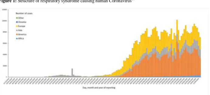
Figure 2: Epidemic curve of confirmed COVID-19 by continent, date of report and WHO region (according to the applied case definition and testing strategies in the affected countries) through 11 May 202036
Taiwan. Several cases of severe acute respiratory syndrome caused by corona and their mortally more than 1000 patient was reported in 2003. This was the black year for scientist. After a deep exercise they conclude and understand the pathogenesis of this disease and discovered it was corona virus. But still total of 8096 patients were confirmed as infected with corona virus. So, in 2004, World health organization and centers for disease control and prevention declared a state emergency. Another study report of Hong Kong confirmed 50 patients of severe acute respiratory syndrome while 30 of them were confirmed as corona virus infected while in 2012, Saudi Arabian reported several infected patient and deaths34-36. COVID-19 was first identified and isolated from pneumonia patient belongs to Wuhan, China.
Epidemiology
As of 25 May 2020, situation report from the WHO showed a total of 5 520 743 confirmed cases of COVID-19 globally36. Since the outbreak of COVID-19 originated from Wuhan City, Hubei province, in China, there has been a steady increase in the daily total number of COVID-19 cases globally, both within and outside China. Regarding new cases of COVID-19, a declining trend was found globally, in China but not outside China, mainly in international conveyance (Japan)
on 11th February 2020. Twenty-eight
Figure 3: The key reservoirs and mode of transmission of Coronaviruses (suspected reservoirs of SARS-CoV-2 are red encircled); only a and b Coronaviruses have the ability to infect humans, the consumption of infected animal as a source of food is the major cause of animal to human transmission of the virus and due to close contact with an infected person, the virus is further transmitted to healthy persons. Dotted black arrow shows the possibility of viral transfer from bat whereas the solid black arrow represents the confirmed transfer47
Figure 4: The life cycle of SARS-CoV-2 in host cells; begins its life cycle when S protein binds to the cellular receptor ACE2. After receptor binding, the conformation change in the S protein facilitates viral envelope fusion with the cell membrane through the endosomal pathway. Then SARS-CoV-2 releases RNA into the host cell. Genome RNA is translated into viral replicase polyproteins pp1a and 1ab, which are then cleaved into small products by viral proteinases. The polymerase produces a series of sub genomic mRNAs by discontinuous transcription and finally translated into relevant viral proteins. Viral proteins and genome RNA are subsequently assembled into virions in the ER and Golgi and then transported via vesicles and released out of the cell. ACE2,
SAR, the United Arab Emirates, Canada, France, the Philippines, the UK, Italy, India, Russia, Finland, Sweden, Sri Lanka, Cambodia, Nepal, Spain and Belgium. China has had the largest number of patients with COVID-19 (n= 42690), followed by Singapore (n = 45). Asia has had most of the reported cases, followed by Europe, North America and Australia, but no cases have been reported in Africa. Within China, Hubei has endured the largest number of infected patients (n = 31,728), followed by Guangdong (n = 1177), Zhejiang (n = 1117) and Henan (n = 1105). A total of 1017 mortalities have been reported globally with only 2 mortalities occurring outside of mainland China, one each in Hong Kong SAR and the Philippines. According to the Taiwan Centers for Disease Control, as of 12th February 2020, there were 45167 cases of COVID-19 reported from 28 countries/region and 1115 (2.5%) of patients had died. Among the 45167 cases, most were found in main-land China (n = 44653) and the reported mortality was 2.5% (n = 1113).
Biochemical
mechanism
of
human
Coronaviruses
All Coronaviruses contain specific genes in ORF1 downstream regions that encode proteins for viral replication, nucleocapsid and spikes formation37. The glycoprotein spikes on the outer surface of Coronaviruses are responsible for the attachment and entry of the virus to host cells (Figure 1). The receptor-binding domain (RBD) is loosely attached among virus, therefore, the virus may infect multiple hosts38, 39. Other Coronaviruses mostly recognize amino peptidases or carbohydrates as a key receptor for entry to human cells while SARS-CoV and MERS-CoV recognize exopeptidases. The entry mechanism of a Coronavirus depends upon cellular proteases which include, human airway trypsin-like protease (HAT), cathepsins and transmembrane protease serine 2 (TMPRSS2) that split the spike protein and establish further penetration changes40,41. MERS-Coronavirus employs dipeptidyl peptidase 4 (DPP4), while HCoV-NL63 and SARS-Coronavirus require angiotensin-converting enzyme 2 (ACE2) as a key receptor42. SARS-CoV-2 possesses the typical Coronavirus structure with spike protein and also expressed other
polyproteins, nucleoproteins, and membrane
proteins, such as RNA polymerase,
3chymotrypsin-like protease, papain-like protease, helicase, glycoprotein, and accessory proteins43,44. The spike protein of SARS-CoV-2 contains a 3-D structure in the RBD region to maintain the van der Waals forces45. The 394 glutamine residues in the RBD region of SARS-CoV-2 is recognized by the critical lysine 31 residue on the human ACE2 receptor46. The entire mechanism of pathogenicity of SARS-CoV-2, from attachment to replication is well mentioned in Figure 4.
Genomic variations in SARS-CoV-2
The genome of the SARS-CoV-2 has been reported over 80% identical to the previous human Coronavirus (SARS-like bat CoV)49. The Structural proteins are encoded by the four structural genes, including spike (S), envelope (E), membrane (M) and nucleocapsid (N) genes. The orf1ab is the largest gene in SARS-CoV-2 which encodes the pp1ab protein and 15 nsps. The orf1a gene encodes for pp1a protein which also contains 10 nsps50-52. It is also reported that Spike glycoprotein of the Wuhan Coronavirus is modified via homologous recombination. The spike glycoprotein of SARS-CoV-2 is the mixture of bat SARS-CoV and a not known Beta-CoV53. In a fluorescent study, it was confirmed that the
SARS-CoV-2 also uses the same ACE2
(angiotensin-converting enzyme 2) cell receptor and mechanism for the entry to host cell which is previously used by the SARS-CoV54,55. The single N501T mutation in SARS-CoV-2 s Spike protein may have significantly enhanced its binding affinity for ACE256.
Spread, control and prevention of COVID-19
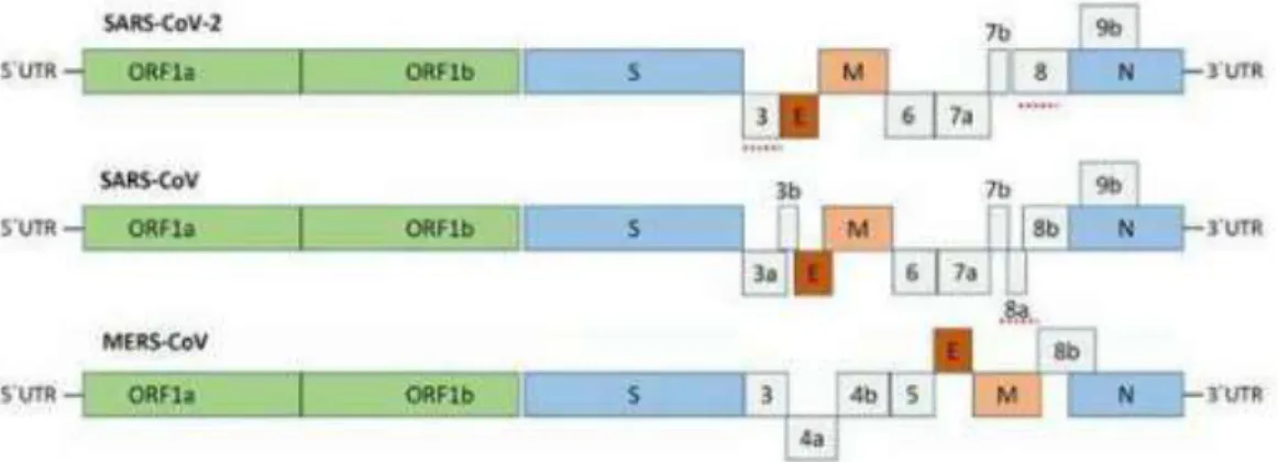
Figure 5: Betacoronaviruses genome organization; The Betacoronavirus for human (SARS-CoV-2, SARS-CoV and MERS-CoV) genome comprises of the 50-untranslated region (50UTR), open reading frame (orf) 1a/b (green box) encoding non-structural proteins (nsp) for replication, structural proteins including spike (blue box), envelop (maroon box), membrane (pink box), and nucleocapsid (cyan box) proteins, accessory proteins (light gray boxes) such as orf 3, 6, 7a, 7b, 8 and 9b in the SARS-CoV-2 genome, and the 30untranslated region (30-UTR). The doted underlined in red are the protein which shows key variation between SARS-CoV-2 and SARS-CoV. The length of nsps and orfs are not drawn in scale48
According to a report published on 24th January 202054, corona virus infected patients had common features such as fever, cough, and fatigue while diarrhea and dyspnea were found to be as uncommon feature. Many of the patient reported bilateral abnormalities. Corona virus was isolated from bronchoalvelor lavage fluid in China in 2020. It is also detected in blood samples. Till now, corona virus was not confirmed in feaces and urine sample of patient.
To decrease the damage associated with COVID-19, public health awareness and infection control measures are urgently required to limit the global spread of the virus59. Experience from the early phase of SARS-CoV-2 pneumonia strongly highlighted that travel history, rather than chest radiography, is of paramount importance for early detection and isolation of SARS-CoV-2 pneumonia cases. It is essential to limit human-to-human transmission in order to reduce secondary infections among close contacts and health-care workers and to prevent transmission amplification events and further international spread from China. Based on previous experience of management of MERS and SARS infections, the WHO recommend infection control interventions to reduce the general risk of transmission of acute respiratory infections, including avoiding close contact with people suffering from acute
respiratory infections, frequent hand-washing especially after direct contact with ill people or their environment, and avoiding unprotected contact with farm or wild animals60. Moreover, people with symptoms of acute respiratory infection should practice cough etiquette, which is to maintain distance, cover coughs and sneezes with disposable tissues or clothing, and wash hands, and within healthcare facilities enhanced standard infection prevention and control practices are recommended in hospitals, especially in emergency departments.
Therapeutic strategies so far
2019-nCoV invades through the respiratory mucosa and infects other cells, inducing cytokine storm systemically61. Some patients may progress rapidly with ARDS, disseminated intravascular coagulation (DIC), septic shock, and eventually multiple organ failure62. Therefore, early identification and timely treatment of critical cases is of crucial importance. Evidence-based therapy and supportive care in ICU (Intensive Care Unit) is the mainstay for the management of severe and life-threatening illness of COVID-19.
management, multi-organ function evaluation,
critical care of the nutritional
assessment/appropriate nutritional support is essential for these patients in ICU. In addition, attention should be paid to bedbound patients to prevent deep vein thrombosis.
Proposed therapeutic measures
At present, there is no drug to treat COVID-19 cases. But the under listed are proposed drugs being tried for the treatment of COVID-19
.
Multiple organ functional support and
nutrition strategy
According to the latest epidemiological report, the incidence for the critically ill patients to develop multiple organ dysfunction syndromes is up to 11%63. COVID-19 may be combined with other organ injuries, including liver injury, cardiac dysfunction, coagulopathy, which may need the routine functional support for critically ill patients in ICU. Moreover, all the critically ill patients with COVID-19 admitted into ICU have negative nitrogen balance and malnutrition64, which has been considered as a contributing factor to the emergence of viral infectious diseases. Therefore, appropriate nutritional strategy is pivotal for the treatment of critical illnesses when necessary.
Antiviral therapy
At present, there is no antiviral treatment with confirmed effectiveness for COVID-19. Available drug options that come from the clinical experience of treating SARS, MERS and other previous influenza virus have been used for the treatment of COVID-19 patients. Although these antiviral drugs are promising in the treatment of COVID-19, it should be kept in mind that: (1) the adverse effects of the drugs need to be monitored, (2) the effects of these drugs on critically ill patients still need to be clarified, (3) the potential mutation of the Coronavirus may lead to the drug resistance of the virus.
Remdesivir
Remdesivir (GS-5734) is a new nucleoside analog and has been recognized as a potential and
promising antiviral drug against a wide array of RNA viruses, including SARS/MERS-CoV. It is currently under clinical development for the treatment of Ebola virus infection56,62. Remdesivir potentially inhibits the RNA-dependent RNA polymerase from MERS-CoV, reduces virus replication, decreases the virus titer in mouse lungs infected with MERS-CoV, and improves the lung tissue damage58. The antiviral activity of Remdesivir and IFN-beta was found to be superior to that of the combination of lopinavir/ritonavir and IFN-beta against MERS-CoV65. A randomized, controlled trial has reported that the prolonged use of Remdesivir in the treatment of Ebola virus disease (EVD) is safe55, and no adverse events have been observed66. As a candidate drug that has not been approved, information about the side effects of Remdesivir has not been reported yet. At present, two randomized, controlled, double-blind clinical trials are ongoing to evaluate the efficacy and safety of Remdesivir (200mg loading dose on Day 1, followed by 100mg I.V. once-daily maintenance dose for 9days) in hospitalized patients with mild/moderate or severe COVID-19 respiratory disease67,68. The results of these clinical trials may open the window for effective antiviral therapy for such an epidemic infectious disease.
Chloroquine and hydroxychloroquine
It is known that angiotensin-converting enzyme-2 (ACE2) as a membrane protein is a functional receptor of SARS-CoV and it can facilitate virus entry into the cells by binding to the spike (S) protein of the virus, which mediates the fusion of viral and host membranes68. Therefore, it may be of importance to block the binding of S protein to ACE2 to treat viral infection, such as SARS-CoV72. Chloroquine is a 9-aminoquin-oline known since 1934, the sulfate and phosphate salts of which have both been commercialized as widely used antimalarial and autoimmune disease drugs69. Chloroquine also shows broad-spectrum antiviral effects73
2019-nCoV infection76. Besides the antiviral activity, Chloroquine has an immune-modulating activity, which may synergistically enhance its antiviral effects in vivo. Recently, Wang et al. have demonstrated that Chloroquine is highly effective in the control of 2019-nCoV infection in vitro and is suggested to be assessed in human patients suffering from COVID-1976
. In addition, the results from more than 100 COVID-19 patients have indicated that Chloroquine phosphate is superior to the control treatment in inhibiting the exacerbation of pneumonia, improving lung imaging, promoting virus negative conversion, and shortening the disease course70. However, attention should be paid to the potential detrimental effects of Chloroquine observed in previous attempts to treat viral diseases. At present, the clinical trial to evaluate the efficacy and safety of Chloroquine in the treatment of COVID-19 is ongoing71. The use of Chloroquine in the treatment of COVID-19 should refer to the most recent announcements if any. In addition, hydroxychloroquine is a 4-aminoquinoline derivative antimalarial drug. Hydroxychloroquine is an immunosuppressive drug with mature clinical application in the treatment of rheumatic immune diseases such as rheumatoid arthritis and systemic lupus erythematosus72, 73. It has been found to be more potent than Chloroquine in
inhibiting 2019-nCoV in vitro.
Hydroxychloroquine sulfate 400mg given twice daily for 1 day, followed by 200 mg twice daily for another 4 days is recommended in the treatment of COVID-1973. At present, the clinical evaluation of hydroxychloroquine in the treatment of COVID-19 is in progress74, which might shortly provide preliminary results about the effectiveness of hydroxychloroquine.
Chinese traditional medicine
In 2003, Chinese traditional medicine was used to prevent and treat SARS75. In 2009, during the pandemic of H1N1 influenza, the Traditional Chinese Medicine of China issued a Chinese traditional medicine prevention program, which included several Chinese herbal medicine formulae for the prevention of infection of adults and children. ShuFengJieDu capsules and Lianhuaqingwen capsules have also played a role
in the prevention and treatment of new respiratory infectious diseases such as influenza A (H1N1)76,77. Some studies have confirmed that Yupingfeng powder has antiviral, anti-inflammatory and immunoregulatory effects77
. A multicenter, large-scale, randomized trial found that Yinqiao powder plus another heat-clearing formula could reduce time for fever resolution in patients with the H1N1 influenza virus infection 78-81
. It is suggested that high-risk populations exposed to COVID-19 patients, including medical staff, family members, and other people who are in close contact with COVID-19 patients, as well as residents living in COVID-19 outbreak areas, might benefit from taking Chinese traditional medicine formulae for prevention. However, the efficacy and safety of these Chinese traditional medicine formulae in COVID-19 need to be further confirmed by clinical trials.
Methods
An extensive search spanning from December 2019 to May 2020 was conducted in Medline, EMBASE, Elsevier, Lancet, BioMed Central, PubMed and Cochrane Library databases. The following search terms were used in all possible combinations: (Coronavirus disease 2019‖ [MeSH] OR ―2019 novel coronavirus‖ [MESH] OR ―SARS-CoV-2‖ [MESH] OR ―COVID-19‖ [MESH] OR ―2019-nCov [MESH], limiting to human subjects. The titles and abstracts of relevant studies were reviewed, and full-text articles were obtained for detailed evaluation which allowed for eligible studies to be included in the systematic review.
Inclusion and exclusion criteria
The inclusion criteria were the following: clinical trials, patients with laboratory-identified
SARS-CoV-2 infection, treatment outcomes,
epidemiological, clinical and laboratory characteristics. The exclusion criteria were absence of clinical characteristics, treatment outcomes, clinical experience and case reports.
Data extraction and quality assessment
remaining articles were screened by their titles and abstracts for relevance. Second, full text articles were reviewed by two of the authors (BOA and EHA) to assess their conformity with the study inclusion criteria. The research domains, publishing dates, journal language, authors‘ affiliations, as well as methodological characteristics were analyzed respectively. All findings and statements regarding the outbreak in this review are based on published information as listed in the references.
Conclusion
The outbreak of COVID-19 has become a clinical threat to the general population and healthcare workers worldwide. However, knowledge about this novel virus remains limited. The effective option of antiviral therapy and vaccination are currently under evaluation and development. What we can do now is aggressively implement infection control measures to prevent the spread of SARS- CoV-2 via human-to-human transmission. Public health authorities should keep monitoring the situation, as the more we learn about this novel virus and its associated outbreaks, the better we can respond.
Contributions of Authors
All authors contributed immensely to this review article. Babatunde O. Adetuyi made substantial contributions to conception and design; Peace A. Olajide and Elukunbi H. Awoyelu participated in acquisition of data and drafting of the manuscript; Oluwatosin A. Adetuyi and Oluwaseun A. Adebisi revised the article critically for important intellectual content; and the Julius K. Oloker gave a final approval of the version to be submitted.
Funding
None
References
1. Lu H, Stratton CW and Tang YW. Outbreak of pneumonia of unknown etiology in Wuhan China: the mystery and the miracle. J Med Virol 2020
Jan 16; [Epub ahead of print]. doi: 10.1002/jmv.25678.
2. Li Q, Guan X, Wu P, Wang X, Zhou L and Tong Y. Early transmission dynamics in Wuhan, China, of novel coronavirus-infected pneumonia. N Engl J Med 2020 Jan 29; [Epub ahead of print]. doi: 10.1056/NEJMoa2001316.
3. Gorbalenya AE, Baker SC, Baric RS, de Groot RJ, Drosten C and Gulyaeva AA. Severe acute respiratory syndrome-related coronavirus: the species and its viruses—a statement of the Coronavirus Study Group. bioRxiv 2020 Feb 11; doi: 10.1101/2020.02.07.937862.
4. Chen N, Zhou M, Dong X, Qu J, Gong F and Han Y. Epidemiological and clinical characteristics of 99 cases of 2019 novel coronavirus pneumonia in Wuhan, China: a descriptive study. Lancet 2020; 395:507–13. doi: 10.1016/ S0140- 6736(20)30211- 7.
5. Huang C, Wang Y, Li X, Ren L, Zhao J and Hu Y. Clinical features of patients infected with 2019 novel coronavirus in Wuhan, China. Lancet 2020; 395:497506. doi: 10.1016/S0140-6736(20)30183-5.
6. Wang C, Horby PW, Hayden FG and Gao GF. A novel coronavirus outbreak of global health concern. Lancet 2020; 395:470–3. doi: 10.1016/S0140-6736(20)30185-9.
7. Holshue ML, DeBolt C, Lindquist S, Lofy KH, Wiesman J and Bruce H. First case of 2019 novel coronavirus in the United States. N Engl J Med 2020 Jan 31; [Epub ahead of print]. doi: 10.1056/NEJMoa2001191.
8. Wang D, Hu B, Hu C, Zhu F, Liu X and Zhang J. Clinical characteristics of 138 hospitalized patients with 2019 novel coronavirus-infected pneumonia in Wuhan, China. JAMA 2020 Feb 7; [Epub ahead of print]. doi: 10.1001/jama.2020. 1585.
9. Chang D, Lin M, Wei L, Xie L, Zhu G and Dela Cruz CS. Epidemiologic and clinical characteristics of novel coronavirus infections involving 13 patients outside Wuhan, China. JAMA 2020 Feb 7; [Epub ahead of print]. doi: 10.1001/ jama.2020.1623.
10. Carlos WG, Dela Cruz CS, Cao B, Pasnick S and Jamil S. Novel Wuhan (2019- nCoV) coronavirus. Am J Respir Crit Care Med 2020; 201: P7–8. doi: 10.1164/ rccm.2014P7.
11. Zhao S, Lin Q, Ran J, Musa SS, Yang G and Wang W. Preliminary estimation of the basic reproduction number of novel coronavirus (2019-nCoV) in China, from 2019 to 2020: a data-driven analysis in the early phase of the outbreak. Int J Infect Dis 2020; 92:214–17. doi: 10.1016/j.ijid.2020.01.050.
January 2020, from the Latin-American Society for Travel Medicine (SLAMVI). Travel Med Infect Dis 2020;101567. doi: 10.1016/j.tmaid.2020.101567.
13. Munster VJ, Koopmans M, van Doremalen N, van Riel D and de Wit E. A novel coronavirus emerging in China—key questions for impact assessment. N Engl J Med 2020 Jan 24; [Epub ahead of print]. doi: 10.1056/NEJMp20 0 0929. 14. Yoo JH. The fight against the 2019-nCoV outbreak: an
arduous march has just begun. J Korean Med Sci 2020; 35:e56. doi: 10.3346/jkms.2020.35. e56. 15. Mailles A, Blanckaert K, Chaud P, van der Werf S, Lina
B and Caro V. First cases of Middle East respiratory syndrome Coronavirus (MERS-CoV) infections in France, investigations and implications for the prevention of human-to-human trans-mission, Euro Surveill. 2013; 18:20502.
16. Buchholz U, Müller MA, Nitsche A, Sanewski A, Wevering N and Bauer-Balci T. Contact investigation of a case of human novel coronavirus infection treated in a German hospital, October-November 2012. Euro Surveill. 2013; 18:20406. 17. Saif LJ. Animal coronaviruses: what can they teach us
about the severe acute respiratory syndrome? Rev Sci Tech. 2004; 23:643–60.
18. Gwaltney JM Jr. Virology and immunology of the common cold. Rhinology. 1985; 23:265.
19. Tyrrell DAJ and Myint SH. Chapter 60: Coronaviruses. In Barson 1 S, editor. Medical microbiology. 4th edition. Galveston: University of Texas Medical Branch at Galveston; 1996.
20. Lee KH, Yoo SG, Cho Y, Kwon DE, La Y and Han SH. Characteristics of community-acquired respiratory viruses infections except seasonal influenza in transplant recipients and non-transplant critically ill patients. J Microbiol Im- munol Infect 2019 Jun 19; [Epub ahead of print]. doi: 10.1016/j.jmii.2019.05.007.
21. Chou CC, Shen CF, Chen SJ, Chen HM, Wang YC and Chang WS. Recommendations and guidelines for the treatment of pneumonia in Taiwan. J Microbiol Immunol Infect 2019; 52:172–99. doi: 10.1016/j.jmii.2018.11.004.
22. Lee JY, Yang PC, Chang C, Lin IT, Ko WC and Cia CT. Community-acquired adenoviral and pneumococcal pneumonia complicated by pulmonary aspergillo- sis in an immunocompetent adult. J Microbiol Immunol Infect 2019; 52:838–9. doi: 10.1016/j.jmii.2019.05.014.
23. Su IC, Lee KL, Liu HY, Chuang HC, Chen LY and Lee YJ. Severe community- acquired pneumonia due to Pseudomonas aeruginosa coinfection in an influenza A(H1N1)pdm09 patient. J Microbiol Immunol Infect 2019; 52:365–6. doi: 10.1016/j.jmii.2018.05.007.
24. Lee SH, Ruan SY, Pan SC, Lee TF, Chien JY and Hsueh PR. Performance of a multiplex PCR pneumonia panel for the identification of
respiratory pathogens and the main determinants of resistance from the lower respiratory tract specimens of adult patients in intensive care units. J Microbiol Immunol Infect 2019; 52:920–8. doi: 10.1016/j.jmii.2019.10.009.
25. Hung HM, Yang SL, Chen CJ, Chiu CH, Kuo CY and Huang KA. Molecular epidemiology and clinical features of rhinovirus infections among hospitalized patients in a medical center in Taiwan. J Microbiol Immunol Infect 2019; 52:233– 41. doi: 10.1016/j.jmii.2018.08.009.
26. Lin GL, Lu CY, Chen JM, Lee PI, Ho SY and Weng KC. Molecular epidemiology and clinical features of adenovirus infection in Taiwanese children, 2014. J Microbiol Immunol Infect 2019; 52:215–24. doi: 10.1016/j.jmii.2018.07.005. 27. Chan JF, Kok KH, Zhu Z, Chu H, To KK and
Yuan S. Genomic characterization of the 2019 novel human-pathogenic coronavirus isolated from a patient with atypical pneumonia after visiting Wuhan. Emerg Microbes Infect 2020; 9:22136. doi: 10.1080/22221751.2020.1719902. 28. Lu R, Zhao X, Li J, Niu P, Yang B and Wu H.
Genomic characterisation and epidemiology of 2019 novel coronavirus: implications for virus origins and receptor binding. Lancet 2020 Jan 30; [Epub ahead of print]. doi: 10.1016/ S0140- 6736(20)30251- 8.
29. Zhu N, Zhang D, Wang W, Li X, Yang B and Song J. A novel coronavirus from patients with pneumonia in China, 2019. N Engl J Med 2020 Jan 24; [Epub ahead of print]. doi: 10.1056/NEJMoa2001017.
30. Jiang S, Du L and Shi Z. An emerging coronavirus causing pneumonia outbreak in Wuhan, China: calling for developing therapeutic and prophylactic strategies. Emerg Microbes Infect
2020; 9:275–7. doi:
10.1080/22221751.2020.1723441.
31. Ren LL, Wang YM, Wu ZQ, Xiang ZC, Guo L and Xu T. Identification of a novel coronavirus causing severe pneumonia in human: a descriptive study. Chin Med J (Engl) 2020 Jan 30; [Epub ahead of print]. doi: 10.1097/CM9. 0 0 0 0 0 0 0 0 0 0 0 0 0722.
32. Chen L, Liu W, Zhang Q, Xu K, Ye G and Wu W. RNA based mNGS approach identifies a novel human coronavirus from two individual pneumonia cases in 2019 Wuhan outbreak. Emerg Microbes Infect 2020; 9:313–19. doi: 10.1080/ 22221751.2020.1725399.
33. Zhou P, Yang XL, Wang XG, Hu B, Zhang L and Zhang W. A pneumonia out- break associated with a new coronavirus of probable bat origin. Nature 2020 Feb 3; [Epub ahead of print]. doi: 10.1038/s41586- 020- 2012- 7.
35. World Health Organization. Coronavirus never before seen in humans is the cause of SARS– update 31. Geneva: The Organization; 2003.
36. World Health Organization Situation report.Available at:
https://www.who.int/emergencies/diseases/novel-coronavirus-2019/situation reports (2020). 37. Peiris JS, Lai ST, Poon LL, Guan Y, Yam LY and Lim
W. Coronavirus as a possible cause of severe acute respiratory syndrome. Lancet. 2003; 361:1319– 25 38. Raj VS, Mou H, Smits SL, Dekkers DH, Müller MA and
Dijkman R. Dipeptidyl peptidase 4 is a functional receptor for the emerging human coronavirus-EMC. Nature 2013; 495(7440):251–4.
39. Perlman S and Netland J. Coronaviruses post-SARS: update on replication and pathogenesis. Nat Rev Microbiol 2009;7(6):439–50.
40. Glowacka I, Bertram S, Müller MA, Allen P, Soilleux E and Pfefferle S. Evidence that TMPRSS2 activates the severe acute respiratory syndrome coronavirus spike protein for membrane fusion and reduces viral control by the humoral immune response. J Virol 2011; 85(9):4122–34.
41. Bertram S, Glowacka I, Müller MA, Lavender H, Gnirss K and Nehlmeier I. Cleavage and activation of the severe acute respiratory syndrome coronavirus spike protein by human airway trypsin-like protease. J Virol 2011; 85 (24):13363–72.
42. Wu F, Zhao S, Yu B, Chen Y-M, Wang W and Song Z- G. A new coronavirus associated with human respiratory disease in China. Nature 2020; 1–5. 43. Zhou P, Yang X, Wang X, Hu B, Zhang L and Zhang W.
A pneumonia outbreak associated with a new coronavirus of probable bat origin. Nature [Internet]. 2020; Feb: 3.
44. Xu X, Chen P, Wang J, Feng J, Zhou H and Li X. Evolution of the novel coronavirus from the ongoing Wuhan outbreak and modeling of its spike protein for risk of human transmission. Science China Life Sciences 2020; 63 (3):457–60.
45. Wan Y, Shang J, Graham R, Baric RS and Li F. Receptor recognition by novel coronavirus from Wuhan: an analysis based on decade-long structural studies of SARS. J Virol 2020.
46. WuA, Peng Y, Huang B, Ding X, Wang X and Niu P. Genome composition and divergence of the novel coronavirus (2019-nCoV) originating in China. Cell Host Microbe 2020.
47. Lu R, Zhao X, Li J, Niu P, Yang B and Wu H. Genomic characterization and epidemiology of 2019 novel coronavirus: implications for virus origins and receptor binding. Lancet 2020; 395(10224):565–74. 48. Chen Y, Liu Q and Guo D. Emerging coronaviruses:
genome structure, replication, and pathogenesis. J Med Virol 2020.
49. Hui DS, I Azhar E, Madani TA, Ntoumi F, Kock R and Dar O. The continuing 2019-nCoV epidemic threat of novel coronaviruses to global health—the latest 2019 novel coronavirus outbreak in Wuhan, China.
International Journal of Infectious Diseases. 2020; 91:264–6.
50. Li B, Si H-R, Zhu Y, Yang X-L, Anderson DE and Shi Z-L. Discovery of Bat Coronaviruses through Surveillance and Probe Capture-Based Next Generation Sequencing. mSphere. 2020; 5(1). 51. Gralinski LE and Menachery VD. Return of the
coronavirus: 2019-nCoV. Viruses 2020; 12(2):135. 52. Xu X, Chen P, Wang J, Feng J, Zhou H and Li X.
Evolution of the novel coronavirus from the ongoing Wuhan outbreak and modeling of its spike protein for risk of human transmission. Sci China Life Sci 2020; 1–4.
53. Sun P, Qie S and Liu Z. Clinical characteristics of 50466 hospitalized patients with 2019-nCoV infection. J
Med Virol. 2020. https
://doi.org/10.1002/jmv.25735.
54. Wang D and Hu B. Clinical characteristics of 138 hospitalized patients with 2019 novel coronavirus-infected pneumonia in Wuhan, China. JAMA. 2020; https ://doi.org/10.1001/jama.2020.1585.
55. Mulangu S, Dodd LE and Davey RT Jr. A randomized, controlled trial of Ebola virus disease therapeutics. N Engl J Med. 2019; 381(24):2293–303.
56. Gordon CJ, Tchesnokov EP and Feng JY. The antiviral compound remdesivir potently inhibits RNA-dependent RNA polymerase from Middle East respiratory syndrome coronavirus. J Biol Chem. 2020; https ://doi.org/10.1074/jbc.AC120 .01305 6. 57. Wit E, Feldmann F and Cronin J. Prophylactic and
therapeutic remdesivir (GS-5734) treatment in the rhesus macaque model of MERS-CoV infection. Proc Natl Acad Sci USA. 2020. https ://doi.org/10.1073/pnas.19220 83117.
58. Sheahan TP, Sims AC and Leist SR. Comparative therapeutic efficacy of remdesivir and combination lopinavir, ritonavir, and interferon beta against MERS-CoV. Nat Communs. 2020; 11(1):222. 59. Holshue ML, DeBolt C and Lindquist S. First case of
2019 novel coronavirus in the United States. N Engl J Med. 2020; 382(10):929–36.
60. Cao B. Mild/moderate 2019-nCoV remdesivir RCT— Full Text View ClinicalTrials.gov. 2020. https ://clinicaltrials.gov/ct2/show/NCT04 25266 4. Accessed 13 Feb 2020.
61. Li W, Moore MJ and Vasilieva N. Angiotensin-
converting enzyme 2 is a functional receptor for the SARS coronavirus. Nature. 2003; 426(6965):450–4. 62. Dimitrov DS. The secret life of ACE2 as a receptor for
the SARS virus. Cell. 2003; 115(6):652–3. 63. Simmons G, Reeves JD and Rennekamp AJ.
Characterization of severe acute respiratory syndrome-associated coronavirus (SARS-CoV) spike glycoprotein-mediated viral entry. Proc Natl Acad Sci USA. 2004; 101(12):4240–5.
is highly effective in treating avian influenza A H5N1 virus infection in an animal model. Cell Res. 2013; 23(2):300–2.
66. Vincent MJ, Bergeron E and Benjannet S. Chloroquine is a potent inhibitor of SARS coronavirus infection and spread. Virol J. 2005; 2:69.
67. Xu H, Zhong L and Deng J. High expression of ACE2 receptor of 2019-nCoV on the epithelial cells of oral mucosa. Int J Oral Sci. 2020; 12(1):8.
68. Wang M, Cao R and Zhang L. Remdesivir and
chloroquine effectively inhibit the recently emerged novel coronavirus (2019-nCoV) in vitro. Cell Res. 2020. https ://doi.org/10.1038/s4142 2-020-0282-0. 69. Gao J, Tian Z and Yang X. Breakthrough: chloroquine
phosphate has shown apparent efficacy in treatment of COVID-19 associated pneumonia in clinical studies. Biosci Trends. 2020; 14(1):72–3.
70. W. S. Chloroquine Prevention of Coronavirus Disease (COVID-19) in the Healthcare Setting (COPCOV). 2020. https ://clinicaltrials.gov/ct2/show/NCT04 30350 7. Accessed 11 Mar 2020.
71. Ponticelli C and Moroni G. Hydroxychloroquine in systemic lupus erythematosus (SLE). Expert Opin Drug Saf. 2017; 16(3):411–9.
72. Schrezenmeier E and Dorner T. Mechanisms of action of hydroxychloroquine and chloroquine: implications for rheumatology. Nat Rev Rheumatol. 2020; 16(3):155–66.
73. Yao X, Ye F and Zhang M. In vitro antiviral activity and projection of optimized dosing design of hydroxychloroquine for the treatment of severe acute respiratory syndrome Coronavirus 2 (SARS-CoV-2). Clin Infect Dis. 2020. https ://doi.org/10.1093/cid/ciaa2 37.
74. Hernandez-Cardenas C. Hydroxychloroquine Treatment
for Severe COVID-19 Pulmonary Infection (HYDRA Trial) (HYDRA). 2020. https ://clinicaltrials.gov/ct2/show/NCT04 31589 6. Accessed 20 Mar 2020.
75. Liu J, Manheimer E and Shi Y. Chinese herbal medicine for severe acute respiratory syndrome: a systematic review and meta-analysis. J Altern Complem Med. 2004; 10(6):1041–51.
76. Ji S, Bai Q and Wu X. Unique synergistic antiviral effects of Shufeng Jiedu Capsule and oseltamivir in influenza A viral-induced acute exacerbation of chronic obstructive pulmonary disease. Biomed Pharmacother. 2020; 121:109652.
77. Ding Y, Zeng L and Li R. The Chinese prescription lianhuaqingwen capsule exerts anti-influenza activity through the inhibition of viral propagation and impacts immune function. BMC Complement Altern Med. 2017; 17(1):130.
78. Du CY, Zheng KY and Bi CW. Yu Ping Feng San, an ancient Chinese herbal decoction induces gene expression of anti-viral proteins and inhibits neuraminidase activity. Phytother Res. 2015; 29(5):656–61.
79. Wang C, Cao B and Liu QQ. Oseltamivir compared with the Chinese traditional therapy maxingshigan-yinqiaosan in the treatment of H1N1 influenza: a randomized trial. Ann Intern Med. 2011; 155(4):217–25. 79.
80. Huang C, Wang Y and Li X. Clinical features of patients infected with 2019 novel coronavirus in Wuhan, China. Lancet. 2020; 395(10223):497–506. 81. Chen N, Zhou M and Dong X. Epidemiological and