Topoisomerase 1 Regulates Gene Expression
in Neurons through Cleavage
Complex-Dependent and -Independent Mechanisms
Angela M. Mabb1,2, Jeremy M. Simon1, Ian F. King1, Hyeong-Min Lee1, Lin-Kun An3, Benjamin D. Philpot1*, Mark J. Zylka1*
1Department of Cell Biology and Physiology, UNC Neuroscience Center, Carolina Institute for
Developmental Disabilities, The University of North Carolina, Chapel Hill, North Carolina, United States of America,2Neuroscience Institute, Georgia State University, Atlanta, Georgia, United States of America, 3School of Pharmaceutical Sciences, Sun Yat-sen University, Guangzhou, China
*[email protected](BDP);[email protected](MJK)
Abstract
Topoisomerase 1 (TOP1) inhibitors, including camptothecin and topotecan, covalently trap TOP1 on DNA, creating cleavage complexes (cc’s) that must be resolved before gene tran-scription and DNA replication can proceed. We previously found that topotecan reduces the expression of long (>100 kb) genes and unsilences the paternal allele ofUbe3ain neurons. Here, we sought to evaluate overlap between TOP1cc-dependent and -independent gene regulation in neurons. To do this, we utilizedTop1conditional knockout mice,Top1 knock-down, the CRISPR-Cas9 system to deleteTop1, TOP1 catalytic inhibitors that do not gener-ate TOP1cc’s, and a TOP1 mutation (T718A) that stabilizes TOP1cc’s. We found that topotecan treatment significantly alters the expression of many more genes, including long neuronal genes, immediate early genes, and paternalUbe3a, when compared toTop1 dele-tion. Our data show that topotecan has a stronger effect on neuronal transcription than
Top1deletion, and identifies TOP1cc-dependent and -independent contributions to gene expression.
Introduction
Topoisomerases are enzymes that resolve DNA supercoils by creating transient single (Type I topoisomerases) or double (Type II topoisomerases) strand breaks [1,2]. These enzymes facili-tate DNA replication, chromosomal segregation, DNA repair, and gene transcription [3]. In postmitotic cells, topoisomerases predominantly regulate gene transcription and DNA repair [4]. Topoisomerase I (TOP1) relieves DNA supercoiling ahead of RNA polymerase to facilitate transcription elongation [5–7]. Although the roles of topoisomerases in dividing cells have been studied extensively, much less is known about their functions in neurons.
Long noncoding RNAs (lncRNA) can act as transcriptional activators or repressors in post-mitotic neurons and other cell types [8,9].Ube3aantisense (Ube3a-ATS) is an extremely long a11111
OPEN ACCESS
Citation:Mabb AM, Simon JM, King IF, Lee H-M, An L-K, Philpot BD, et al. (2016) Topoisomerase 1 Regulates Gene Expression in Neurons through Cleavage Complex-Dependent and -Independent Mechanisms. PLoS ONE 11(5): e0156439. doi:10.1371/journal.pone.0156439
Editor:Michal Hetman, University of Louisville, UNITED STATES
Received:February 23, 2016
Accepted:May 14, 2016
Published:May 27, 2016
Copyright:© 2016 Mabb et al. This is an open access article distributed under the terms of the
Creative Commons Attribution License, which permits unrestricted use, distribution, and reproduction in any medium, provided the original author and source are credited.
Data Availability Statement:RNA-seq data were deposited in the GEO database (accession no. GSE79951).
lncRNA (>1 Mb) and is expressed exclusively from the paternal allele in most neurons during development and throughout adulthood. Paternal expression ofUbe3a-ATSsilences the pater-nal copy ofUbe3avia a transcriptional collision mechanism [10–12].
We previously found that TOP1 and TOP2 inhibitors unsilence the paternal allele ofUbe3a in postmitotic neurons by reducing expression ofUbe3a-ATS[13]. Mutations that reduce or increase UBE3A function are linked to Angelman syndrome (AS) and autism, respectively [14–21]. In addition to downregulatingUbe3a-ATS, TOP1 and TOP2 inhibitors also downre-gulate the expression of other long (generally>100 kb) genes in neurons, many of which are associated with neurotransmission and synaptic function [22]. Consistent with reduced expres-sion of long synaptic genes, inhibition of TOP1 with topotecan disrupts excitatory and inhibi-tory synaptic transmission in cortical neuron cultures, an effect that is reversible following inhibitor washout [23]. TOP1 inhibitors also reduce expression of long genes in non-neuronal cell types [24,25].
Topotecan binds at the interface between TOP1 and DNA, creating a TOP1-DNA enzyme intermediate known as a TOP1 cleavage complex (TOP1cc) [26]. Given this unique mecha-nism of inhibition, we sought to determine the extent to which TOP1 and TOP1cc formation contribute to neuronal gene expression andUbe3aregulation. To answer these questions, we generated aTop1conditional knockout mouse to genetically deleteTop1from cultured neu-rons. We also utilized the CRISPR-Cas9 system to deleteTop1, used short hairpin (sh)RNAs to knock-downTop1, compared TOP1 catalytic inhibitors that do not generate TOP1cc’s to topo-tecan, and utilized a TOP1 (T718A) mutation that stabilizes TOP1cc’s. Surprisingly, we found that topotecan affected the expression of many more genes when compared to deletion ofTop1 —the molecular target of topotecan. Taken together, our findings reveal TOP1cc-dependent and -independent control of gene expression andUbe3aregulation in neurons. Our findings also have implications for cancer therapies that target TOP1 via these distinct mechanisms.
Materials and Methods
Knockout first ES cells targeting theTop1gene were acquired from the KOMP Repository Knockout Mouse Project (Project ID: CSD36970,Top1tm1a(KOMP)Wtsi). ES cells were microin-jected into albino C57BL/6 blastocysts by the UNC Animal Models Core Facility. Two chimeric lines were bred for germline transmission. Successful germline transmitted mice were then crossed to aFLP1recombinase deleter mouse B6.Cg-Tg(ACTFLPe)9205Dym/J (Jackson Labo-ratory) to excise thelacZ/neomycincassette (removal confirmed by PCR), then backcrossed further to C57Bl/6 mice to remove the Flp transgene. To distinguish genotypes forTop1cKO mice, the following primers flanking the LoxP site and within theTop1gene were used: geno 2, 5’-GAGTTTCAGGACAGCCAGGA-3’and geno 3,5’-GGACCGGGAAAAGTCTAAGC-3’.
Neuronal Cultures
Embryonic day E13.5–15.5 mouse cortical neuron cultures were prepared by cervical disloca-tion of adult C57BL6/J wild-type females as described [13]. Animals were kept on a 12-hour light-dark cycle and givenad libitumaccess to food and water. All experimental animal proce-dures were carried out according to the NIHGuide for the Care and Use of Laboratory Animals and were approved by the Institutional Animal Care and Use Committee at the University of North Carolina at Chapel Hill. For immunostaining, dissociated neurons were plated in 24-well dishes containing poly-D-lysine (0.1 mg/ml) coated 12 mm coverslips at a density of 2.5 x 10⁵cells/well. For biochemistry, dissociated neurons were seeded on poly-D-lysine coated 12-well dishes at a density of 5 x 10⁵cells/well.
J.M.S.), and by the National Natural Science Foundation of China (No. 8137357) to L.K.A. A.M.M. was supported by the Joseph E. Wagstaff Postdoctoral Research Fellowship from the Angelman Syndrome Foundation. The confocal imaging core was funded by grants from National Institute of Neurological Disorders and Stroke and National Institute of Child Health and Human Development (P30NS045892; P30HD03110). The funders had no role in study design, data collection and analysis, decision to publish, or preparation of the manuscript.
Western Blotting
Lentiviruses harboring pLenti-CaMKIIα-tdTomato and pLenti-CamKIIα -tdTomato-P2A-CRE based vectors were prepared by the UNC Lentiviral Core. LentiviralTop1shRNA was generated as previously described [22]. Briefly, cortical neurons were transduced at DIV 3 with lentivirus at a multiplicity of infection of at least two to maximize the number of trans-duced cells (around 85–90% transduction efficiency). Media containing lentivirus was removed 24 hours later and replaced with conditioned media. The CaMKIIαpromoter limited tdTo-mato expression to neurons and was detectable without antibody amplification 3–4 days post transduction. Neurons were then treated at DIV 15 with vehicle (0.003% DMSO, Neurobasal medium) or 300 nM topotecan (Molcan Corporation; in 0.003% DMSO, Neurobasal medium) and harvested 3 days later.
For western blot experiments, cells were harvested and lysed in RIPA buffer (50 mM Tris-HCl, 150 mM NaCl, 0.5% sodium deoxycholate, 1% Triton X-100, and 0.1% SDS, pH 7.4) with 1 mM DTT, 1μg/mL aprotinin, 2μg/mL leupeptin, and 0.1 mM PMSF. Total protein (25– 40μg) was run on a 4–15% gradient SDS-PAGE gel (Bio-RAD). Proteins were then transferred to nitrocellulose membrane, blocked overnight in Odyssey Blocking Buffer (LI-COR), and immunoblotted overnight using the following antibodies: rabbit anti-UBE3A (1:1,000; Bethyl Laboratories, A300-352A), mouse anti-UBE3A (1:1,000; BD Biosciences), mouse anti-NLGN1 (1:500; Synaptic Systems, 129 111), mouse anti-NRXN1 (1:500; BD Biosciences, 611882), mouse anti-CNTNAP2 (1:1,000; NeuroMab, 75–075), mouse anti-β-actin (1:5,000; Millipore, MAB1501R), rabbit anti-TOP1 (1:10,000; GeneTex, GTX63013), or mouse anti-TOP1 (1:250; Santa Cruz, sc-271285). The GeneTex rabbit monoclonal antibody was raised against the N-terminus of human TOP1. The Santa Cruz mouse monoclonal antibody was raised against the C-terminus (amino acids 685–765) of human TOP1. Both antibodies are predicted to react with mouse TOP1. Membranes were washed three times with water at room temperature and the appropriate IRDye secondary antibodies (Li-COR) were added at a dilution of 1:15,000– 1:20,000 for 1 hour at room temperature. Blots were then washed two times in Tris-buffered saline containing 0.1% Tween-20 and two times with water. Membranes were dried in the dark and imaged using the ODYSSEY CLx Infrared Imaging System (LI-COR). Equivalent amounts of protein per sample were loaded and loading controls were used to ensure equiva-lent loading between samples. Experiments were performed on a minimum of three indepen-dent culture sets.
Immunocytochemistry
with 1:1,500 dilution of mouse anti-TOP1-DNA covalent complex antibody (Millipore, clone 1.1A) in 3% NGS in PBS overnight at 4°C.
Images were acquired using a Zeiss LSM 710 upright microscope with a 20X/0.8 NA objec-tive. Images were acquired with identical settings (gain, contrast, pinhole) for UBE3A, TOP1, and TOP1 DNA covalent complexes. Treatment and transfected conditions were interleaved during each imaging session. The intensity of UBE3A, TOP1, and TOP1 DNA covalent com-plexes was quantified from maximum intensity projections in FIJI following thresholding at least 2 standard deviations above background. For the CRISPR-Cas9Ube3aunsilencing experi-ments, regions of interest (ROIs) were identified by outlining the soma of tdTomato positive neurons manually. ROIs were then transposed on both the UBE3A and TOP1 channel to mea-sure the integrated density. Note that for theTop1CrispR-Cas9 experiments, we excluded cells where TOP1 was not deleted, and average TOP1 integrated density was at or above the average TOP1 intensity in untransfected neurons. For experiments usingAS::Top1fl/flneuron cultures, ROIs were selected automatically in FIJI. Briefly, DAPI images were thresholded and nuclei were separated using the Watershed Tool. ROIs were then outlined using the Analyze Particles tool with a setting size of 8μm to infinity. The integrated density of each ROI was then trans-posed to a thresholded (two standard deviations above background) UBE3A channel where the integrated density was measured. ROIs were then transposed onto the GFP channel to identify transfected and untransfected neurons.
Cloning
To generate the pLenti-CamKIIα-tdTomato and pLenti-CamKIIα-tdTomato-P2A-CRE con-structs, tdTomato and tdTomato-P2A-CRE fragments were PCR cloned into the pLenti-Cam-KIIα-ChR2-mCherry vector (http://www.everyvector.com/sequences/show/20437). Briefly, ChR2-mCherry was excised and replaced with tdTomato or td-Tomato-P2A-CRE usingAgeI andBsrGIsites for tdTomato and BamHI and EcoRI sites for tdTomato-P2A-CRE. Human GFP-TOP1 was PCR cloned into the the pLenti-CamKIIα-ChR2-mCherry vector (modified from Karl Deisseroth’s laboratory) usingAgeIandEcoRIrestriction sites. The PCR template for human TOP1 was a kind gift from Stefan Weger from the Intitut fur Virologie in Berlin, Germany. The GFP-TOP1 cleavable complex mimetic (T718A) was created using site directed mutagenesis using the following primer sets:5’-AAACAGATTGCCCTGGGAGCCTCCAAACTC AATTATC-3’and5’-GATAATTGAGTTTGGAGGCTCCCAGGGCAATCTGTTT-3’. CRISPR-Cas9 targeting ofTop1was accomplished by annealingTop1sgRNAs into the lentiCRISPR v1 vector backbone (Addgene) using the suggested cloning strategy ( http://www.genome-engineering.org/crispr/?page_id=23). A total of fourTop1sgRNA targets were designed using the E-CRISP design tool (http://www.e-crisp.org/E-CRISP/). The following primer sets were used to clone into the lentiCRISPR v1 backbone:Clone #1:5’-CACCGCCGGGGCTTTTCCGA
GGCCG-3’and5’-AAACCGGCCTCGGAAAAGCCCCGGC-3’Clone #2:5’-CACCGATCGG
AAATCCGCTTCGATC-3’and5’-AAACGATCGAAGCGGATTTCCGATC-3’Clone #3:5’
-CACCGTCGGAAATCCGCTTCGATCT-3’and5’-AAACAGATCGAAGCGGATTTCCGAC-3’
Clone #4:5’-CACCGAGATCGAGAACACCGGCATA-3’and5’-AAACTATGCCGGTGTTCT CGATCTC-3’. Each individual clone was tested forTop1loss by immunostaining for TOP1 protein in neurons. Clone #1 and #4 were deemed the most efficient; clone #4 was used for sub-sequent experiments.
RNA-seq
RNA yield and quality was determined with a Nanodrop 1000 Spectrophotometer (Thermo Scientific). Samples were further assessed for quality using either an Agilent Bioanalyzer 2100 or TapeStation 2200 to obtain a RNA integrity number (RIN). RIN values exceeding 7 were used for sequencing. RNA samples were used to generate and barcode cDNA libraries using the TruSeq RNA Library Preparation Kit at the UNC High Throughput Sequencing Facility. Pools of 24 multiplexed samples were sequenced per lane in a HiSeq 2500 sequencer using 50 bp paired-end reads.
RNA-Seq Data Processing
RNA-seq reads were filtered using TagDust and aligned to the reference mouse genome (mm9) with TopHat using default parameters. Reads aligning to rRNA genes were removed. Tran-script abundance was estimated by computing RPKM using RefSeq gene models aggregated by gene symbol. For differential expression analyses, raw counts over RefSeq exons were used and compared across samples using EdgeR. RNA-seq data were deposited in the GEO database (accession no. GSE79951).
Synthesis of TOP1 Catalytic Inhibitors
TOP1 catalytic inhibitors were synthesized and characterized as described [28–30].
Results
TOP1-Dependent Control of Neuronal Genes
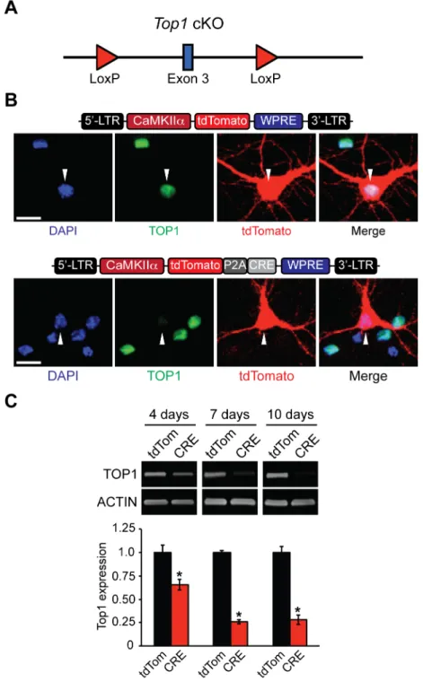
The TOP1 inhibitor topotecan suppresses expression of long genes and unsilences the paternal copy ofUbe3ain neurons [13,22]. To determine if these transcriptional effects could be recapit-ulated by deletion ofTop1, we generated aTop1conditional knockout mouse (cKO), as homo-zygous deletion ofTop1is embryonic lethal with failure occurring between the 4 and 16-cell stages [31]. TheTop1cKO allele contains two LoxP sites flanking exon 3 (Fig 1A) such that Cre-mediated excision is predicted to facilitate nonsense-mediated decay ofTop1mRNA and thus disrupt TOP1 protein levels. To confirm thatTop1can be deleted in these mice, we cre-ated tdTomato (control) and CRE-dependent lentiviral constructs driven by the neuron-spe-cific CamKIIαpromoter (Fig 1B). Transfection of CRE, but not tdTomato, reduced TOP1 protein levels inTop1cKO neurons (Fig 1B). Infection ofTop1fl/flcultured neurons with tdTo-mato or CRE lentivirus resulted in a transduction efficiency ranging from 85–90% (data not shown). TOP1 levels were maximally decreased 7 days post infection with CRE compared to tdTomato control neurons (Fig 1C). Additionally, no lower molecular weight TOP1-reactive bands were detected, indicating that truncated products of TOP1 are not generated in neuronal cultures fromTop1cKO mice (S1A Fig). The residual levels of TOP1 most likely originate from uninfected neurons and/or non-neuronal cells in the cultures.
Fig 1. Generation and validation ofTop1cKO mouse.(A) Schematic of theTop1cKO allele. LoxP sites flankexon 3. (B) Schematic of tdTomato (top) and tdTomato-P2A-CRE lentiviral plasmids (bottom).Top1fl/fl neurons were transfected with tdTomato or tdTomato-P2A-CRE plasmids. Neurons were fixed and immunostained with an anti-TOP1 antibody. Scale bar, 10μm. (C) Cortical neurons were infected with tdTomato or tdTomato-P2A-CRE lentivirus at DIV 3 and then were harvested at DIV 7, DIV 10, and DIV 13. Representative immunoblots and quantification of TOP1 protein expression normalized to ACTIN (bottom). Mean±s.e.m., unpaired student’s t-test;*p<0.05, n = 3 cultures.
suggesting the transcriptional effects of topotecan depend onTop1and are thus molecularly on-target.
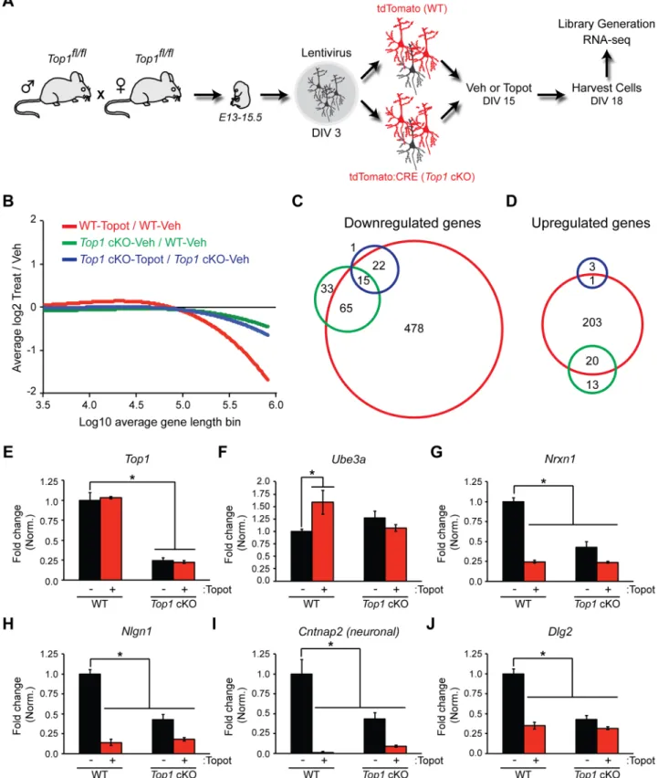
Relative to WT-Veh, we found multiple downregulated (Fig 2C) and upregulated (Fig 2D) genes (FDR of<0.01) in WT-Topot,Top1cKO-Veh, andTop1cKO-Topot cortical neuron cultures. A total of 580 genes were significantly decreased in WT-Topot cells, whereas 113 genes were significantly decreased inTop1cKO-Veh cells. Eighty of these 113 genes were downregulated in both WT-Topot andTop1cKO-Veh cells (S1 Table). Based on Gene Ontol-ogy, downregulated genes in WT-Topot andTop1cKO-Veh cells were functionally annotated to common biological processes such as synaptic transmission and cell adhesion (S2 Table). There was no statistically identifiable functional annotation forTop1cKO-Topot cells in the downregulated gene set (S2 Table). We also looked at the expression of individual genes that were reduced in WT-Topot cells but not inTop1 cKO-Veh cells. Strikingly, a large proportion of immediate early genes (IEGs) were decreased in WT-Topot but not inTop1cKO-Veh cells (S2 Fig). Moreover, relative toTop1cKO-Veh, we did not detect a decrease in IEG expression inTop1cKO-Topot cells, suggesting the change in IEG expression isTop1-dependent. These findings indicate that topotecan reduces expression of IEGs in a TOP1-dependent manner, and that deletion of TOP1 alone does not reduce expression of IEGs. Collectively, these data indicate that the transcriptional effects of topotecan are significantly greater than the effects of TOP1 deletion, consistent with the fact that topotecan generates TOP1cc’s and does not simply inhibit TOP1.
We found that 224 genes were upregulated in WT-Topot cells, whereas only 33 genes were upregulated inTop1cKO-Veh cells. Additionally,Top1cKO-Topot cells had a significant increase in 4 genes, of which 1 overlapped with WT-Topot cells (Fig 2DandS1 Table). Based on Gene Ontology, upregulated genes in WT-Topot cells were functionally annotated to axon guidance and cell motion processes whereTop1cKO-Veh upregulated genes were functionally annotated to eye lens development (S3 Table).
As expected,Top1transcript levels were reduced inTop1cKO cells (Fig 2E). Moreover, we observed elevated expression ofUbe3ain WT-Topot but not inTop1cKO-Veh cells (Fig 2F). Several long synaptic adhesion genes were also downregulated in WT-Topot andTop1 cKO--Veh cells (Fig 2G–2J), consistent with our previous findings [22]. Taken together, these data indicate that topotecan-treatment or TOP1 deletion reduces expression of a subset of long genes, which include extremely long synaptic adhesion genes.
Top1
Deletion Reduces Synaptic Adhesion Protein Levels
Previously, we found that topotecan downregulates synaptic proteins and dampens synaptic transmission [23]. Here we found that conditional deletion ofTop1in cortical neuron cultures reduced the expression of synaptic adhesion proteins to a similar extent as in topotecan-treated WT cells (Fig 3A and 3B). Addition of topotecan to CRE-infected cells did not further decrease protein expression, indicating thatTop1deletion occluded additional effects of topotecan on these synaptic adhesion proteins (Fig 3A and 3B). Using an independent genetic approach, we employed aTop1-specific lentiviral shRNA to reduce TOP1 (S3A and S3B Fig), which also reduced the expression of two long synaptic adhesion proteins, NEUREXIN-1 and NEUROLI-GIN-1 (S3A and S3B Fig).
Top1cKO (Top1cKO-Veh, green), and topotecan-treatedTop1cKO (Top1cKO-Topot, blue) cortical neuron cultures relative to vehicle-treated WT (WT-Veh) cells. The FDR was set at a value of<0.01. (D) Venn diagram showing the number of significantly upregulated genes. (E—J) Representative transcript level changes from RNA-seq analysis. Normalized RPKM values (relative to WT-Veh) in WT-Veh, WT-Topot,Top1cKO-Veh, andTop1cKO-Topot. Mean±s.e.m., FDR<0.1, n = 3 cultures.
Topotecan, but Not
Top1
Deletion, Unsilences
Ube3a
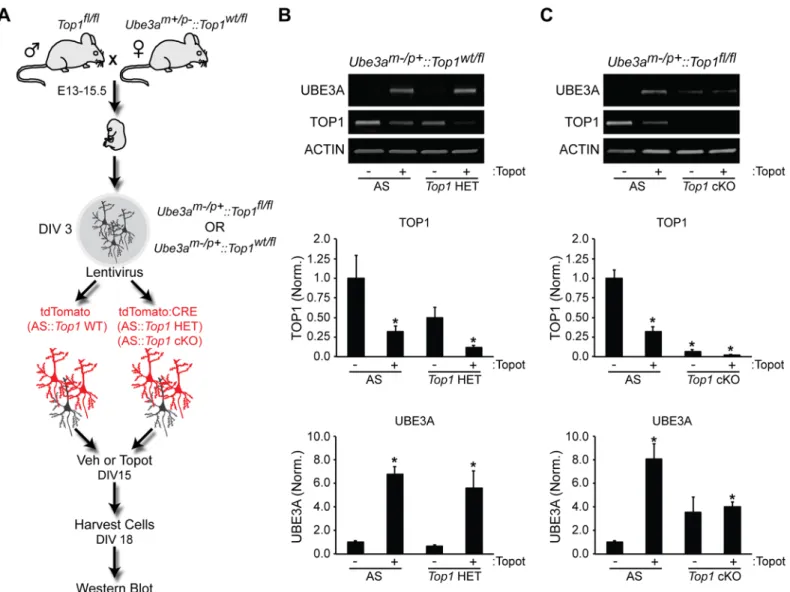
We next sought to determine if genetic reduction or deletion ofTop1could unsilenceUbe3ain neurons. First, we cultured cortical neurons lacking the maternal copy ofUbe3am-/p+(AS) and transduced them with lentiviralTop1shRNA (S3C and S3D Fig). This manipulation reduced TOP1 protein levels but was not sufficient to unsilenceUbe3a, as demonstrated by the lack of detectable paternal UBE3A protein (S3C and S3D Fig). TOP1 protein levels were reduced ~50% in these knockdown experiments, raising the possibility that residual levels of TOP1 might maintainUbe3a-ATStranscription and hence maintain repression of paternalUbe3a. To examine this possibility, we crossedTop1cKO mice with AS mice, prepared cortical neuron cultures, and transduced cells with either tdTomato control (WT) or CRE lentivirus to delete Top1(Top1cKO) (Fig 4A). Neurons were then treated with vehicle or topotecan to test for the ability to unsilenceUbe3a. As expected, treatment ofAS::Top1wt/flneurons with topotecan led to unsilencing of paternalUbe3ain WT andTop1heterozygous mutant neurons (Fig 4B). However, complete deletion ofTop1did not significantly increase UBE3A levels inAS::Top1fl/fl neurons (Fig 4C). Moreover, compared to WT neurons,Top1cKO neurons exhibited blunted Ube3aunsilencing after treatment with topotecan (Fig 4C).
To replicate our findings using a different genetic approach, we utilized the CRISPR-Cas9 system to deleteTop1in wildtype (WT) and AS cortical neuron cultures. WT cells were trans-fected with Cas9 alone (control) or Cas9 with an sgRNA directed toTop1. We observed a near-complete loss of TOP1 using a sgRNA targeted toTop1relative to controls (Fig 5A and 5B, S4A Fig). Consistent with ourTop1cKO studies above,Top1CRISPR-mediated deletion did not increase UBE3A expression (Fig 5A and 5B). In contrast, UBE3A levels were increased in topotecan-treated neurons (Fig 5A and 5B), but not in topotecan-treatedTop1deficient
Fig 3. TOP1 deletion reduces synaptic adhesion protein levels.(A)Top1fl/flneuron cultures were infected with tdTomato (WT) or tdTomato-P2A-CRE (Top1cKO) lentivirus at DIV 3. Cells were then treated at DIV 15 with vehicle (DMSO) or 300 nM topotecan for 72 hours. Shown are representative immunoblots with antibodies to NRXN1, NLGN1, CNTNAP2, TOP1, and ACTIN. (B) Quantification of fold change in TOP1, NRXN1, NLGN1, and CNTNAP2 protein expression normalized to ACTIN. Mean±s.e.m., unpaired student’s t-test;*p<0.05, n = 3 cultures.
neurons. To examine changes in paternal UBE3A expression, we transfected Cas9 and the sgRNA targetingTop1into AS neurons. Consistent with ourTop1cKO studies above, CRISPR-mediated deletion ofTop1was not sufficient to increase UBE3A in AS cortical neu-rons (Fig 5C and 5D). Moreover, the increase in paternal UBE3A expression in topotecan-treated AS neurons was attenuated in topotecan-topotecan-treatedTop1deficient neurons (Fig 5C and 5D).
Formation of TOP1 Cleavage Complexes Unsilences
Ube3a
Since deletion ofTop1did not unsilenceUbe3a, whereas topotecan (which forms TOP1cc’s) did unsilenceUbe3a, we hypothesized that TOP1cc’s may be required to unsilenceUbe3ain neurons. To test this hypothesis, we compared topotecan to a series of TOP1 catalytic inhibi-tors that inhibit TOP1 without forming TOP1cc’s (S5A Fig) [28,29]. As previously found [13], topotecan unsilenced the paternalUbe3a-YFP alleleinUbe3am+/pYFPcortical cultures
Fig 4.Top1deletion does not efficiently unsilence the paternalUbe3aallele.(A) Schematic of the experimental setup used to assessUbe3a unsilencing. (B,C) Representative immunoblots and quantification of indicated protein levels normalized to ACTIN in neurons fromUbe3am-/p+::Top1wt/fl mice (B) orUbe3am-/p+::Top1fl/flmice (C). Mean±s.e.m., unpaired student’s t-test;*p<0.05, n = 3–4 cultures.
(S5B and S5C Fig). However, paternalUbe3a-YFPwas not unsilenced after treating with four different TOP1 catalytic inhibitors, including CYB-L10 [29], over a range of doses (S5B and S5C Fig). CYB-L10 did significantly reduce expression of synaptic adhesion molecules in wild-type cells (S5D and S5E Fig), suggesting the drug can enter cells and reduce expression of genes that are affected byTop1deletion.
We further tested the importance of TOP1cc’s in the mechanism ofUbe3aunsilencing by evaluating how a TOP1 cleavage complex mimetic (T718A) affectedUbe3aexpression. This TOP1 T718A point mutation slows the DNA religation rate of TOP1 and was previously used to address the functional relevance of TOP1cc’s [32,33]. In yeast, this point mutation is lethal [32], but we found that postmitotic cortical neurons tolerated expression for at least 7 days (S6A Fig). In cultured neurons, the TOP1 T718A mutant increased TOP1-DNA covalent com-plexes compared to GFP or WT TOP1 (S6A and S6B Fig). We co-transfectedAS::Top1fl/fl corti-cal neurons with CRE to selectively deleteTop1and with plasmids expressing GFP,
GFP-TOP1, or GFP-TOP1 T718A (Fig 6A) and then monitored changes in UBE3A protein levels. We found that overexpression of the T718A point mutation upregulated paternal UBE3A, whereas GFP and GFP-TOP1 alone had no effect (Fig 6A and 6B). Taken together, these data suggest that TOP1cc formation can unsilence the paternal copy ofUbe3a.
Discussion
Topoisomerases have been extensively studied in the cancer field [2,4], but their contribution to nervous system function is only beginning to emerge. Here, we created aTop1cKO mouse to elucidate the mechanisms governing TOP1-dependent gene regulation in postmitotic neu-rons. Surprisingly, we found that deletion ofTop1results in down- and upregulation of only a fraction of genes compared to treatment with the TOP1 inhibitor, topotecan (Fig 2C and 2D). Topotecan does not further reduce transcript levels inTop1cKO neurons, suggesting that the transcriptional effects of topotecan are dependent on TOP1. We also found that genetic dele-tion ofTop1reduced the expression of a subset of long genes, as has been demonstrated previ-ously [22]; however the consequences of genetic deletion were smaller than the effects of topotecan treatment (Fig 2B). Like topotecan,Top1deletion decreased protein levels of synap-tic adhesion molecules such as NEUREXIN-1, NEUROLIGIN-1, and CNTNAP2 [23], all of which are encoded by extremely long genes. However, unlike in topotecan-treated neurons, we found thatTop1deletion was not sufficient to unsilenceUbe3a, nor was it sufficient to decrease the expression of IEGs. Using TOP1 catalytic inhibitors that block TOP1 unwinding activity but do not create TOP1cc’s [28,29], we observed decreased expression of synaptic adhesion proteins but noUbe3aunsilencing, even at the highest doses tested. However, expression of a TOP1 cleavage complex mimetic (T718A) was sufficient to unsilenceUbe3a. Taken together, our findings strongly indicate that TOP1cc’s contribute to repression of theUbe3a-ATS, and unsilencing of the paternalUbe3aallele in neurons, and may be critical for repression of a mul-titude of neuronal genes (Fig 6C).
Here we identified two mechanisms underlying TOP1-dependent dysregulation of gene expression in neurons (Fig 6C). 1) Expression of TOP1cc-dependent genes are affected follow-ing topotecan treatment but not changed followfollow-ingTop1knockout (Fig 6C, left). Most of these differentially expressed genes (n = ~500) require the formation of TOP1cc’s and are long
sgRNA directed toTop1at DIV 3. Neurons were then treated with vehicle (DMSO) or 300 nM topotecan for 72 hours. Scale bar, 20μm. (D) Quantification of TOP1 (top) or UBE3A (bottom) fluorescence. Values represent raw integrated density values divided by a value of 1000. Mean±s.e.m., unpaired student’s t-test relative to vehicle-treated Ctrl.;*p<0.05, n = 3 cultures.N.D. = Not detected.
(318 kb on average). An analog of topotecan (camptothecin) likewise forms TOP1cc’s and reduces expression of numerous long genes in mammalian cell lines [24]. 2) Expression of TOP1cc-independent genes are affected inTop1cKO neurons but are not affected in WT neu-rons treated with topotecan (Fig 6C, right). These genes tend to be much smaller in size (~43 kb). Additionally, a third group of genes are sensitive to both of these mechanisms: TOP1 levels or TOP1cc’s. These genes are altered inTop1cKO neurons and in topotecan-treated WT neu-rons, and tend to be exceptionally long (~440 kb, 80 in total) (S4 Table). This gene list contains synaptic adhesion molecules such asNlgn1,Nrxn1, andCntnap2.
Fig 6.Ube3aunsilencing is TOP1cc-dependent.(A)Ube3am-/p+::Top1fl/fl(AS) cortical neuron cultures were transfected with tdTomato-P2A-CRE and GFP, GFP-TOP1, or the TOP1 cleavage complex mimetic GFP-TOP1 T718A at DIV 6. Cells were then fixed at DIV 13. (B) Quantification of UBE3A immunostaining. Values are normalized to UBE3A intensity in the GFP control. Mean±s.e.m., unpaired student’s t-test;*p<0.05, n = 5 culture sets. (C) Model depicting TOP1 regulation of gene transcription in neurons. Most genes (n = 500) that are downregulated following TOP1 disruption are TOP1cc-dependent and long (on average ~318 kb), while a minority of genes (n = 33) are TOP1cc-inTOP1cc-dependent and short (on average ~43 kb). Expression of some genes (n = 80), are sensitive to TOP1cc-dependent or -independent mechanisms and are exceptionally long (on average ~444 kb). Listed are potential factors that may coordinate with TOP1 to allow for these distinct mechanisms of TOP1-dependent gene regulation.
Using three different genetic approaches (conditional knockout, CRISPR-Cas9 deletion, shRNA knockdown), we found thatTop1deletion does not significantly increaseUbe3a expression (Figs2F,4Cand5,S3 Fig). In contrast, overexpression of TOP1 T718A, a mutant that stabilizes TOP1cc’s in neurons, did unsilence paternalUbe3a(Fig 6A and 6B). And, topo-tecan, an inhibitor that forms TOP1cc’s, unsilenced paternalUbe3a. Inhibitors that do not form TOP1cc’s, including CYB-L10, did not unsilence paternalUbe3a. These data strongly suggest TOP1cc formation, and not loss of TOP1, drives paternalUbe3aunsilencing. However, other mechanisms besides TOP1cc formation may promote reactivation of paternalUbe3a. For example, TOP2 inhibitors unsilence paternal UBE3A [13], although whether these inhibi-tors stabilize TOP1cc’s in neurons is unknown.
Additional mechanisms are known to participate in TOP1-dependent gene regulation. For example, TOP1 promotes efficient transcription by resolving DNA supercoiling, which mini-mizes R-loop (DNA:RNA hybrids) formation [4]. Deletion ofTop1leads to R-loop formation and impairment of gene transcription [34]. TOP1 inhibitors that form cleavable complexes increase R-loops in neurons [35,36], and R-loop formation is implicated in unsilencing the paternalUbe3aallele [35]. One could envisage a model where excessive R-loops created by stalled TOP1cc’s shut down transcription in neurons. Whether more R-loops are formed fol-lowing TOP1cc formation relative toTop1deletion is unknown. Although, given thatTop1 deletion did not unsilenceUbe3a, our data suggest that any R-loops that are formed following Top1deletion may not be sufficient to fully block long gene transcription andUbe3a-ATS. TOP1cc’s may also be required to facilitate this downregulation.
Topoisomerase cleavage complexes can be converted into DNA double strand breaks and in some cases, serve as a mechanism to initiate transcription [37,38]. In neurons, inhibition of Top2βwith etoposide increases the expression of IEGs by generating DNA double strand breaks and recruiting transcriptional coactivators [39]. In our present study, and in previous work [40], we found that topotecan decreased IEG expression in neuronal cultures. This is the opposite of what was observed followingTop2βinhibition. Moreover, we found that deletion ofTop1is not sufficient to decrease IEG expression, suggesting that the formation of TOP1cc’s downregulate IEG expression. Alternatively, decreased IEG expression might reflect an indirect consequence of reduced spontaneous neuronal activity, which occurs following topotecan treatment [23].
Intriguingly, the transcriptome of neurons is biased for longer genes relative to non-neuro-nal cell types [41–43], and this length bias is more pronounced in some brain regions like pre-frontal cortex and amygdala over other regions [41]. Moreover, these long genes are involved in neurotransmission and synaptic function—processes that are uniquely important to neu-rons. Our findings raise the possibility that neurons might be particularly vulnerable to tran-scriptional deficits that originate from TOP1cc’s or TOP1 deletion.
Stalled TOP1cc’s can recruit factors that physically remove TOP1 from DNA [37,44–46]. These factors include ATM, a master DNA repair protein, and DNA-PK, which both regulate ubiquitin-dependent turnover of TOP1 [45,47]. In the absence of these two factors, TOP1cc’s accumulate in neurons. Misregulation of TOP1 has been observed in neurodegenerative disor-ders [45,48,49] and missense mutations and disruptions of genes that regulate TOP1 have been identified in individuals with autism spectrum disorders [22,50–52]. Thus, changes in
TOP1cc’s and TOP1 levels could contribute to a multitude of neurological disorders.
Supporting Information
immunoblots for rabbit anti-TOP1 and mouse anti-TOP1. ACTIN was used as a loading con-trol. Molecular weight markers are shown on the right.
(TIF)
S2 Fig. Topotecan reduces expression of immediate early genes (IEGs) in aTop1-dependent manner but deletion ofTop1alone is not sufficient to reduce expression of IEGs.(A—H) Quantification of transcript level changes from RNA-seq. Normalized RPKM values relative to WT-Veh. Mean ± s.e.m. FDR<0.1, n = 3 cultures.
(TIF)
S3 Fig. TOP1 depletion by shRNA reduces synaptic adhesion protein expression but does not unsilenceUbe3a.(A) Cortical neuron cultures were infected with scrambled (Scr) control orTop1-shRNA lentiviruses at DIV 3. Neurons were harvested at DIV 10. Representative immunoblots for NRXN1, NLGN1, UBE3A, TOP1, and ACTIN. (B) Quantification of fold change in protein expression normalized to ACTIN. Mean ± s.e.m., unpaired student’s t-test;
p<0.05, n = 4 cultures. (C)Ube3am-/p+(AS) cortical neuron cultures were infected with
either Scr control orTop1-shRNA at DIV 3. Neurons were harvested at DIV 10. Representative immunoblots for UBE3A, TOP1, and ACTIN. (D) Quantification of fold change in protein expression normalized to ACTIN. Mean ± s.e.m., unpaired student’s t-test;p<0.05, n = 3 cultures.
(TIF)
S4 Fig. Top1 deletion is not sufficient to unsilenceUbe3a.(A) Zoomed in images of WT (top) and AS (bottom) cortical neuron cultures were transfected with tdTomato and Cas9 alone (Ctrl.) or Cas9 and a sgRNA directed toTop1. Scale bar, 10μm.
(TIF)
S5 Fig. TOP1 catalytic inhibitors do not unsilenceUbe3abut do reduce expression of syn-aptic adhesion proteins.(A) Structures of TOP1 catalytic inhibitors used to testUbe3a unsi-lencing. (B)Ube3awt/YFPcortical neuron cultures were treated with Vehicle (Veh), the catalytic TOP1 inhibitor CYB-L10, or topotecan at DIV 7 for 72 hours. Scale bar, 100μm. (C) Dose response curve for UBE3A-YFP paternal unsilencing following treatment with topotecan, CY08C, CY13B, CYB-L01, or CYB-L10. (D) Cortical neuron cultures were treated with Vehicle (Veh), the catalytic TOP1 inhibitor CYB-L10, or topotecan at DIV 7 for 72 hours. Representa-tive immunoblots for NRXN1, NLGN1, CNTNAP2, UBE3A, and ACTIN. (E) Quantification of fold change in protein expression normalized to ACTIN. Mean ± s.e.m., unpaired student’s t-test;p<0.05, n = 3.
(TIF)
S6 Fig. TOP1 T718A point mutation increases TOP1cc’s in primary cortical neurons.(A) WT cortical neuron cultures were transfected with tdTomato and GFP, GFP-TOP1, or the TOP1 cleavage complex mimetic GFP-TOP1 T718A at DIV 6. Cells were then fixed at DIV 13. TOP1cc intensity is shown using the Fire Lookup Table in FIJI. Scale bar, 50μm. Zoomed inset scale bar, 10μm. (B) Quantification of TOP1cc immunostaining. Mean ± s.e.m., unpaired stu-dent’s t-test;p<0.05, n = 9 cells per condition.
(TIF)
S2 Table. Functional annotation of significant downregulated genes in each condition using DAVID analysis.
(XLSX)
S3 Table. Functional annotation of significant upregulated genes in each condition using DAVID analysis.
(XLSX)
S4 Table. List of Top1cc-dependent, Top1cc-independent, and Top1cc-dependent or -inde-pendent TOP1 downregulated genes and their functional annotations.Graph of average gene length of downregulated genes in the three classes listed above.
(XLSX)
Acknowledgments
We thank Margaret Twomey and Brandon Pearson for technical assistance with the RNA-seq studies, Piotr Mieczkowski at the UNC High Throughput Sequencing Facility for advice and assistance with Illumina library preparation and sequencing, Tal Kafri, Lalaine Santiago, and Ping Zhang at the UNC Lentiviral Core for assistance with the preparation of lentiviral vectors, Jayalakshmi Miriyala, Megumi Aita, and Eric McCoy for technical assistance.
Author Contributions
Conceived and designed the experiments: AMM BDP MJZ. Performed the experiments: AMM IFK HML. Analyzed the data: AMM JMS HML. Contributed reagents/materials/analysis tools: LKA. Wrote the paper: AMM JMS BDP MJZ.
References
1. Wang JC (2002) Cellular roles of DNA topoisomerases: a molecular perspective. Nature reviews Molecular cell biology 3: 430–440. PMID:12042765
2. Pommier Y (2006) Topoisomerase I inhibitors: camptothecins and beyond. Nat Rev Cancer 6: 789– 802. PMID:16990856
3. Vos SM, Tretter EM, Schmidt BH, Berger JM (2011) All tangled up: how cells direct, manage and exploit topoisomerase function. Nature reviews Molecular cell biology 12: 827–841. doi:10.1038/nrm3228 PMID:22108601
4. Ashour ME, Atteya R, El-Khamisy SF (2015) Topoisomerase-mediated chromosomal break repair: an emerging player in many games. Nat Rev Cancer 15: 137–151. doi:10.1038/nrc3892PMID: 25693836
5. Tsao YP, Wu HY, Liu LF (1989) Transcription-driven supercoiling of DNA: direct biochemical evidence from in vitro studies. Cell 56: 111–118. PMID:2535966
6. Wu HY, Shyy SH, Wang JC, Liu LF (1988) Transcription generates positively and negatively super-coiled domains in the template. Cell 53: 433–440. PMID:2835168
7. Zhang H, Wang JC, Liu LF (1988) Involvement of DNA topoisomerase I in transcription of human ribo-somal RNA genes. Proc Natl Acad Sci U S A 85: 1060–1064. PMID:2829214
8. Ng SY, Lin L, Soh BS, Stanton LW (2013) Long noncoding RNAs in development and disease of the central nervous system. Trends Genet 29: 461–468. doi:10.1016/j.tig.2013.03.002PMID:23562612 9. Rudenko A, Tsai LH (2014) Epigenetic modifications in the nervous system and their impact upon
cog-nitive impairments. Neuropharmacology 80: 70–82. doi:10.1016/j.neuropharm.2014.01.043PMID: 24495398
10. Rougeulle C, Cardoso C, Fontes M, Colleaux L, Lalande M (1998) An imprinted antisense RNA over-laps UBE3A and a second maternally expressed transcript. Nat Genet 19: 15–16. PMID:9590281 11. Horsthemke B, Wagstaff J (2008) Mechanisms of imprinting of the Prader-Willi/Angelman region. Am J
12. Meng L, Person RE, Huang W, Zhu PJ, Costa-Mattioli M, Beaudet AL (2013) Truncation of Ube3a-ATS unsilences paternal Ube3a and ameliorates behavioral defects in the Angelman syndrome mouse model. PLoS Genet 9: e1004039. doi:10.1371/journal.pgen.1004039PMID:24385930
13. Huang HS, Allen JA, Mabb AM, King IF, Miriyala J, Taylor-Blake B, et al. (2012) Topoisomerase inhibi-tors unsilence the dormant allele of Ube3a in neurons. Nature 481: 185–189.
14. Kishino T, Lalande M, Wagstaff J (1997) UBE3A/E6-AP mutations cause Angelman syndrome. Nat Genet 15: 70–73. PMID:8988171
15. Matsuura T, Sutcliffe JS, Fang P, Galjaard RJ, Jiang YH, Benton CS, et al. (1997) De novo truncating mutations in E6-AP ubiquitin-protein ligase gene (UBE3A) in Angelman syndrome. Nat Genet 15: 74– 77. PMID:8988172
16. Yi JJ, Berrios J, Newbern JM, Snider WD, Philpot BD, Hahn KM, et al. (2015) An Autism-Linked Muta-tion Disables PhosphorylaMuta-tion Control of UBE3A. Cell 162: 795–807. doi:10.1016/j.cell.2015.06.045 PMID:26255772
17. Noor A, Dupuis L, Mittal K, Lionel AC, Marshall CR, Scherer SW, et al. (2015) 15q11.2 Duplication Encompassing Only the UBE3A Gene Is Associated with Developmental Delay and Neuropsychiatric Phenotypes. Hum Mutat 36: 689–693. doi:10.1002/humu.22800PMID:25884337
18. Smith SE, Zhou YD, Zhang G, Jin Z, Stoppel DC, Anderson MP (2011) Increased gene dosage of Ube3a results in autism traits and decreased glutamate synaptic transmission in mice. Science transla-tional medicine 3: 103ra197.
19. Schroer RJ, Phelan MC, Michaelis RC, Crawford EC, Skinner SA, Fender D, et al. (1998) Autism and maternally derived aberrations of chromosome 15q. Am J Med Genet 76: 327–336. PMID:9545097 20. Jiang YH, Armstrong D, Albrecht U, Atkins CM, Noebels JL, Eichele G, et al. (1998) Mutation of the
Angelman ubiquitin ligase in mice causes increased cytoplasmic p53 and deficits of contextual learning and long-term potentiation. Neuron 21: 799–811. PMID:9808466
21. Mabb AM, Judson MC, Zylka MJ, Philpot BD (2011) Angelman syndrome: insights into genomic imprinting and neurodevelopmental phenotypes. Trends Neurosci 34: 293–303. doi:10.1016/j.tins. 2011.04.001PMID:21592595
22. King IF, Yandava CN, Mabb AM, Hsiao JS, Huang HS, Pearson BL, et al. (2013) Topoisomerases facil-itate transcription of long genes linked to autism. Nature 501: 58–62. doi:10.1038/nature12504PMID: 23995680
23. Mabb AM, Kullmann PH, Twomey MA, Miriyala J, Philpot BD, Zylka MJ (2014) Topoisomerase 1 inhibi-tion reversibly impairs synaptic funcinhibi-tion. Proc Natl Acad Sci U S A.
24. Solier S, Ryan MC, Martin SE, Varma S, Kohn KW, Liu H, et al. (2013) Transcription poisoning by Topo-isomerase I is controlled by gene length, splice sites, and miR-142-3p. Cancer research 73: 4830– 4839. doi:10.1158/0008-5472.CAN-12-3504PMID:23786772
25. Teves SS, Henikoff S (2014) Transcription-generated torsional stress destabilizes nucleosomes. Nat Struct Mol Biol 21: 88–94. doi:10.1038/nsmb.2723PMID:24317489
26. Pommier Y (2009) DNA topoisomerase I inhibitors: chemistry, biology, and interfacial inhibition. Chemi-cal reviews 109: 2894–2902. doi:10.1021/cr900097cPMID:19476377
27. Patel AG, Flatten KS, Peterson KL, Beito TG, Schneider PA, Perkins AL, et al. (2016) Immunodetection of human topoisomerase I-DNA covalent complexes. Nucleic Acids Res 44: 2816–2826. doi:10.1093/ nar/gkw109PMID:26917015
28. Wu N, Wu XW, Agama K, Pommier Y, Du J, Li D, et al. (2010) A novel DNA topoisomerase I inhibitor with different mechanism from camptothecin induces G2/M phase cell cycle arrest to K562 cells. Bio-chemistry 49: 10131–10136. doi:10.1021/bi1009419PMID:21033700
29. Yu LM, Zhang XR, Li XB, Yang Y, Wei HY, He XX, et al. (2015) Synthesis and biological evaluation of 6-substituted indolizinoquinolinediones as catalytic DNA topoisomerase I inhibitors. Eur J Med Chem 101: 525–533. doi:10.1016/j.ejmech.2015.07.007PMID:26188908
30. Cheng Y, An LK, Wu N, Wang XD, Bu XZ, Huang ZS, et al. (2008) Synthesis, cytotoxic activities and structure-activity relationships of topoisomerase I inhibitors: indolizinoquinoline-5,12-dione derivatives. Bioorg Med Chem 16: 4617–4625. doi:10.1016/j.bmc.2008.02.036PMID:18296054
31. Morham SG, Kluckman KD, Voulomanos N, Smithies O (1996) Targeted disruption of the mouse topo-isomerase I gene by camptothecin selection. Molecular and cellular biology 16: 6804–6809. PMID: 8943335
32. Fiorani P, Amatruda JF, Silvestri A, Butler RH, Bjornsti MA, Benedetti P (1999) Domain interactions affecting human DNA topoisomerase I catalysis and camptothecin sensitivity. Molecular pharmacology 56: 1105–1115. PMID:10570037
34. Santos-Pereira JM, Aguilera A (2015) R loops: new modulators of genome dynamics and function. Nat Rev Genet 16: 583–597. doi:10.1038/nrg3961PMID:26370899
35. Powell WT, Coulson RL, Gonzales ML, Crary FK, Wong SS, Adams S, et al. (2013) R-loop formation at Snord116 mediates topotecan inhibition of Ube3a-antisense and allele-specific chromatin decondensa-tion. Proceedings of the National Academy of Sciences of the United States of America 110: 13938– 13943. doi:10.1073/pnas.1305426110PMID:23918391
36. Sordet O, Redon CE, Guirouilh-Barbat J, Smith S, Solier S, Douarre C, et al. (2009) Ataxia telangiecta-sia mutated activation by transcription- and topoisomerase I-induced DNA double-strand breaks. EMBO Rep 10: 887–893. doi:10.1038/embor.2009.97PMID:19557000
37. Puc J, Kozbial P, Li W, Tan Y, Liu Z, Suter T, et al. (2015) Ligand-dependent enhancer activation regu-lated by topoisomerase-I activity. Cell 160: 367–380. doi:10.1016/j.cell.2014.12.023PMID:25619691 38. Ju BG, Lunyak VV, Perissi V, Garcia-Bassets I, Rose DW, Glass CK, et al. (2006) A topoisomerase
IIbeta-mediated dsDNA break required for regulated transcription. Science 312: 1798–1802. PMID: 16794079
39. Madabhushi R, Gao F, Pfenning AR, Pan L, Yamakawa S, Seo J, et al. (2015) Activity-Induced DNA Breaks Govern the Expression of Neuronal Early-Response Genes. Cell 161: 1592–1605. doi:10. 1016/j.cell.2015.05.032PMID:26052046
40. Pearson BL, Simon JM, McCoy ES, Salazar G, Fragola G, Zylka MJ (2016) Identification of chemicals that mimic transcriptional changes associated with autism, brain aging and neurodegeneration. Nat Commun 7: 11173. doi:10.1038/ncomms11173PMID:27029645
41. Zylka MJ, Simon JM, Philpot BD (2015) Gene length matters in neurons. Neuron 86: 353–355. doi:10. 1016/j.neuron.2015.03.059PMID:25905808
42. Gabel HW, Kinde B, Stroud H, Gilbert CS, Harmin DA, Kastan NR, et al. (2015) Disruption of DNA-methylation-dependent long gene repression in Rett syndrome. Nature 522: 89–93. doi:10.1038/ nature14319PMID:25762136
43. Sugino K, Hempel CM, Okaty BW, Arnson HA, Kato S, Dani VS, et al. (2014) Cell-type-specific repres-sion by methyl-CpG-binding protein 2 is biased toward long genes. J Neurosci 34: 12877–12883. doi: 10.1523/JNEUROSCI.2674-14.2014PMID:25232122
44. Desai SD, Liu LF, Vazquez-Abad D, D'Arpa P (1997) Ubiquitin-dependent destruction of topoisomer-ase I is stimulated by the antitumor drug camptothecin. The Journal of biological chemistry 272: 24159–24164. PMID:9305865
45. Katyal S, Lee Y, Nitiss KC, Downing SM, Li Y, Shimada M, et al. (2014) Aberrant topoisomerase-1 DNA lesions are pathogenic in neurodegenerative genome instability syndromes. Nat Neurosci 17: 813– 821. doi:10.1038/nn.3715PMID:24793032
46. Desai SD, Zhang H, Rodriguez-Bauman A, Yang JM, Wu X, Gounder MK, et al. (2003) Transcription-dependent degradation of topoisomerase I-DNA covalent complexes. Mol Cell Biol 23: 2341–2350. PMID:12640119
47. Cristini A, Park JH, Capranico G, Legube G, Favre G, Sordet O (2015) DNA-PK triggers histone ubiqui-tination and signaling in response to DNA double-strand breaks produced during the repair of transcrip-tion-blocking topoisomerase I lesions. Nucleic Acids Res.
48. Takashima H, Boerkoel CF, John J, Saifi GM, Salih MA, Armstrong D, et al. (2002) Mutation of TDP1, encoding a topoisomerase I-dependent DNA damage repair enzyme, in spinocerebellar ataxia with axonal neuropathy. Nat Genet 32: 267–272. PMID:12244316
49. El-Khamisy SF, Saifi GM, Weinfeld M, Johansson F, Helleday T, Lupski JR, et al. (2005) Defective DNA single-strand break repair in spinocerebellar ataxia with axonal neuropathy-1. Nature 434: 108– 113. PMID:15744309
50. Iossifov I, Ronemus M, Levy D, Wang Z, Hakker I, Rosenbaum J, et al. (2012) De novo gene disrup-tions in children on the autistic spectrum. Neuron 74: 285–299. doi:10.1016/j.neuron.2012.04.009 PMID:22542183
51. Neale BM, Kou Y, Liu L, Ma'ayan A, Samocha KE, Sabo A, et al. (2012) Patterns and rates of exonic de novo mutations in autism spectrum disorders. Nature 485: 242–245. doi:10.1038/nature11011PMID: 22495311


![Triethylammonium bis{2 [(2 oxido 5 nitrobenzylidene)amino]benzoato}ferrate(III) monohydrate](data:image/gif;base64,R0lGODlhAQABAIAAAP///wAAACH5BAEAAAAALAAAAAABAAEAAAICRAEAOw==)