Received 18 Oct 2016
|
Accepted 26 May 2017
|
Published 30 Aug 2017
Small nucleoli are a cellular hallmark of longevity
Varnesh Tiku
1,2
, Chirag Jain
1
, Yotam Raz
3
, Shuhei Nakamura
4
, Bree Heestand
5
, Wei Liu
6
, Martin Spa
¨th
2
,
H. Eka. D. Suchiman
3
, Roman-Ulrich Mu
¨ller
2,7
, P. Eline Slagboom
3
, Linda Partridge
1,2
& Adam Antebi
1,2
Animal lifespan is regulated by conserved metabolic signalling pathways and specific
transcription factors, but whether these pathways affect common downstream mechanisms
remains largely elusive. Here we show that NCL-1/TRIM2/Brat tumour suppressor extends
lifespan and limits nucleolar size in the major
C. elegans
longevity pathways, as part of a
convergent mechanism focused on the nucleolus. Long-lived animals representing distinct
longevity pathways exhibit small nucleoli, and decreased expression of rRNA, ribosomal
proteins, and the nucleolar protein fibrillarin, dependent on NCL-1. Knockdown of fibrillarin
also reduces nucleolar size and extends lifespan. Among wildtype
C. elegans
, individual
nucleolar size varies, but is highly predictive for longevity. Long-lived dietary restricted fruit
flies and insulin-like-peptide mutants exhibit small nucleoli and fibrillarin expression, as do
long-lived dietary restricted and IRS1 knockout mice. Furthermore, human muscle biopsies
from individuals who underwent modest dietary restriction coupled with exercise also display
small nucleoli. We suggest that small nucleoli are a cellular hallmark of longevity and
metabolic health conserved across taxa.
DOI: 10.1038/ncomms16083
OPEN
1Max Planck Institute for Biology of Ageing, Joseph Stelzmann Strasse 9b, 50931 Cologne, Germany.2Cologne Excellence Cluster on Cellular Stress Responses in Aging-Associated Diseases (CECAD), University of Cologne, 50674 Cologne, Germany.3Section of Molecular Epidemiology, Leiden University Medical Center, 2300 RC Leiden, The Netherlands.4Department of Genetics, Graduate School of Medicine, Osaka University 2-2 Yamadaoka,
O
ver the last several decades, studies in model genetic
organisms have revealed that animal lifespan is plastic and
regulated by evolutionarily conserved signalling pathways.
These pathways include reduced insulin/IGF and mTOR signalling,
reduced mitochondrial function, dietary restriction mediated
longevity, and signals from the reproductive system, which act
through specific constellations of transcription factors to extend
life
1. Whether they converge on common regulators or shared
downstream processes, however, has remained largely an open
question. One process universally required across the major
longevity pathways is autophagy, the turnover of cellular
components through lysosomal degradation
2,3. Accordingly, a key
transcriptional regulator of autophagy, HLH-30/TFEB has been
shown to be responsible to extend life in various
C. elegans
longevity pathways
4. More recently, we and others have shown the
Mondo complexes to also do so, as part of an extensive HLH
transcriptional network together with HLH-30/TFEB
5,6. However,
the full extent of this regulatory tier and the precise relationship to
downstream processes remain poorly understood.
A related question is whether there are common causal
biomarkers of aging. Considerable efforts have been invested to
identify biomarkers predictive of biological age, including
physiologic readouts, metabolic parameters, glycomic profiles
and others
7. Nevertheless, markers with strong predictive power,
and those proximal to the process of aging have remained elusive.
More recently, the discovery of a DNA methylation clock, which
monitors changes in hundreds of sites across the genome, has
been used to robustly predict human chronological age, as well as
aspects of biological age, but the functional and physiologic
significance of this marker still remains obscure
8.
The nucleolus is a nuclear subcompartment where ribosomal
RNA is synthesized and assembled into ribosomal subunits
9. It is
a dynamic organelle subject to inputs from growth signalling
pathways, nutrients, and stress, whose size correlates with rRNA
synthesis
10. The nucleolus is also a production site for other
ribonucleoprotein particles, including various splicing factors, the
signal recognition particle, stress granules and the siRNA
machinery. It thus can be thought of as a central hub of
protein and RNA quality control and assembly.
Here we report the discovery of the nucleolus as a convergent
point of regulation of major longevity pathways across species.
Our studies reveal that several
C. elegans
longevity pathways
impinge on regulators of nucleolar function, including NCL-1,
a homologue of BRAT/TRIM2, which inhibits production of
FIB-1/fibrillarin, a nucleolar protein involved in the regulation
and maturation of rRNA. Our work suggests that small nucleoli
are a visible cellular hallmark of longevity and metabolic health,
and that molecules associated with nucleolar function might serve
as predictive, causal biomarkers of life expectancy.
Results
ncl-1
mediates DR and other forms of longevity
. We identified
the conserved B-box protein NCL-1 in genetic screens for
novel mediators of DR induced longevity
11. NCL-1 is an ortholog
of the TRIM2/BRAT tumour suppressor, which inhibits
rRNA transcription and protein synthesis
12. Consistent with a
role in ribosome biogenesis, NCL-1 regulates nucleolar size and
ncl-1
mutants have larger nucleoli especially in neuronal, muscle
and hypodermal cells
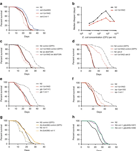
13. We found that whereas
ncl-1
loss had
little effect on wildtype lifespan, it potently suppressed the
longevity of
eat-2
mutants, a genetic model of DR (Fig. 1a and
Supplementary Fig. 1a).
ncl-1
mutation also abrogated longevity
across a wide range of bacterial food dilutions, revealing a
function in the nutrient response to dietary restriction (Fig. 1b
and Supplementary Fig. 1b).
We next asked if
ncl-1
also modulates longevity in other known
longevity models. Reduced TOR signalling is partly responsible
for lifespan extension under DR conditions
14. Accordingly,
ncl-1
mutation abrogated longevity induced by
let-363/TOR RNAi
knockdown (Fig. 1c and Supplementary Fig. 1c), suggesting that
ncl-1
mediates lifespan extension on TOR down-regulation.
Reduced insulin/IGF signalling potently promotes longevity
across taxa, and knockdown of
daf-2, the
C. elegans
insulin/IGF
receptor, doubles the lifespan
15;
ncl-1
mutation partially
suppressed
daf-2
longevity as well (Fig. 1d and Supplementary
Fig. 1d). Furthermore,
ncl-1
loss abolished lifespan extension in
long-lived germlineless
glp-1
mutants
16(Fig. 1e) and partially
suppressed longevity triggered by mutation of the iron sulfur
protein
isp-1, which reduces mitochondrial function
17(Fig. 1f).
A modest reduction in translation is known to extend lifespan in
different organisms
18.
C. elegans
harbouring loss-of-function
mutations in
ife-2
or
ifg-1, which encode translation initiation
factors, have reduced translation and extended lifespan
14,19,20.
Similarly
rsks-1
codes for the ribosomal protein S6 kinase (S6K),
which is a known downstream target of the TOR kinase whose
deficiency reduces protein synthesis and extends lifespan in
multiple species
21. Loss of
ncl-1
by RNAi largely abolished the
longevity phenotype of
ife-2, ifg-1
and
rsks-1
mutant worms
(Fig. 1g and Supplementary Fig. 1e,f). Altogether these findings
reveal that
ncl-1
works in major longevity pathways to affect
lifespan, as part of a convergent mechanism.
To investigate the role of
ncl-1
further, we generated
extra-chromosomal transgenic lines expressing wildtype
ncl-1
fused to
gfp.
Arrays restored normal nucleolar size and extended
lifespan in
eat-2;ncl-1
double mutants, demonstrating that the
transgene is functional (Supplementary Fig. 1g–j). The fusion
protein was found to reside in multiple tissues including neurons,
body wall muscle, pharynx, seam cells and vulva (Supplementary
Fig. 1k). Consistent with an instructive role,
ncl-1
over-expression
in the wildtype background was sufficient to reduce nucleolar
size (Supplementary Fig. 1g,h) and increase lifespan in two
independent transgenic lines (Fig. 1h). No further increase of
lifespan of
eat-2
on
ncl-1
over-expression was seen, indicating
an overlapping mechanism (Supplementary Fig. 1l).
Nucleolar size inversely correlates with longevity
. Since
ncl-1
affects both longevity and nucleolar size, we wondered if nucleolar
size also changes in long-lived genotypes. To address this issue, we
measured the nucleolar size of superficial hypodermal cells on the
first day of adulthood. As previously shown,
ncl-1
mutants had
enlarged nucleoli compared to wildtype (Fig. 2a and Supplementary
Fig. 2a). We further observed that
eat-2
mutants had smaller nucleoli
(Fig. 2a,c), and accordingly found that reducing nutrient levels
through bacterial dilution diminished nucleolar size (Fig. 2b).
Nucleolar size was enlarged in
eat-2;ncl-1
double mutants, revealing
that
ncl-1
is epistatic to
eat-2
for both nucleolar size and longevity
(Fig. 2a and Supplementary Fig. 2b). These intriguing observations
led us to ask whether other longevity pathways more generally
affect nucleolar size. Surprisingly, reduced insulin/IGF signalling
(daf-2), reduced mTOR (let-363), reduced mitochondrial function
(isp-1), reduced translation (rsks-1,
ife-2,
ifg-1), and germlineless
animals (glp-1) all displayed smaller nucleoli in several tissues
(Fig. 2c,d and Supplementary Fig. 2d,e).
ncl-1
mutation variously
suppressed nucleolar size in these backgrounds (Supplementary
Fig. 2b–d). The FOXO homolog
daf-16, which promotes
daf-2
longevity, was also required for small nucleolar size of
daf-2
mutants,
others as late as day 30, despite culture in a uniform environment.
The basis of this variability however has remained elusive. We
also found that wild-type animals showed variability in nucleolar
size, and therefore wondered if these differences associate with
lifespan in wild-type populations. To address this question, we
imaged the nucleoli of age-matched worms on the first day of
adulthood, recovered them on single plates and monitored their
lifespan individually (Fig. 2e). We found a striking inverse
correlation (Pearson correlation coefficient, 0.61–0.93) between
nucleolar size and longevity, where animals with smaller nucleoli
lived considerably longer than the ones with larger nucleoli
(Fig. 2f). Thus nucleolar size could be a source of variability in
longevity and may predict
C. elegans
life expectancy.
Longevity mutants have reduced ribosome biogenesis
. To
unravel molecular mechanisms, we examined how
ncl-1
and
various longevity mutants affected nucleolar functions. Loss of
ncl-1
has been previously shown to up-regulate the nucleolar
protein FIB-1/fibrillarin
22,23, which serves as a methyltransferase
for pre-rRNA processing and modification, and regulates histone
modification
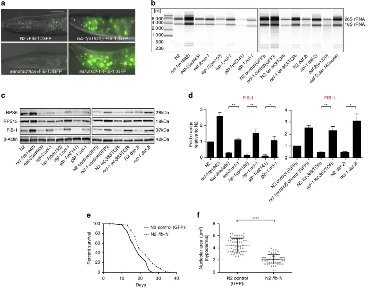
9,24. In accord with this, we also observed increased
levels of FIB-1::GFP as well as endogenous FIB-1 in
ncl-1
mutants
(Fig. 3a,c and Supplementary Fig. 3a,b). Conversely
ncl-1
over-expression down-regulated FIB-1 (Supplementary Fig. 3b). We
next asked if FIB-1 expression was affected in various longevity
mutants. Indeed both FIB-1::GFP and endogenous FIB-1 were
significantly reduced in
eat-2, daf-2, glp-1, isp-1
mutants and on
TOR knockdown, and loss of
ncl-1
reversed this effect (Fig. 3a,c,d
and Supplementary Fig. 3a,c), revealing that these pathways
converge on FIB-1 expression. We further asked if FIB-1 is a
passive marker or a causal factor for longevity. Consistent with
the latter,
fib-1
RNAi knockdown reduced nucleolar size and
extended lifespan of wild-type worms (Fig. 3e,f). RNAi
knockdown of another gene involved in nucleolar function,
rrn-3, which encodes TIF1A that assists in rRNA transcription
0 10 20 30 40 50
0 20 40 60 80
100 N2
eat-2(ad465)
eat-2;ncl-1 ncl-1(e1942)
Days
Percent survival
0 20 40
60 N2
Median lifespan (days)
0 10 20 30 40
0 20 40 60 80 100
ncl-1(e1942) control (GFP)i N2 control (GFP)i
Days
Percent survival
0 10 20 30 40 50
0 20 40 60 80 100
Percent survival
Days
N2 ncl-1(e1942) glp-1(e2141) glp-1;ncl-1
0 20 40 60 80
0 20 40 60 80 100
N2 control (GFP)i ncl-1(e1942) control (GFP)i N2 daf-2i
ncl-1(e1942) daf-2i
Days
Percent survival
N2 ncl-1(e1942) isp-1(qm150) isp-1;ncl-1
0 10 20 30 40 50
0 20 40 60 80 100
Percent survival
Days
N2
N2+ncl-1::gfp(dhEx1007) N2+ncl-1::gfp(dhEx1008)
a
b
c
d
e
f
g
0 20 40 60
0 20 40 60 80 100
Percent survival
Days
ncl-1(e1942)
h
0 10 20 30 40
0 20 40 60 80 100
Percent survival
Days
N2 control (GFP)i
N2 ncl-1i
ife-2(ok306) control (GFP)i
ife-2(ok306) ncl-1i
E. coli concentration (CFU per ml) 106 107 108 109 1010
N2 let-363/TORi ncl-1(e1942) let-363/TORi
Figure 1 |ncl-1mediates DR and other forms of longevity.(a) Longevity ofeat-2(ad465)is abolished with the loss ofncl-1(e1942)(Po0.0001, three
mediated by RNA Polymerase I
9, had little observable effect on
longevity, perhaps because achieving a balance where benefits
outweigh deleterious effects is difficult (Supplementary Fig. 3j).
Nucleoli are the cellular site of ribosome biogenesis. We
therefore examined the expression levels of rRNA and ribosomal
proteins. Mutation of
ncl-1
increased rRNA and ribosomal
protein levels (Fig. 3b,c and Supplementary Fig. 3f–h). These
molecules were also reduced in worms over-expressing
ncl-1
(Supplementary Fig. 3d,e). Notably long-lived
eat-2, daf-2, glp-1,
isp-1
and TOR RNAi knockdown worms exhibited reduced levels
of rRNA and ribosomal proteins RPS6 and RPS15, suggesting
down-regulated ribosome biogenesis associates with longevity
(Fig. 3b,c and Supplementary Fig. 3f–h). Loss of
ncl-1
variously
suppressed these phenotypes in double mutant backgrounds
(Fig. 3b,c and Supplementary Fig. 3f–h). Similarly,
daf-16
mutation restored the reduced rRNA and ribosomal proteins
levels seen in
daf-2
mutants back to wild-type levels (Fig. 3b and
Supplementary Fig. 3f,i). Taken together, these results suggest
that smaller nucleoli, reduced fibrillarin and ribosome biogenesis,
are signatures of long life.
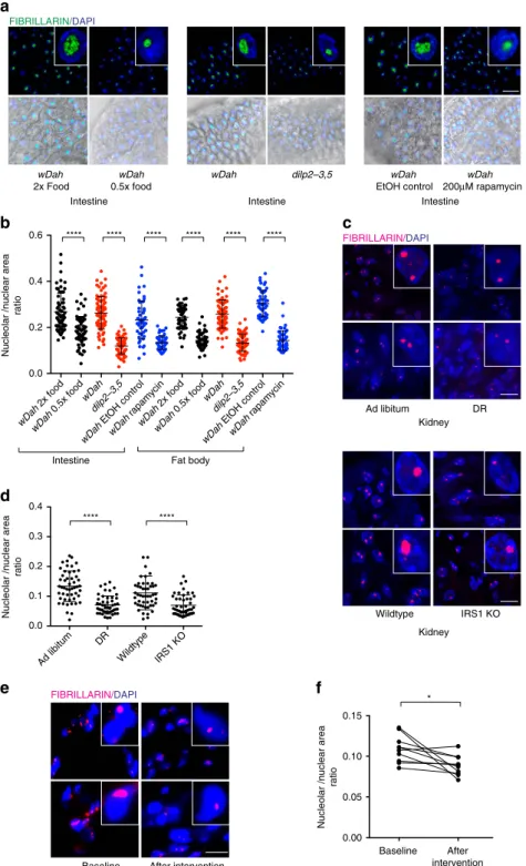
Smaller nucleoli associate with longevity in higher organisms
.
Given our results in
C. elegans, we wondered if these findings
hold true in long-lived models in other species. Remarkably we
found that long-lived
Drosophila melanogaster
undergoing DR,
exposed to the mTOR inhibitor rapamycin, or harbouring
dele-tion of the insulin-like peptides
ilp-2-3,5, all had smaller nucleoli
in the fat body and intestine (Fig. 4a,b). Furthermore, they
showed reduced levels of fibrillarin and ribosomal proteins,
although RPS6 and RPS15 levels did not significantly change on
Rapamycin treatment in flies unlike worms (Supplementary
Fig. 4a–d). Age-matched mice undergoing DR and long-lived
IRS1 knockout mice also exhibited smaller nucleoli in kidney,
liver and whole brain sections compared to controls (Fig. 4c,d and
Supplementary Fig. 4e–g). Finally, we also observed an overall
trend towards reduction of nucleolar size in muscle biopsies of
d
N2
eat-2(ad465) N2
let-363/TORiisp-1(qm150)glp-1(e2141)daf-2(e1370)
daf-2;daf-16(mu86)
** * **** * ** ns
Median lifespan (days)
Median lifespan Nucleolar area
a
b
c
e
f
N2 eat-2(ad465) ncl-1(e1942) eat-2;ncl-1
Age synchronized worms
Single worm lifespan
6.5x10⁸ 5.85x10⁹ 1.95x10⁹
Nucleolar area (
μ
m
2)
(hypodermis)
Nucleolar area (
μ
m
2)
(pharyngeal muscle)
Nucleolar area (
μ
m
2)
(hypodermis)
Nucleolar area (
μ
m
2)
(hypodermis)
Image hypodermal nucleoli of randomly picked worms on day 1
Lifespan (days)
8 12 16 20 24 28 32 36
0 2 4 6 8
0 20 40 60
0 1 2 3 4 5
0 5 10 15
0 4 8 12 ****
ns
***
0 1 2 3 4
E. coli concentration (CFU per ml)
N2
eat-2(ad465)
N2 let-363/TORi
isp-1(qm150)daf-2(e1370)
daf-2;daf-16(mu86) N2 at 25°C
glp-1(e2141)
Recover worms after imaging on single dishes
R2 = 0.93
Figure 2 | Nucleolar size inversely correlates with longevity.(a)eat-2(ad465)animals have smaller nucleoli whilencl-1(e1942)andeat-2;ncl-1animals
elderly human volunteers who underwent a regime reducing
caloric intake by 12.5% combined with moderate increase in
exercise by 12.5% (Fig. 4e,f).
Discussion
Altogether our studies reveal that multiple longevity pathways
strikingly reduce nucleolar size, and diminish expression of the
nucleolar protein FIB-1, ribosomal RNA, and ribosomal proteins
across different species. A trend towards nucleolar size reduction
is also seen with interventions that improve metabolic health in
humans, thus revealing a reversible process linking metabolic
state to a simple cellular readout. Conversely a parallel study
reported that fibroblasts derived from Hutchinson-Gilford
progeria syndrome patients show enlarged nucleolar size and
elevated ribosome biogenesis and protein synthesis
25. These
markers are not simply molecular correlates, however, but likely
responsible in part for prolonged life. Notably,
C. elegans
FIB-1 is
regulated by multiple molecular pathways, and its
down-regulation is sufficient to extend lifespan. Although knockdown
of the nucleolar RRN-3/TIF1A had little effect on lifespan,
conceivably other nucleolar functions could play a role. Evidently
NCL-1 is critical to regulating nucleolar size and inhibiting FIB-1
expression, thereby affecting lifespan in multiple pathways. How
cytosolic NCL-1 impacts nucleolar function remains unclear,
although evidence hints that it regulates FIB-1 in part via its
3
0UTR
23. NCL-1 itself is not visibly regulated by longevity
pathways (V. Tiku, personal communication); further studies
should help unravel the mechanism of NCL-1 and FIB-1 action.
Our studies are among the first to reveal that nucleolar functions
work pervasively across many longevity pathways. If small nucleoli
are a hallmark for longevity, what are the proximal mechanisms
responsible for extended life? Reduced ribosome biogenesis
and protein synthesis are the most obvious candidates: These
energetically costly processes consume considerable resources, and
a modest reduction of ribosomal proteins or translational
regulators in model organisms prolongs life
14,20,26,27. Notably,
transcriptomic analyses typically exclude ribosomal RNA, thus
overlooking the very molecules that could well affect longevity. If
protein synthesis alone were rate limiting, then knockdown of this
ncl-1(e1942)+FIB-1::GFP
eat-2;ncl-1+FIB-1::GFP eat-2(ad465)+FIB-1::GFP
N2+FIB-1::GFP
N2
ncl-1(e1942)eat-2(ad465)isp-1(qm150)eat-2;ncl-1 isp-1;ncl-1glp-1(e2141)glp-1;ncl-1
N2 let-363
/TORi
ncl-1 let-363/ TORi
N2 daf-2i
ncl-1 daf-2i
b
d
FIB-1 RPS15 RPS6
β-Actin
28kDa
18kDa
37kDa
42kDa
26S rRNA 18S rRNA
N2
ncl-1(e1942)eat-2(ad465) isp-1(qm150)eat-2;ncl-1 isp-1;ncl-1glp-1(e2141)glp-1;ncl-1
200 500 1,000 2,000 4,000 6,000 [nt]
N2 control(GFP)iN2 let-363
/TORi
ncl-1 let-363/ TORi
N2 daf-2i
ncl-1 daf-2i
ncl-1
control(GFP)i daf-2(e1370)
daf-2;daf-16(mu86)
N2 control(GFP)i ncl-1
control(GFP)i
FIB-1
Fold change relative to N2
N2 control (GFP)i N2 fib-1i
f
e
****Nucleolar area (
μ
m
2)
(hypodermis)
0 1 2 3
N2
ncl-1(e1942)eat-2(ad465)eat-2;ncl-1isp-1(qm150)isp-1;ncl-1glp-1(e2141)glp-1;ncl-1
** *
FIB-1
0 1 2 3 4
N2 control (GFP)i
ncl-1(e1942) control (GFP)iN2 let-363
/TORi
ncl-1 let-363 /TORi
N2 daf-2i
ncl-1 daf-2i
0 10 20 30 40
0 20 40 60 80 100
Days
Percent survival
0 2 4 6 8
** ** *
N2 control (GFP)i
N2 fib-1i
a
c
Figure 3 | Longevity mutants have reduced ribosome biogenesis.(a) FIB-1::GFP is strongly down-regulated ineat-2(ad465)animals but up-regulated in
process should restore longevity to
ncl-1
mutants. Surprisingly, it
did not. Our genetic analysis reveals that mutants that reduce
protein synthesis, namely
rsks-1,
ife-2, and
ifg-1, prolong life in a
ncl-1
dependent manner, suggesting that NCL-1 works largely
downstream or parallel to protein synthesis. This raises the
interesting prospect that NCL-1 perturbs protein synthesis
independently of these other factors, or that other cellular
processes might be involved. Reduced protein synthesis
per se
may represent only one aspect of longevity, since lifespan extension
by ribosomal inhibition triggers various aspects of the stress
response
28,29. In yeast recombination at rDNA repeats has been
implicated in aging
30. The nucleolus is also the site for assembly of
other ribonucleoprotein particles including splicing complexes,
telomerase, the signal recognition particle, stress granules and
microRNA machinery, and regulates processes involved in genome
integrity, nuclear architecture, stress signalling, cell cycle, and
growth
9. Conceivably these other nucleolar processes could also
contribute.
wDah
2x Food
wDah
0.5x food
dilp2–3,5 wDah
FIBRILLARIN/DAPI
FIBRILLARIN/DAPI
a
b
c
d
Ad libitum DR
Nucleolar /nuclear area
ratio
****
wDah
EtOH control
wDah
200μM rapamycin
Nucleolar /nuclear area
ratio
wDah
0.5x food
0.0 0.2 0.4 0.6
Intestine Fat body
Wildtype IRS1 KO
****
0.0 0.1 0.2 0.3 0.4
Kidney
Kidney
Intestine Intestine Intestine
0.00 0.05 0.10 0.15
Nucleolar /nuclear area
ratio
e
Baseline After intervention *
Baseline After intervention
f
FIBRILLARIN/DAPI wDah
2x food
wDah
0.5x food
wDah dilp2–3,5
wDah
EtOH control
wDah
rapamycin
wDah
2x food wDah
dilp2–3,5
wDah
EtOH control
wDah
rapamycin
Ad libitum DR
Wildtype IRS1 KO
**** **** **** **** **** ****
Figure 4 | Smaller nucleoli associate with longevity in higher organisms.(a,b) DR,dilp2-3,5and Rapamycin treatedD. melanogasterpossess small
Our work suggests that nucleolar size is a highly predictive
marker for wild-type
C. elegans
longevity. Other markers that can
approximate life expectancy and health status have been reported
in
C. elegans
31,32. Our study opens up the exciting prospect that
nucleolar size can predict life expectancy in higher organisms. If
so, quantification of nucleolar size could be used as a single-cell
readout of metabolic changes to study biological heterogeneity in
aging and longevity, or to assess how various environmental and
pharmacologic interventions impact health. We also imagine that
there may well be exceptions or conditions, in which downstream
processes uncouple nucleolar size from lifespan;
ncl-1
mutants
themselves have enlarged nucleoli but near normal lifespan.
In the future it will be fascinating to dissect the mechanisms
underlying NCL-1 and FIB-1 action, the proximal nucleolar
functions critical for longevity, and to further explore nucleolar
functions as biomarkers of health and lifespan.
Methods
C. elegansstrains
.
All the worm strains were grown using standard procedures at 20°C unless otherwise noted33. Strains carryingglp-1(e2141)mutation weremaintained at 15°C and shifted to 25°C for inducing germlineless phenotype. The strains used for the experiments were: N2 (wild type),eat-2(ad465), ncl-1(e1865), ncl-1(e1942), eat-2(ad465);ncl-1(e1865), eat-2(ad465);ncl-1(e1942), isp-1(qm150), isp-1(qm150);ncl-1(e1942), glp-1(e2141), glp-1(e2141);ncl-1(e1942), daf-2(e1370), daf-2(e1370);daf-16(mu86), cguIs001(FIB-1::GFP)34, eat-2(ad465)þcguIs001, ncl-1(e1942)þ cguIs001andeat-2(ad465);ncl-1(e1942)þcguIs001, ife-2(ok306),
ifg-1(cxTi9279)andrsks-1(sv31). dhEx1007anddhEx1008 ncl-1extra-chromosomal
transgenic strains were generated by injecting fosmid DNA WRM0611AC10 (ncl-1::TY1 EGFP) (30 ngml1) and a co-injectable marker (myo-2::mcherryat
10 ngml1) in N2 strain and further crossed intoeat-2(ad465),eat-2(ad465); ncl-1(e1865)andeat-2(ad465);ncl-1(e1942)backgrounds. The transgenic worms were maintained by selecting the worms showing the expression of the co-injected marker.
Lifespan analyses
.
All the lifespan analyses experiments were performed in atleast three independent biological replicates at 20°C. Animals that crawled off the plates, burst due to a ruptured vulva or had internal hatching of the eggs were censored from the experiment. RNAi lifespan analysis experiments were carried out following previously described protocol35. All RNAi treatments were
performed throughout development and adulthood exceptlet-363/TOR andfib-1, which were initiated on the first day of adulthood. For BDR lifespan analyses, the method followed was the same as described previously36. 90 worms were used for
each bacterial concentration to be tested and the worms were scored every 3–4 days. The worms were transferred to freshly prepared bacterial conditions on each day of scoring. BDR medium containing FUdR (1mg mll) was used for the first
two weeks of the experiment to prevent progeny production. All the lifespan experiments were performed in a blinded manner and repeated at least three times except forife-2(ok306)(performed twice), rsks-1(sv31)(performed twice),
ifg-1(cxTi9279)(performed once) andrrn-3RNAi (performed once). Mantel-Cox
Log Rank method was used for statistical analysis (Supplementary Table 1).
rRNA analysis
.
Age-matched day 1 adult worms were collected in TRIzol(Invitrogen) and snap-frozen in liquid nitrogen. RNA extraction was performed using RNeasy Mini kit (QIAGEN). Levels of rRNA were analysed by running total RNA, extracted from the same number of worms on Agilent 4200 TapeStation System following High Sensitivity RNA ScreenTape System protocol (Agilent). rRNA levels were also examined by running total RNA extracted from the same number of worms on agarose gels. NorthernMax Kit protocol was followed for running RNA gels. The gels were imaged with Alpha Innotech MultiImage II.
(For RNA extraction:n¼100 worms/replicate, 3 independent replicates)
Western blotting
.
Day 1 adult worms (50) were collected in Laemmli lysisbuffer and snap-frozen in liquid nitrogen. The samples were then boiled at 95°C for 5 min, ultrasonicated for 10 cycles and loaded on 4–15% Mini-PROTEAN TGX Precast Protein Gels. After separation, proteins were blotted on a nitrocellulose membrane and probed with the following antibodies against: RPS-6 (Abcam ab70227, 1:1,000), RPS-15 (antibodies-online.com ABIN503870, 1:1,000), Fibrillarin (Novus Biologicals NB300-269, 1:1,000) andb-Actin (Abcam ab8224, 1:5,000). The uncropped western blotting images are shown in Supplementary Fig. 5.
(For all western blots:n¼50 worms/replicate, 3 independent replicates)
ForDrosophilawestern blots, 5 females were homogenized in 100ml of RIPA
lysis buffer carrying 1X Complete mini protease inhibitor (EDTA free) (Roche). Extracts were cleared by centrifugation and protein content determined with BCA assay. 30mg of total protein was loaded on precast gels (Bio-Rad Any KD, Mini-PROTEAN TGX). The proteins were transferred to nitrocellulose membranes
and probed with the same antibodies as above. The uncropped western blotting images are shown in Supplementary Fig. 5. (For all western blots: n¼5 flies/replicate, 3 independent replicates).
Immunofluorescence
.
Immunofluorescence was performed on 10mm thickcryo-sections of mouse tissues derived from kidney, liver and brain. The samples were fixed with 4% Paraformaldehyde (PFA) for 15 min at room temperature (RT) followed by three washes with PBS at RT. The samples were then blocked with 5% Normal Donkey Serum in PBS with 0.1% Triton-X for 30 min at RT followed by an over-night incubation at 4°C with the primary antibody against Fibrillarin (Abcam ab166630, 1:200). After three subsequent washes with PBS, the samples were then probed with the secondary anti-rabbit antibody at RT for one hour followed by three more washes with PBS. The samples were mounted with ProLong Gold Mounting Medium (ThermoFisher Scientific). Immunofluorescence quantification represents three independent biological replicates with each replicate representing 3 mice (DR) and 2 mice (IRS1 KO). Imaging and quantification of the experiments were performed in a blinded manner.
Drosophilaguts and fat bodies were dissected out in PBS followed by immediate fixation with 4% PFA in PBS and permeabilization for 10 min at RT with 0.3% Triton X-100 in PBS (PBST). Blocking, primary and secondary antibody incubation were done in 5% BSA in PBST using Fibrillarin (Novus Biologicals NB300-269, 1:250) as the primary antibody and goat anti-mouse conjugated to Alexa Fluor 488 (Invitrogen, Inc., 1:1,000) as the secondary antibody. Hoechst 33342 was applied at 1:1,000 for staining nuclei. Tissues were extensively washed with PBST after antibody treatments and finally mounted on glass slides with 80% glycerol in PBS. The quantification represents three independent biological replicates with each replicate representing 5 dissected flies. Imaging and quantification of the experiments were performed in a blinded manner.
For staining human muscle biopsies, samples were thawed at RT. Then the samples were blocked with 5% milk in PBS with 0.05% Tween (PBST) for 30 min at RT, followed by three washes with PBST. The primary antibody, Rabbit-anti-Fibrillarin (Abcam ab166630, 1:600 in PBST), was incubated overnight at 4°C. After three washes with PBST, samples were incubated with the secondary goat-anti-rabbit-conjugated-Alexa647 antibody (Molecular Probes, 1:1,000 in PBST) for 1 h at RT, followed by three washes in PBST and one wash in PBS containing DAPI (0.5mg mll, Sigma-Aldrich, Saint Louis, Missouri, USA). Slides were mounted with Aqua Poly-Mount (Polysciences Inc, Niles, Illinois, USA). All samples were stained on the same day with the same antibody mixes.
Imaging and quantification
.
DIC microscopy was used to perform all thenucleolar imaging. Hypodermal, germ cell and pharyngeal muscle nucleoli of age-matched day 1 adults were imaged using 100X magnification with Axio Imager Z1 (Zeiss). Nucleolar area was quantified manually with the freehand tool using Fiji software. Details of the nucleolar size analysis are given in Supplementary Table 2. Worms carrying FIB-1::GFP and NCL-1::GFP transgenes were imaged using 63X magnification with Axio Imager Z1 (Zeiss). Immunofluorescent images were acquired using a laser-scanning confocal microscope (TCS SP5-X; Leica), equipped with a white light laser, a 405- diode UV laser, and a 100 objective lens (HCX Plan-Apochromat CS 100 oil, 1.46 NA). For human muscle biopsies, a total 15 representative fields with a 63X objective from each muscle sample were obtained, using the DM5500 fluorescent microscope (Leica) and the LAS AF software (version 2.3.6, Leica). Anti-Fibrillarin was detected with the Y5 cube, and nuclei were detected with the A4 cube. The area of the nucleolar and nuclear regions was quantified manually with the freehand tool, and subsequently the ratio of nucleolar/nuclear area was calculated. For the human samples, the average ratio of nucleolar/nuclear area (from an average of 100.4 (±28.9) nuclei) per sample was
used for the analyses.
Drosophila melanogaster experiments
.
DR inDrosophila melanogasterwasperformed by feeding a total of 50 hatched flies with 0.5x SYA food compared to ad libitum food supply of 2SYA for 10 days37. Rapamycin treatment was
performed by dissolving Rapamycin in absolute ethanol and mixing it with SYA food at a final concentration of 200mM and fed to a total of 50 age-matched flies. For control food, ethanol alone was added. Both DR and Rapamycin treatment were performed for 10 days before harvesting the flies for experiments. The treatments were performed separately in 3 different vials serving as 3 independent biological replicates. Long-liveddilp2-3,5(ref. 38) and controlwDahflies were harvested on day 1 of adulthood. The flies were dissected and immunofluorescence was performed on the dissected tissues as described above.
DR and IRS1 KO mice
.
Mouse experiments were performed according to themice were killed at the age of 14 weeks along with the ad libitum fed controls to perform cryo-sectioning for the analysis of nucleoli. The tissues sampled with sectioning were kidney and liver.
C57BL/6 IRS1 KO male39and WT control male mice were maintained
similarly on SC diet. The animals were killed at the age of 12 months to perform cryo-sectioning for the analysis of nucleoli. The tissues sampled with sectioning were kidney and brain. C57BL/6 IRS1 KO mice were originally obtained from Prof. Dominic Withers’ lab (Imperial College, London) and were bred on-site at the mouse facility in Max Planck Institute for Biology of Ageing, Cologne.
For both the experiments cryo-sectioning was performed horizontally across the entire tissue. This nature of processing aided in observing the effect of the treatments across different cell types in each tissue.
Dietary restriction and exercise intervention in human volunteers
.
Samples fornucleolar staining were obtained from the biomaterial collected in the Growing Old Together Study, a 13-weeks lifestyle intervention in older adults, consisting of 12.5% caloric restriction and 12.5% increase in physical activity, resulting in an average weight loss of 3.3 kg. The study design, inclusion and exclusion criteria, and changes in metabolic parameters have been described previously40. For the current study we used samples from 5 men and 5 women selected based on the greatest weight loss due to the intervention and the availability of muscle tissue from before and after the lifestyle intervention. This subgroup had an average age of 62.4 years (±4.1) and lost an average of 6.8 kg (±1.3) due to the intervention. Characteristics
of this subgroup are detailed in Supplementary Table 3.
All participants signed a written informed consent for participating in this study. All experiments were performed in accordance with the relevant regulations and guidelines. The medical ethical committee of the Leiden University Medical Center approved this study. This trial (NTR3499) was registered at the Dutch Trial Register (www.trialregister.nl).
Muscle biopsies and sectioning
.
Muscle biopsies were collected from thevastuslateralismuscle before and after the lifestyle intervention. Biopsies were collected 40–45 min following a standardized liquid meal (Nutridrink, Nutricia Advanced Medical Nutricion, Zoetermeer, The Netherlands) in the morning after at least 10 h of fasting. Under local anaesthesia, an incision was made 10 cm cranial of the patella on the lateral side of the upper leg. A biopsy needle (3 mm thick) was inserted to obtain the muscle biopsy. The muscle biopsy was immediately frozen in liquid nitrogen and stored at80°C before cryosectioning. Cryosections of 16mm were made with the CM3050-S cryostat (Leica, Wetzlar, Germany), pasted on SuperFrost Plus slides (Menzel-Gla¨ser, Braunschweig, Germany) and stored at
20°C before staining.
Blinding of experiments
.
All the lifespan analysis experiments were performed ina blinded manner. For blinding, the strain names were concealed during scoring, analysing and plotting the data. Nucleolar imaging and quantification were also performed with concealed strain names.
Drosophilanucleolar size analysis was performed in a blinded manner. Two
different people were involved in performing the experiment. One individual carried out fly feeding and mutant strain maintenance and the samples were passed on blinded for imaging and quantification to the second experimenter.
Mouse nucleolar size analysis was also carried out blinded. Three different people were involved in performing the experiments. One experimenter maintained the mice while carrying out the feeding/treatments and killed the mice for sectioning. The experiment was blinded henceforth. The sectioning was carried out blinded by the second experimenter. The blinded sections were stained, imaged and quantified by the third experimenter.
Two different experimenters performed human muscle biopsy staining. The whole experiment including staining, imaging and image quantification were performed completely blinded.
Data availability
.
The authors declare that all the data and the methods used inthis study are available within this article, its Supplementary Information files, the peer-review file, or are available from the corresponding author on request.
References
1. Kenyon, C. J. The genetics of ageing.Nature464,504–512 (2010).
2. Hansen, M.et al.A role for autophagy in the extension of lifespan by dietary restriction in C. elegans.PLoS Genet.4,e24 (2008).
3. Melendez, A.et al.Autophagy genes are essential for dauer development and life-span extension inC. elegans.Science301,1387–1391 (2003).
4. Lapierre, L. R.et al.The TFEB orthologue HLH-30 regulates autophagy and modulates longevity in Caenorhabditis elegans.Nat. Commun.4,2267 (2013).
5. Nakamura, S.et al.Mondo complexes regulate TFEB via TOR inhibition to promote longevity in response to gonadal signals.Nat. Commun.7,10944 (2016).
6. Johnson, D. W.et al.The Caenorhabditis elegans Myc-Mondo/Mad complexes integrate diverse longevity signals.PLoS Genet.10,e1004278 ð2014Þ:
7. Burkle, A.et al.MARK-AGE biomarkers of ageing.Mech. Ageing Dev.151, 2–12 (2015).
8. Horvath, S. DNA methylation age of human tissues and cell types.Genome Biol.14,R115 (2013).
9. Grummt, I. The nucleolus-guardian of cellular homeostasis and genome integrity.Chromosoma122,487–497 (2013).
10. Boulon, S., Westman, B. J., Hutten, S., Boisvert, F. M. & Lamond, A. I. The nucleolus under stress.Mol. Cell40,216–227 (2010).
11. Heestand, B. N.et al.Dietary restriction induced longevity is mediated by nuclear receptor NHR-62 in Caenorhabditis elegans.PLoS Genet.9,e1003651 (2013).
12. Frank, D. J. & Roth, M. B. ncl-1 is required for the regulation of cell size and ribosomal RNA synthesis inCaenorhabditis elegans.J. Cell Biol.140, 1321–1329 (1998).
13. Hedgecock, E. M. & Herman, R. K. The ncl-1 gene and genetic mosaics of Caenorhabditis elegans.Genetics141,989–1006 (1995).
14. Hansen, M.et al.Lifespan extension by conditions that inhibit translation in Caenorhabditis elegans.Aging Cell6,95–110 (2007).
15. Kenyon, C., Chang, J., Gensch, E., Rudner, A. & Tabtiang, R. A C. elegans mutant that lives twice as long as wild type.Nature366,461–464 ð1993Þ:
16. Arantes-Oliveira, N., Apfeld, J., Dillin, A. & Kenyon, C. Regulation of life-span by germ-line stem cells inCaenorhabditis elegans.Science295, 502–505 (2002).
17. Feng, J., Bussiere, F. & Hekimi, S. Mitochondrial electron transport is a key determinant of life span in Caenorhabditis elegans.Dev. Cell1,633–644 (2001).
18. Kaeberlein, M. & Kennedy, B. K. Hot topics in aging research: protein translation and TOR signaling, 2010.Aging Cell10,185–190 (2011). 19. Syntichaki, P., Troulinaki, K. & Tavernarakis, N. eIF4E function in somatic
cells modulates ageing inCaenorhabditis elegans.Nature445,922–926 ð2007Þ:
20. Pan, K. Z.et al.Inhibition of mRNA translation extends lifespan in Caenorhabditis elegans.Aging Cell6,111–119 (2007).
21. Kapahi, P.et al.With TOR, less is more: a key role for the conserved nutrient-sensing TOR pathway in aging.Cell Metab.11,453–465 (2010).
22. Lee, L. W., Lee, C. C., Huang, C. R. & Lo, S. J. The nucleolus of Caenorhabditis elegans.J. Biomed. Biotechnol.2012,601274 (2012).
23. Yi, Y. H.et al.A genetic cascade of let-7-ncl-1-fib-1 modulates nucleolar size and rRNA pool inCaenorhabditis elegans.PLoS Genet.11,e1005580 (2015).
24. Tessarz, P.et al.Glutamine methylation in histone H2A is an RNA-polymerase-I-dedicated modification.Nature505,564–568 (2014).
25. Buchwalter, A. & Hetzer, M. Nucleolar expansion and elevated protein translation in premature aging.Nat. Commun.8,doi: 10.1038/s41467-017-00322-z (2017). 26. Kaeberlein, M. & Kennedy, B. K. Large-scale identification in yeast of conserved
ageing genes.Mech. Ageing Dev.126,17–21 (2005).
27. Syntichaki, P., Troulinaki, K. & Tavernarakis, N. Protein synthesis is a novel determinant of aging in Caenorhabditis elegans.Ann. NY Acad. Sci.1119, 289–295 (2007).
28. Steffen, K. K.et al.Yeast life span extension by depletion of 60s ribosomal subunits is mediated by Gcn4.Cell133,292–302 (2008).
29. Rogers, A. N.et al.Life span extension via eIF4G inhibition is mediated by posttranscriptional remodeling of stress response gene expression inC. elegans.
Cell Metab.14,55–66 (2011).
30. Sinclair, D. A. & Guarente, L. Extrachromosomal rDNA circles--a cause of aging in yeast.Cell91,1033–1042 (1997).
31. Shen, E. Z.et al.Mitoflash frequency in early adulthood predicts lifespan in Caenorhabditis elegans.Nature508,128–132 (2014).
32. Hahm, J. H.et al.C. elegans maximum velocity correlates with healthspan and is maintained in worms with an insulin receptor mutation.Nat. Commun.6, 8919 (2015).
33. Brenner, S. The genetics of Caenorhabditis elegans.Genetics77,71–94 (1974). 34. Lee, L. W., Lo, H. W. & Lo, S. J. Vectors for co-expression of two genes in
Caenorhabditis elegans.Gene455,16–21 (2010).
35. Kamath, R. S., Martinez-Campos, M., Zipperlen, P., Fraser, A. G. & Ahringer, J. Effectiveness of specific RNA-mediated interference through ingested double-stranded RNA in Caenorhabditis elegans.Genome Biol.2,RESEARCH0002 (2001).
36. Panowski, S. H., Wolff, S., Aguilaniu, H., Durieux, J. & Dillin, A. PHA-4/Foxa mediates diet-restriction-induced longevity of C. elegans.Nature447,550–555 (2007).
38. Gronke, S., Clarke, D. F., Broughton, S., Andrews, T. D. & Partridge, L. Molecular evolution and functional characterization ofDrosophilainsulin-like peptides.PLoS Genet.6,e1000857 (2010).
39. Selman, C.et al.Evidence for lifespan extension and delayed age-related biomarkers in insulin receptor substrate 1 null mice.FASEB J.22,807–818 (2008). 40. van de Rest, O.et al.Metabolic effects of a 13-weeks lifestyle intervention in
older adults: The Growing Old Together Study.Aging (Albany NY)8,111–126 (2016).
Acknowledgements
We thank S.J. Lo (Chang Gung University) for the FIB-1::GFP strain and the
CaenorhabditisGenetics Center for other strains. We also thank Joana Goncalves and Kathrin Riehl for their assistance with mouse tissue sectioning, Dr Yidong Shen (Chinese Academy of Science) for his help in generatingncl-1transgenic lines, Dr Julia Noack and Dr Marian Beekman for help with data analysis. This work was supported by the Max Planck Society, CECAD/Deutsche Forschungsgemeinschaft (DFG), and Cologne Graduate School of Ageing Research doctoral scholarship (V.T.). The Growing Old Together Study received funding from the European Union’s Seventh Framework Program (FP7/2007–2011), grant agreement number 259679.
Author contributions
V.T., S.N. and A.A. designed experiments. V.T., C.J., B.H., W.L. and M.S. performed the experiments. Y.R. and H.E.D.S. performed stainings on human muscle biopsies. R.-U.M., P.E.S. and L.P. provided useful ideas in designing experiments. V.T. and Y.R. performed data analysis. V.T. and A.A. wrote the manuscript.
Additional information
Supplementary Informationaccompanies this paper at http://www.nature.com/ naturecommunications
Competing interests:The authors declare no competing financial interests.
Reprints and permissioninformation is available online at http://npg.nature.com/ reprintsandpermissions/
How to cite this article:Tiku, V.et al.Small nucleoli are a cellular hallmark of longevity.
Nat. Commun.8,16083 doi: 10.1038/ncomms16083 (2017).
Publisher’s note: Springer Nature remains neutral with regard to jurisdictional claims in published maps and institutional affiliations.
Open Access This article is licensed under a Creative Commons Attribution 4.0 International License, which permits use, sharing, adaptation, distribution and reproduction in any medium or format, as long as you give appropriate credit to the original author(s) and the source, provide a link to the Creative Commons license, and indicate if changes were made. The images or other third party material in this article are included in the article’s Creative Commons license, unless indicated otherwise in a credit line to the material. If material is not included in the article’s Creative Commons license and your intended use is not permitted by statutory regulation or exceeds the permitted use, you will need to obtain permission directly from the copyright holder. To view a copy of this license, visit http://creativecommons.org/ licenses/by/4.0/