IDENTIFICATION OF NOVEL TRANSCRIPTION FACTORS REGULATING RECOVERY OF THE ENDOTHELIAL LINEAGE IN AVASCULAR MUTANTS
Christopher E. Schmitt
A dissertation submitted to the faculty of the University of North Carolina at Chapel Hill in partial fulfillment of the requirements for the degree of Doctor of Philosophy in
the Curriculum in Genetics and Molecular Biology.
Chapel Hill 2012
Approved by:
ii
ABSTRACT
CHRISTOPHER SCHMITT: Identification of Novel Transcription Factors Regulating
Recovery of the Endothelial Lineage in Avascular Mutants (Under the direction of Dr. Suk-Won Jin)
Multiple mesodermal tissues are known to give rise to endothelial cells during development. Furthermore, markers for functionally distinct endothelium are well established. Therefore, it is likely that multiple distinct populations of endothelial cells exist during development and into adulthood. Two zebrafish mutants, cloche and groom of cloche provide a unique opportunity to interrogate the
heterogeneous origin of the endothelial lineage. Homozygous mutant embryos lack the majority of the endothelial lineage at early developmental stages, however, generate rudimentary vessels at later stages, indicating that they retain the ability to generate endothelial cells despite this initial lack of early
endothelial progenitors. To delineate the developmental source of the endothelial cells in these avascular mutant embryos, we first performed lineage tracing from early gastrula to determine the fate of mesoderm. Consistent with their phenotype, we found an increase of kdrl- unspecified mesoderm, indicating that much of the mesoderm apportioned to become angioblasts fails to become specified. Conversely, endothelial differentiation from the tailbud, a proposed secondary source of endothelial cells during development, was largely unperturbed. Consistent with this finding, the majority of the early kdrl+ cells found in avascular mutant embryos are specified from tailbud. To better elucidate the molecular basis of endothelial recovery in avascular mutant embryos, we analyzed the gene expression profile using microarray analysis on isolated mutant endothelial cells. We find that the expression of the genes characteristic of other mesodermal lineages are substantially elevated in kdrl+ cells isolated from
iii
are expressed in the vasculature and knockdown of either gene results in a vascular phenotype. Additionally, we found that the function of Pax9 appears to be evolutionarily conserved. Taken together, our analyses illustrate a complex regulation of endothelial specification and differentiation during
vertebrate development. Furthermore, we have identified two new key transcription factors involved in vascular development.
iv
ACKNOWLEDGEMENTS
I would like to thank:
My advisor, Suk-Won Jin
The Jin Lab, past and present members:
Hyeseon Kang, Samantha Lee, Jose Cardona-Costa, Will Dunworth, Jun-Dae Kim, Sophie Dal-Pra
My Committee Members Vicki Bautch Frank Conlon
John Rawls Bob Goldstein
v
TABLE OF CONTENTS
ABSTRACT ... iii
LIST OF FIGURES ... viii
LIST OF ABBREVIATIONS ... xi
Chapter I. INTRODUCTION ... 1
Emergence of the Vascular System ... 1
Genetic and Molecular Mechanisms of Vascular Development ... 4
cloche and groom of cloche, Two Avascular Zebrafish Mutants ... 6
Research presented in this dissertation ... 9
Figures ... 12
References ... 15
II. Mutant-Specific Gene Expression Profiling Identifies SRY-Related HMG Box 11b (Sox11b) as a Novel Regulator of Vascular Development in Zebrafish ... 19
Summary ... 19
Introduction ... 20
vi
Results ... 24
Conclusion ... 28
Figures ... 30
References ... 34
III. A Paired-Box Homeodomain Transcription Factor pax9 as a Novel Regulator for the Migration of Endothelial Cells during Vascular Morphogenesis ... 38
Summary ... 38
Highlights ... 39
Results and Discussion ... 39
Experimental Methods ... 46
Figures ... 51
References ... 68
IV. Conclusions ... 71
Introduction ... 71
Chapter 2 ... 71
Chapter 3 ... 74
Conclusion ... 77
References ... 78
V. Visualizing Vascular Networks in Zebrafish: An Introduction to Microangiography ... 79
Summary ... 79
Introduction ... 79
vii
General Considerations and Experimental Design ... 81
Materials ... 82
Method ... 84
Notes ... 86
Figures ... 88
References ... 92
VI. LRP1-Dependent Endocytic Mechanism Governs the Signaling Output of the Bmp System in Endothelial Cells and in Angiogenesis ... 94
Summary ... 94
Introduction ... 94
Methods ... 96
Results ... 97
Discussion ... 109
Figures ... 115
References ... 131
VII. Vascular Endothelial Growth Factor Signaling Regulates the Segregation of Artery and Vein via ERK Activity During Vascular Development ... 134
Summary ... 134
Introduction ... 134
Experimental Methods ... 137
Results and Discussion ... 139
Figures ... 144
viii
LIST OF FIGURES
Figure
1.1 Emergence of the endothelial lineage in zebrafish embryos ... 12 1.2 An example of endothelial heterogeneity ... 13 1.3 Model incorporating data from this thesis ... 14 2.1 Avascular mutant embryos generate endothelial cells
at later stages ... 30 2.2 Expression profile of kdrl+ cells isolated from
avascular mutant embryos ... 31 2.3 sox11b expression is elevated in kdrl+ cells isolated
from avascular mutant embryos ... 32 2.4 sox11b regulates sprouting angiogenesis during development. ... 33 3.1 Failure of the majority of endothelial cells to
specify early on in avascular mutants ... 55
3.2 Recovery of the endothelial lineage via
tailbud-dervied angioblasts ... 57 3.3 pax9 expression in the vasculature and
cell-autonomous function in vascular
morphogenesis ... 60 3.4 Conservation of the role of pax9 in zebrafish
and human endothelium ... 62 3.5 Phenotypic analysis: expression of
hemato-vascular genes in grc ... 65 3.6 Cloning of grc mutation ... 67 3.7 Expression profiling of pax9 within endothelial
cells ... 68 3.8 pax9 does not affect vascular morphogenesis
ix
3.9 pax9 expression is decreased by WNT agonist BIO ... 70
3.10 Efficacy of pax9 splice-blocking morpholinos ... 71
5.1 Schematic drawings of microangiography ... 88
5.2 Commonly used microinjectors for microangiography ... 89
5.3 An exemplary result of microangiography ... 90
6.1 LDL receptor-related protein 1 (LRP1) associates with bone morphogenetic protein endothelial cell precursor-derived regulator (Bmper) ... 115
6.2 LDL receptor-related protein 1 (LRP1) is required for bone morphogenetic protein endothelial cell precursor-derived regulator (Bmper) endocytosis. ... 116
6.3 The association of LDL receptor-related protein 1 (LRP1) and activin-like kinase receptor (ALK)6 ... 118
6.4 LDL receptor-related protein 1 (LRP1)-mediated endocytosis is required for the bone morphogenetic protein endothelial cell precursor-derived regulator (Bmper)-dependent regulation of bone morphogenetic protein (Bmp)4 downstream signaling ... 119
6.5 LDL receptor-related protein 1 (LRP1) is required for cardiovascular development in zebrafish. ... 121
6.6 A schematic model shows how LDL receptor-related protein 1 (LRP1) is required for bone morphogenetic protein (Bmp)4/bone morphogenetic protein endothelial cell precursor-derived regulator (Bmper) signaling ... 122
6.7 LRP1 is associated with Bmper ... 131
6.8 LRP1 is required for Bmper endocytosis ... 123
6.9 ALK2, 3 and 6 are required for Smad1/5/8 activation induced by Bmp4 ... 126
x
6.11 LRP1 is required for vascular development in
zebrafish ... 129 7.1 Attenuation of Vegf-A signaling components
causes defects in axial vessel segregation as observed in
kdrls828 mutants ... 144 7.2 Kdrl functions as the main receptor for Vegf-A
signaling during the segregation of axial vessels. ... 145 7.3 Attenuation of Vegf-A downstream effectors
caused defects in segregation of axial vessels ... 146 7.4 Activation of PKC or Erk activity can rescue
the defects in segregation of axial vessels in
embryos with compromised Vegf-A signaling ... 147 7.5 Photoactivation of IP3 does not rescue the
xi
LIST OF ABBREVIATIONS
BMP bone morphogenetic protein
BrdU bromodeoxyuridine
BSA bovine serum albumin
cas casanova
clo cloche
CVP caudal vein plexus
EDTA Ethylenediaminetetraacetic acid
eGFP enhanced green fluorescent protein (GFP)
EG-VEGF endocrine-gland-derived vascular endothelial growth factor
etv2 ets variant 2. The zebrafish homolog of mouse ER71
FACS fluorescence-activated cell sorting FBS fetal bovine serum
FGF fibroblast growth factor
gata1 GATA binding protein 1
gata2 GATA binding protein 2
grc groom of cloche
HBSS Hank’s balanced salt solution
hlx-1 H2.0-like homeobox 1
HPF hours post-fertilization
HUVEC Human umbilical vein endothelial cells
xii
kdrl kinase insert domain receptor like. The functional zebrafish ortholog of VEGFR2.
msx1 msh homeobox 1
pax9 paired box 9
PBS phosphate buffered saline
PCR polymerase chain reaction
pu.1(spi1) spleen focus forming virus (SFFV) proviral integration oncogene qRT-PCR quantitative reverse transcription-polymerase chain reaction RIN RNA integrity number
RT-PCR reverse transcription-polymerase chain reaction SAM Significance analysis of microarrays
sox11b SRY-related HMG-box transcription factor 11b
sox17 SRY-related HMG-box transcription factor 17
tal1 T-cell acute lymphocytic leukemia protein 1
tbx20 T-box protein 20
TUNEL terminal deoxynucleotidyl transferase mediated dUTP nick end labeling
VEGF vascular endothial growth factor
WNT wingless-type MMTV integration site family
Chapter 1
Introduction
Emergence of the vascular system.
The vascular system is comprised of multiple cell types that allow for
circulation of blood, nutrients, water and waste. Vascular smooth muscle cells,
mural cells, and endothelial cells make up this complex system that is essential
for the development, growth, and survival of a vertebrate organism. Endothelial
cells line the vessels and mediate the exchange of oxygen between blood and
surrounding tissues. In addition to diseases directly involving the vasculature
such as thrombosis, numerous pathologies are exacerbated by aberrant vascular
patterning or regulation [1]. During development, endothelial cells undergo
morphogenetic events, which can be separated into two different processes:
vasculogenesis and angiogenesis. Vasculogenesis is characterized by the
formation of new blood vessels de novo, during mid-stages of somitogenesis,
while subsequent formation of blood vessels from pre-existing vessels is
collectively referred to as angiogenesis.
During development, endothelial cells are specified from the mesoderm
during early stages of gastrulation in close proximity to hematopoietic cells [2]. In
endothelial cells [3], as is T-cell acute lymphocytic leukemia protein 1 (TAL1) [4].
Similarly, in zebrafish, it has been shown that prior to kdrl expression,
angioblasts express the ets-domain transcription factor, etv2 within the lateral
plate mesoderm [5]. Subsequently, angioblasts express kdrl and migrate into the
midline. These angioblasts coalesce and form a primitive vascular cord in the
midline of the embryo [6, 7]. During development, the vertebrate endothelial
lineage arises from mesodermal tissues. It has been reported that diverse
mesodermal tissues including lateral plate mesoderm[8], blood islands within the
yolk sac[9, 10], allantois[11], somitic mesoderm[12], as well as placenta[13, 14],
can produce endothelial cells during development. Moreover, the entire
mesoderm excluding notochord and prechordal mesoderm can serve as sources
for endothelial cells[15], suggesting that angiogenic potential might be one of the
intrinsic properties of the developing mesoderm. In addition, almost every cell in
a 16 or 32-cell stage Xenopus embryos was fate-mapped and shown to give rise
to endothelial cells, indicating the diverse origin of endothelial cells [16]. In
addition, lineage tracing studies have indicated that a small portion of the
endothelial lineage comes from hemangioblast, a common progenitor of both
hematopoietic and vascular lineages [17]. Subsequently, endothelial cells further
differentiate as arterial, venous or lymphatic endothelial cells, each of which
possesses unique molecular and cellular characteristics.
Not only do endothelial cells originate from spatially distinct populations,
they emerge during separate waves of differentiation during development. For
3
migrate from the lateral plate mesoderm at multiple time points during
development [6], suggesting that the differentiation of endothelial progenitors is
also regulated in a temporal manner. During development, lateral plate
mesoderm-derived angioblasts have been shown to give rise to the majority of
trunk vasculature [6]. In zebrafish, it has been shown that the tailbud also
contributes to the endothelial lineage in a WNT-dependant manner [18]. That is,
Wnt signaling regulates the formation of the endothelial lineage within the tailbud
mesoderm, without obvious effects on the lateral plate mesoderm [18]. In fact
the majority of caudal vasculature is derived from tailbud (Schmitt et al,
submitted).
Subsequent processes, termed angiogenesis include arterio-venous
specification and formation of secondary vessels. Conventional wisdom is that
the majority of new vessels form by angiogenesis after early development. After
early developmental stages, there is evidence for continued vasculogenesis
mediated by circulating endothelial progenitor cells, which are thought to repair
vascular damage and contribute to angiogenesis [19, 20]. Nonetheless,
pathological models have indicated that de novo angioblast specification may
occur in adult tissues. For instance, neuroblastoma stem cells have been shown
to have the capacity to give rise to endothelial and these cultured cells are wholly
derived from ectoderm [21]. Despite recent progress, the molecular and cellular
mechanisms that regulate the specification and differentiation of endothelial
lineage remain elusive. Given their diverse function and morphology, it is
4
of progenitors during development [22]. And by delineating novel transcriptional
programs directing vasculogenesis and angiogenesis, therapeutics for a wide
variety of diseases can be created.
Genetic and Molecular Mechanisms of Vascular Development
As previously mentioned specification and differentiation of the endothelial
lineage is regulated by multiple arrays of signaling pathways and transcription
factors. Many of these pathways play key roles during angiogenesis. Previous
research has identified key signaling pathways that modulate the patterning and
proliferation of endothelial cells, including Vascular Endothelial Growth Factor
(VEGF)[23, 24], Fibroblast Growth Factor (FGF)[25, 26], Wnt[27], and Bone
Morphogenetic Protein (BMP)[28], as well as essential transcription factors such
as ETS transcription factor family member, Etv2/ER71 and FLI1 [29].
Molecular and cellular analyses have led to the characterization of many
markers of arterial, venous and lymphatic endothelium [30]. There have been
many genes characterized which mark only subsets of endothelial cells [30]. For
instance, it is well established that ephrinB2, notch1b, and tbx20 are
preferentially expressed within arterial endothelial cells, while ephB4, disabled2,
and nr2f2 (COUP-TFII in mouse) are selectively expressed within venous
endothelial cells during development [30, 31]. Even endothelial cells residing
within the same vessel display high level of heterogeneity. For instance, a
subset of endothelial cells within the dorsal aorta differentiate as a hemogenic
endothelium and express hematopoietic stem cell markers runx1 and c-myb,
5
certain markers such as sox17 appear to be expressed at varying levels in cells
within the same vessel (Fig. 1.3). Therefore, multiple lines of evidence support
the prevalent heterogeneity within the endothelial lineage.
Consistent with the observation that the developmental origin of the
endothelial lineage is diverse, subtypes of endothelial cells appear to possess
distinct molecular and cellular properties. Indeed, it has been shown that
subtypes of endothelial cells distinctively respond to extracellular signaling.
EG-VEGF was shown to promote proliferation of endocrine gland endothelium
selectively over other types of endothelial cells [35]. It was further shown that
this mitogen, EG-VEGF, elicited very little response from endothelial cells in
other tissues, such as cornea or skeletal muscle, indicating that endothelial cells
from different tissues may have distinct molecular identities [35]. We have
recently reported that BMP2 signaling selectively activates venous endothelial
cells without very little influence arterial endothelial cells [36]. This effect was
shown to be important in multiple venous tissues such as caudal vein plexus and
sub-intestinal vein [36]. While markers of arterial or venous identity have been
well established, selective response to angiogenic cues is relatively new.
Expression of genes in the Notch pathway [37] as well as a transcription factor,
hlx-1 [38], have been shown to determine tip versus stalk cell phenotype in the
developing vasculature. Therefore, even transient changes in expression levels
of genes within endothelial cells can greatly affect behavior and phenotype.
Taken together, the vertebrate endothelial lineage is comprised of a
6
programs. Therefore, identification of additional factors that regulate
specification and differentiation of the endothelial lineage will help to further study
the genetic mechanisms of vascular development.
cloche and groom of cloche, two avascular zebrafish mutants
Zebrafish has been extensively used to study cardiovascular development
and is an excellent model to delineate the cellular and molecular mechanisms
that contribute to heterogeneity within the endothelial lineage. Since embryos
are transparent[39] and begin circulation by 24 hours post fertilization (hpf)[40],
the zebrafish allows for short completion of experiments. A high fecundity allows
for the collection of data with a large sample size [41]. Additionally, the embryos
are small enough that development can proceed in the absence of heartbeat,
induced chemically or genetically [42]. Furthermore, zebrafish vascular
development is remarkably similar to mammalian development. The stepwise
timing of events such as vascular cord coalescence, arterio-venous specification,
onset of circulation, intersegmental vessel angiogenesis, and lymphangiogenesis
recapitulate that which is observed in mammalian systems [39].
Previous attempts have been made to harness the attributes of zebrafish
as a model system to better understand development of higher vertebrate
species. For instance, several forward genetic screens have identified many
mutations with altered development of the endothelial lineage [43]. One
mutation, cloche (clo), is particularly interesting. Homozygous clo mutant
embryos lack kdrl+ endothelial cells as well as gata1+ or gata2+ hematopoietic
7
because of the cardiac morphology of mutant resembles a bell (“cloche” in
French). Lineage tracing studies have determined that the increase in
myocardial lineage is at the expense of most hematopoietic, endocardial and
endothelial lineages, indicating common progenitors for these lineages in
development [45]. Although the nature of the locus that is affected by clo
mutation is currently unknown, it appears to be the one of the earliest genes
involved in endothelial differentiation since over-expression of several known
factors alleviate the phenotype of homozygous mutant embryos. Nevertheless,
clo homozygotes display a dramatic early absence of scl+, gata1+, pu.1+, and
kdrl+ lineages, leading to embryonic lethality at around 7dpf [46].
To identify additional factors involved in vascular development, a large
scale forward genetic screen was performed using Tg(kdrl:eGFP)s843 transgenic
zebrafish, which labels all endothelial cells with eGFP.[25] From the screen, we
have identified a novel mutant, groom of cloche (grc), which lacks the majority of
the endothelial lineage at early stage[47], which is reminiscent of previously
isolated mutant, cloche (clo) that lacks both endothelial and hematopoietic
lineages[44]. Additionally, we found that other mesodermal lineages are not
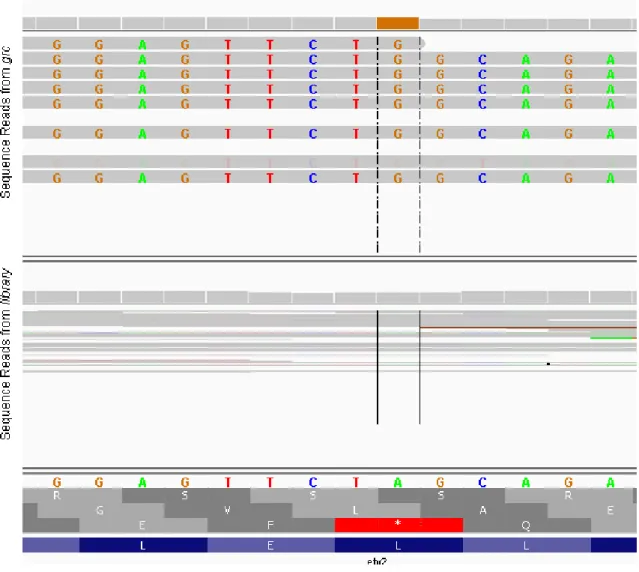
perturbed using in situ hybridization. Using high-throughput genome and
transcriptome sequencing, we determined that grc-/- embryos have a point
mutation causing a leucine to proline amino acid change in the etv2 gene, also
known as etsrp/er71. Mutations in this gene with similar phenotypes have also
been reported as it appears to function to specify the majority of endothelium
8
missing or delayed in these mutants, a crude vascular network does form in both
grc and clo homozygotes. Despite the lack of endothelial cells at early stages,
these avascular mutant embryos can generate endothelial cells at later
stages[47], suggesting that distinct molecular mechanisms may be used to
modulate the emergence of the endothelial lineage in these embryos.
Nevertheless, both mutants retain a small population of kdrl+ progenitors
which subsequently undergo vasculogenesis and angiogenesis, such as
formation of intersegmental vessels. Additionally, grc are distinct from clo in that
homozygotes have been raised to adulthood and are viable. I have hypothesized
that the majority of kdrl+ cells in both mutants are derived from tailbud and that
grc is able to specify some angioblasts from the lateral plate mesoderm (Figure
1.1 Panel A). Using the Tg(kdrl:EGFP) transgenic line, we found that kdrl+ cells
appear in homozygous grc and clo embryos as early as 18.5 hpf (shown in
Figure 1.1 panel C). While kdrl+ cells found in homozygous clo embryos are
clustered within the intermediate cell mass (ICM), which is located in the
posterior region of the embryos, those found in homozygous grc embryos are
also clustered in this region with sparse kdrl+ cells along the entire body axis,
indicating that some lateral plate-derived angioblasts are specified in grc.
Interestingly, kdrl+ cells in grc or clo homozygous embryos fail to express cdh5
(VE-Cadherin), suggesting that these cells are not fully differentiated endothelial
cells. Therefore, the kdrl+ cells found in these embryos appear to be a unique
population, and might be distinct in their molecular and cellular characteristics.
9
hypothesized that the existing kdrl+ cells are able to be specified despite their clo
or grc genetic backgrounds. Furthermore, this apparent resiliency may indicate a
unique transcriptional program or developmental origin.
As seen in Figure 1.1 Panel B (white arrows), GFP+ cells arise in the
caudal region of the embryo, posterior to the yolk-extension. Vasculature in
these mutants is aberrant. This may be due to a developmental delay where
kdrl+ cells are specified at a later time and therefore miss their proper migratory
cues or are intrinsically unable to pattern properly.
Research presented in this dissertation
The majority of this thesis is based upon a pilot experiment that involved
the transcriptional profiling of kdrl+ cells in the early mutant embryos to elucidate
alternative mechanisms of vasculogenesis and/or angiogenesis. In Chapter 2,
we performed Significance analysis of microarrays (SAM) [49] to reveal the
molecular profile of avascular mutant kdrl+ cells. We isolated kdrl+ cells using
FACS sorting and profiled their transcriptome using microarray analyses and
found that the relative levels of thousands of mRNAs in both mutants compared
to wild-type was significantly different. Screening these lists for transcription
factors, we identified 43 transcription factors that were greater than two-fold
upregulated in avascular mutant kdrl+ cells. Using morpholino-mediated
knockdown, the function of one transcription factor, SRY-related HMG Box 11b
(sox11b), was interrogated and found to be necessary for proper vascular
10
In Chapter 3, I focused on another validated transcription factor, pax9,
which was found to preferentially affect avascular mutant secondary vessel
morphogenesis. I characterized the novel mutation, grc, which lacks a majority
of endothelial cells by evaluating the phenotype, the mode of action of grc and
cloning of the genetic lesion. Using lineage tracing in early gastrula, I determined
that much of the mesodermal lineage defaults to an unspecified
mesodermal-subtype in the absence of clo or etv2 function. Additionally, using tailbud lineage
tracing found that the majority of kdrl+ cells in these mutants are derived from the
tailbud as opposed to lateral plate mesoderm.
Using multifaceted approaches, we validated a subset of transcription
factors indicated to be upregulated by the array and found that two, pax9 and
sox11b, were involved in vascular morphogenesis. Taken together, the research
presented in this dissertation includes multiple novel findings. Foremost, kdrl+
cells in clo or grc have a unique transcriptional profile. These endothelial cells
are dependent on pax9 for proper morphogenesis (summarized in Figure1.3).
Much of the avascular mutant endothelial cells come from tailbud mesoderm as
opposed to lateral plate mesoderm. Our model has allowed for the discovery of
new genes involved in vascular development. Our results demonstrate that
developmental ontogeny of the endothelial lineage is far more complex than
previously thought. The research in this thesis expands our current
understanding on how spatial, developmental and molecular heterogeneity within
the endothelial lineage is a real phenomenon and is regulated during
11
associated with the onset and subsequent progression of various pathological
conditions, including cancer, retinopathy, and diabetes, knowledge obtained from
the thesis research present here may help us to develop anti- or pro-angiogenic
12
Figures:
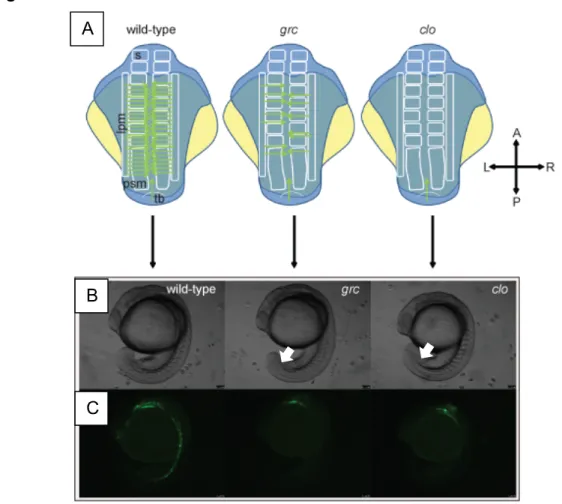
Fig. 1.1. Emergence of the endothelial lineage in zebrafish embryos.
(A) Schematic diagram illustrating the location of emerging endothelial cells in wild-type (left), grc/etv2 (middle), and clo (right) embryos. The top row is a depiction of a dorsal view of 12-15 somite embryos. Lateral plate mesoderm (lpm), Somite (s), Pre-somitic mesoderm (psm) and tailbud (tb) are all depicted in the wild-type embryo. Green arrows indicate angioblast migration into the
midline. (B) Brightfield micrographs of 18.5 hpf wild-type (left), grc/etv2 (middle), and clo (right) embryos. (C) epifluorescent micrographs of 18.5 hpf wild-type
A
B
13
(left), grc/etv2 (middle), and clo (right) embryos in Tg(kdrl:eGFP) transgenic background. The only apparent kdrl+ lineage in the avascular mutants is the
pharyngeal endoderm at 18.5 hpf, though kdrl+ cells are present in the midline
(Data not shown).
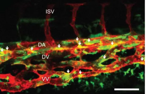
Fig. 1.2. An example of endothelial heterogeneity.
Flattened confocal z-stacks image series taken from the caudal vein plexus of a 72 hpf Tg(sox17:eGFP;kdrl:mCherry) embryo. Lateral view; anterior left and posterior right. Intersegmental vessels (ISV), dorsal aorta (DA), dorsal vein (DV), and ventral vein (VV) are labeled. White arrows indicate double positive cells. Scale bar= 30μM.
ISV
VV DA
14
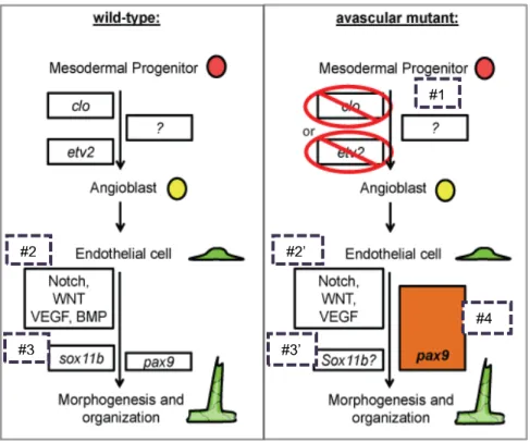
Fig. 1.3.Model incorporating data from this thesis.
Vasculogenesis and angiogenesis in wild-type (left) and avascular mutant (right) embryos during development. 1) In the absence of clo or etv2, alternative mechanism(s) may facilitate specification of angioblasts to compensate the initial deficit of the endothelial lineage. 2) While Notch, WNT and VEGF modulation all seemed to affect vascular morphogenesis in avascular mutants, BMP appears to be less important. 3) sox11b only affected wild-type vascular morphogenesis. 4) pax9 is dispensable for wild-type angiogenesis, yet essential for avascular mutant angiogenesis.
#1
#2
#3 #3’
#2’
15
REFERENCES
1. Carmeliet, P., Angiogenesis in life, disease and medicine. Nature,
2005. 438(7070): p. 932-6.
2. Saha, M.S., E.A. Cox, and C.W. Sipe, Mechanisms regulating the origins of the vertebrate vascular system. J Cell Biochem, 2004. 93(1):
p. 46-56.
3. Rasmussen, T.L., et al., ER71 directs mesodermal fate decisions during embryogenesis. Development, 2011. 138(21): p. 4801-12.
4. Visvader, J.E., Y. Fujiwara, and S.H. Orkin, Unsuspected role for the T-cell leukemia protein SCL/tal-1 in vascular development. Genes Dev,
1998. 12(4): p. 473-9.
5. Sumanas, S. and S. Lin, Ets1-related protein is a key regulator of vasculogenesis in zebrafish. PLoS Biol, 2006. 4(1): p. e10.
6. Jin, S.W., et al., Cellular and molecular analyses of vascular tube and lumen formation in zebrafish. Development, 2005. 132(23): p.
5199-209.
7. Noden, D.M., Interactions and fates of avian craniofacial mesenchyme. Development, 1988. 103 Suppl: p. 121-40.
8. Pardanaud, L., et al., Two distinct endothelial lineages in ontogeny, one of them related to hemopoiesis. Development, 1996. 122(5): p.
1363-71.
9. Ferkowicz, M.J. and M.C. Yoder, Blood island formation: longstanding observations and modern interpretations. Exp Hematol, 2005. 33(9): p.
1041-7.
10. Risau, W. and I. Flamme, Vasculogenesis. Annu Rev Cell Dev Biol,
1995. 11: p. 73-91.
11. Caprioli, A., et al., Hemangioblast commitment in the avian allantois: cellular and molecular aspects. Dev Biol, 2001. 238(1): p. 64-78.
12. Wilting, J., et al., Angiogenic potential of the avian somite. Dev Dyn,
1995. 202(2): p. 165-71.
13. Demir, R., Y. Seval, and B. Huppertz, Vasculogenesis and
16
14. Yamaguchi, T.P., et al., flk-1, an flt-related receptor tyrosine kinase is an early marker for endothelial cell precursors. Development, 1993. 118(2): p. 489-98.
15. Noden, D.M., Embryonic origins and assembly of blood vessels. Am
Rev Respir Dis, 1989. 140(4): p. 1097-103.
16. Mills, K.R., D. Kruep, and M.S. Saha, Elucidating the origins of the vascular system: a fate map of the vascular endothelial and red blood cell lineages in Xenopus laevis. Dev Biol, 1999. 209(2): p.
352-68.
17. Vogeli, K.M., et al., A common progenitor for haematopoietic and endothelial lineages in the zebrafish gastrula. Nature, 2006. 443(7109): p. 337-9.
18. Martin, B.L. and D. Kimelman, Canonical Wnt signaling dynamically controls multiple stem cell fate decisions during vertebrate body formation. Dev Cell, 2012. 22(1): p. 223-32.
19. Asahara, T., et al., Isolation of putative progenitor endothelial cells for angiogenesis. Science, 1997. 275(5302): p. 964-7.
20. Drake, C.J., Embryonic and adult vasculogenesis. Birth Defects Res C
Embryo Today, 2003. 69(1): p. 73-82.
21. Pezzolo, A., et al., Tumor origin of endothelial cells in human neuroblastoma. J Clin Oncol, 2007. 25(4): p. 376-83.
22. Liao, E.C., et al., Hereditary spherocytosis in zebrafish riesling illustrates evolution of erythroid beta-spectrin structure, and function in red cell morphogenesis and membrane stability.
Development, 2000. 127(23): p. 5123-32.
23. Leung, D.W., et al., Vascular endothelial growth factor is a secreted angiogenic mitogen. Science, 1989. 246(4935): p. 1306-9.
24. Keck, P.J., et al., Vascular permeability factor, an endothelial cell mitogen related to PDGF. Science, 1989. 246(4935): p. 1309-12.
25. Gospodarowicz, D., J. Cheng, and M. Lirette, Bovine brain and pituitary fibroblast growth factors: comparison of their abilities to support the proliferation of human and bovine vascular endothelial cells. J Cell
17
26. Abraham, J.A., et al., Nucleotide sequence of a bovine clone encoding the angiogenic protein, basic fibroblast growth factor. Science, 1986. 233(4763): p. 545-8.
27. Ishikawa, T., et al., Mouse Wnt receptor gene Fzd5 is essential for yolk sac and placental angiogenesis. Development, 2001. 128(1): p. 25-33.
28. Yamashita, H., et al., Growth/differentiation factor-5 induces angiogenesis in vivo. Exp Cell Res, 1997. 235(1): p. 218-26.
29. Wernert, N., et al., c-ets1 proto-oncogene is a transcription factor expressed in endothelial cells during tumor vascularization and other forms of angiogenesis in humans. Am J Pathol, 1992. 140(1): p.
119-27.
30. De Val, S. and B.L. Black, Transcriptional control of endothelial cell development. Dev Cell, 2009. 16(2): p. 180-95.
31. Aranguren, X.L., et al., Transcription factor COUP-TFII is
indispensable for venous and lymphatic development in zebrafish and Xenopus laevis. Biochem Biophys Res Commun, 2011. 410(1): p.
121-6.
32. North, T., et al., Cbfa2 is required for the formation of intra-aortic hematopoietic clusters. Development, 1999. 126(11): p. 2563-75.
33. North, T.E., et al., Runx1 expression marks long-term repopulating hematopoietic stem cells in the midgestation mouse embryo.
Immunity, 2002. 16(5): p. 661-72.
34. Bertrand, J.Y., et al., CD41+ cmyb+ precursors colonize the zebrafish pronephros by a novel migration route to initiate adult
hematopoiesis. Development, 2008. 135(10): p. 1853-62.
35. LeCouter, J., et al., Identification of an angiogenic mitogen selective for endocrine gland endothelium. Nature, 2001. 412(6850): p. 877-84.
36. Wiley, D.M. and S.W. Jin, Bone Morphogenetic Protein functions as a context-dependent angiogenic cue in vertebrates. Semin Cell Dev
Biol, 2011. 22(9): p. 1012-8.
37. Hellstrom, M., et al., Dll4 signalling through Notch1 regulates
formation of tip cells during angiogenesis. Nature, 2007. 445(7129): p.
18
38. Herbert, S.P., J.Y. Cheung, and D.Y. Stainier, Determination of
Endothelial Stalk versus Tip Cell Potential during Angiogenesis by H2.0-like Homeobox-1. Curr Biol, 2012. 22(19): p. 1789-94.
39. Lawson, N.D. and B.M. Weinstein, In Vivo Imaging of Embryonic Vascular Development Using Transgenic Zebrafish. Dev Biol, 2002. 248(2): p. 307-318.
40. Winata, C.L., et al., The role of vasculature and blood circulation in zebrafish swimbladder development. BMC Dev Biol, 2010. 10: p. 3.
41. Mullins, M.C., et al., Large-scale mutagenesis in the zebrafish: in search of genes controlling development in a vertebrate. Curr Biol,
1994. 4(3): p. 189-202.
42. Isogai, S., et al., Angiogenic network formation in the developing vertebrate trunk. Development, 2003. 130(21): p. 5281-90.
43. Stainier, D.Y., et al., Mutations affecting the formation and function of the cardiovascular system in the zebrafish embryo. Development,
1996. 123: p. 285-92.
44. Stainier, D.Y., et al., Cloche, an early acting zebrafish gene, is required by both the endothelial and hematopoietic lineages.
Development, 1995. 121(10): p. 3141-50.
45. Schoenebeck, J.J., B.R. Keegan, and D. Yelon, Vessel and blood specification override cardiac potential in anterior mesoderm. Dev
Cell, 2007. 13(2): p. 254-67.
46. Thompson, M.A., et al., The cloche and spadetail genes differentially affect hematopoiesis and vasculogenesis. Dev Biol, 1998. 197(2): p.
248-69.
47. Jin, S.W., et al., A transgene-assisted genetic screen identifies
essential regulators of vascular development in vertebrate embryos.
Dev Biol, 2007. 307(1): p. 29-42.
48. Palencia-Desai, S., et al., Vascular endothelial and endocardial progenitors differentiate as cardiomyocytes in the absence of Etsrp/Etv2 function. Development, 2011. 138(21): p. 4721-32.
49. Tusher, V.G., R. Tibshirani, and G. Chu, Significance analysis of microarrays applied to the ionizing radiation response. Proc Natl
CHAPTER 2
Mutant-Specific Gene Expression Profiling Identifies SRY-Related HMG Box 11b (Sox11b) as a Novel Regulator of Vascular Development in Zebrafish
Summary
Previous studies have identified two zebrafish mutants, cloche and groom of
cloche, which lack the majority of the endothelial lineage at early developmental
stages. However, at later stages, these avascular mutant embryos generate
rudimentary vessels, indicating that they retain the ability to generate endothelial
cells despite this initial lack of endothelial progenitors. To further investigate
molecular mechanisms that allow the emergence of the endothelial lineage in these
avascular mutant embryos, we analyzed the gene expression profile using
microarray analysis on isolated endothelial cells. We find that the expression of the
genes characteristic of other mesodermal lineages are substantially elevated in the
kdrl+ cells isolated from avascular mutant embryos. Subsequent validation and
analyses of the microarray data identifies Sox11b, a zebrafish ortholog of
SRY-related HMG box 11 (SOX11), which has not previously implicated in vascular
development. We further define the function of sox11b during vascular
development, and find that Sox11b function is essential for developmental
angiogenesis in zebrafish embryos, specifically regulating sprouting angiogenesis.
Taken together, our analyses illustrate a complex regulation of endothelial
20
Introduction
Endothelial cells are a major component of the vascular system, which is
essential for the development, growth, and survival of an individual. Failures in
regulating the development of endothelial lineage contribute to a wide variety of
pathological conditions, including cancer, psoriasis, arthritis, congenital or inherited
diseases, as well as heart and brain ischemia, neurodegeneration, and osteoporosis
[1]. During development, the endothelial lineage arises from mesodermal tissues. It
has been reported that diverse mesodermal tissues including lateral plate mesoderm
[2], blood islands within the yolk sac [3,4], allantois [5], somitic mesoderm [6], as well
as placenta [7,8], can produce endothelial cells during development. Moreover, the
entire mesoderm excluding notochord and prechordal mesoderm can serve as
sources for endothelial cells [9], suggesting that angiogenic potential might be one of
the intrinsic properties of the developing mesoderm. Subsequently, endothelial cells
further differentiate as arterial, venous or lymphatic endothelial cells, each of which
possesses unique molecular and cellular characteristics.
Specification and differentiation of the endothelial lineage are regulated by
arrays of signaling pathways and transcription factors. Many pathways that regulate
the emergence and organization of the endothelium have been characterized such
as receptor tyrosine kinases [10,-13], G-protein signaling pathways [14],
serine-threonine kinase [15], as well as transcription factors [16-17]. Overall, many of these
pathways are preferentially required for the proper development of a subset of
vasculature, such as cranial versus trunk or arterial versus venous endothelium.
21
cells. One of the most striking examples is that of BMP2 signaling, which is
preferentially required for venous endothelial development in zebrafish [18].
Similarly, Wnt signaling regulates the formation of the endothelial lineage within the
tailbud mesoderm, without obvious effects on the lateral plate mesoderm [19].
Therefore, identification of additional factors that regulate specification and
differentiation of the endothelial lineage will help us to further delineate the
heterogeneity of endothelial cells.
From a forward genetic screen [20], we have isolated a mutant, named groom
of cloche (grc), which lacks the majority of early Tg(kdrl:eGFP)+ cells [21], which is
reminiscent of another mutant, cloche (clo) that lack displays a severe deficiency of
both hematopoietic and endothelial lineages [22]. Nevertheless, both mutant
embryos recover [21]. This indicates that alternative mechanisms for the emergence
of the endothelial lineage.
In this report, we performed microarray analysis using the endothelial cells
isolated from late stage avascular mutant embryos and compared the expression
profile of transcription factors with endothelial cells isolated from wild-type embryos.
We find that the expression level of 43 transcription factors is significantly
up-regulated in endothelial cells isolated from avascular mutant embryos. The majority
of transcription factors we identified in our microarray have not been implicated in
vascular development. We further analyze the function of one of these transcription
factors, SRY-related HMG Box 11b (Sox11b), in endothelial differentiation and
subsequent vascular patterning. We find that Sox11b is expressed in endothelial
22
Our results demonstrate that developmental ontogeny of the endothelial lineage is
far more complex than previously thought.
Experimental Methods Zebrafish Husbandry
Zebrafish (Danio Rerio) embryos were raised as previously described [23].
The following transgenic and mutant fish lines were utilized: Tg(kdrl:eGFP)s843 [20],
cloche (clo)s5 [22], Casanova (cas)s4 [24], groom of cloche (grc)s635 [21].
Florescent Activated Cell Sorting (FACS) and RNA Isolation
18.5 hpf Tg(kdrl:eGFP)s843 embryos were dissociated in HBSS with 5% FBS
and subsequently incubated with 100μg/ml Liberase solution (Roche) for 15 minutes
at 37°C. Embryos were then triturated and the resulting suspension was pushed
through a 40μM cell culture filter (BD Biosciences) and the reaction was stopped
using 5mM EDTA, pH 8.0 in HBSS minus Ca2+ and Mg2+. Gates for flow cytometry
were selected based on the Phycoerythrin versus FITC plot. Double sorts indicated
an enrichment to >95% GFP+ cells. RNA was extracted from isolated cells using
Trizol (Invitrogen) and the accompanying protocol. Multiple rounds of flow cytometry
were performed and RNA for each biological replicate was pooled.
Microarray Analyses and quantitative RT-PCR
The WT ovation Pico Kit was used to amplify the RNA samples to satisfactory
RNA integrity score (RIN) score [25]. Otherwise, gene expression profiling was
performed as previously described [26] using an Agilent Zebrafish array version 2.
23
clo and cas was analyzed. We disregarded genes whose expression was
down-regulated in cas, which would represent genes expressed in pharyngeal endoderm.
Genes highly significantly up-regulated (q=0, fold change > 2) in both grc and clo
mutants were further analyzed.
For qRT-PCR, RNA was extracted using the QIagen RNeasy mini kit and
accompanying protocol opting to add 300ng of carrier RNA to each sample. The
iScript cDNA synthesis kit (Bio-Rad) was used to transcribe entire RNA extracts,
immediately after RNA extraction. cDNA samples were then diluted to a volume of
300μl. Using 2X Power syber mastermix, 640nM of each primer, and 8μl of cDNA in
a 25μl reaction, amplification of transcript amplicon was monitored on a Bio-Rad
cfx96 system. Gene expression was normalized to either 18S rRNA or B-actin
housekeeping genes. Melting curve analysis was performed on all reactions. Ct
versus cDNA concentration plots were also used to determine that there was a linear
ratio of amplification of housekeeping genes to gene-of-interest at a particular cDNA
concentration. Data was analyzed using the 2-∆∆CT method [27]. At least three
biological replicates of three technical replicates were performed for each conditon.
Primers used were:
18s rRNA (5'-CACTTGTCCCTCTAAGAAGTTGCA-3' and
5'-GGTTGATTCCGATAACGAACGA-3'), sox11b
(5'-CGAGTTCCCGGACTATTGCA-3' and 5' TCTCCCGCGATCATCTCACT-3'),
zfhx4 (5'-CTCCTTTGTGTGGGAAGCAT-3' and
5'-CCCTGAATGTGGAACAGCAT-3'), and klf5l
24 5'-ATCCATCTCCATCCGTGTCTGAGC-3').
in situ probe synthesis
Probes were synthesized using the SP6/T7 DIG-UTP labeling kit (Roche)
from linearized template. RNA was quantified, monitored by agarose gel
electrophoresis for a singular product, diluted in in situ hybridization solution to
100ng/μl and stored at -20°C.
Morpholino knockdown of sox11b
Previously reported and validated sox11b morpholino
(5'-CATGTTCAAACACACTTTTCCCTCT-3'), which blocks peptide synthesis, and
control morpholino
(5'-CCTCTTACCTCAGTTACAATTTATA-3') were used [28]. All embryos were
injected with 4.6nL of injection mix containing 5μM HEPES, pH 7.6 and 10% Phenol
red as a tracer.
Results
As previously reported, both clo and grc homozygous mutant embryos lack
endothelial cells at 18 hpf [21,22] (data not shown). However, at 72 hpf, kdrl+ cells
were present in these avascular mutant embryos (Figure 1A-1C). Interestingly,
counterstaining with DAPI in this experiment also showed that the midline region
where are exclusively populated by kdrl+ cells in wild-type embryos also contains a
substantial number of kdrl- cells in avascular mutant embryos (Figure 1A yellow
asterisks), alluding that vascular progenitors in these embryos may fail to undergo
25
To better understand molecular mechanisms underlying the recovery of
endothelial lineage, we analyzed the transcriptional profile of kdrl+ cells in wild-type
and avascular mutant embryos by microarray analyses (Figure 2A). Since kdrl, a
zebrafish ortholog of Vascular Endothelial Growth Factor Receptor 2 (VEGFR2) [29],
is also expressed in pharyngeal endoderm [20], it is possible that a significant
portion of kdrl+ cells isolated in avascular mutant embryos may represent
non-endothelial lineage. Therefore, we used homozygous cas embryos wherein the
entire presumptive endoderm fails to specify with little apparent effect on the
vasculature [20]. Genes down-regulated in kdrl+ cells isolated from homozygous cas
mutant embryos were discarded prior to further analyses and validation.
We found that the expression level of endothelial-enriched genes were largely
unaltered in kdrl+ cells of homozygous grc mutant embryos when compared to those
seen in wild-type. In contrast, the majority of these genes were down-regulated in
the same population from homozygous clo mutant embryos (Figure 2A), suggesting
that a locus affected by grc mutation may be only required in a subset of endothelial
cells. A small subset of endothelial-enriched genes was markedly down-regulated in
both homozygous grc and clo mutant embryos. For instance, we found that an
arterial specific marker, tbx20 [30], as well as a putative zebrafish ortholog of
mammalian Platelet Endothelial Cell Adhesion Molecule (PECAM),
ENSDART00000084729, was significantly down-regulated in kdrl+ cells isolated from
both homozygous grc and clo mutant embryos (Figure 2A). Interestingly, we found
26
other mesodermal, non-endothelial lineages such as somite, blood, or kidney (Figure
2B). For instance, we found protein kinase c delta a (prkcda), which are expressed
in blood and somitic lineages [31], and adenosine kinase a (adka), which are
expressed in blood and pronephric lineages [32], was up-regulated in kdrl+ cells from
avascular mutant embryos. Taken together, our microarray data suggest that kdrl+
cells found in avascular mutant embryos may retain more mesodermal
characteristics than those from wild-type embryos.
To better understand molecular characteristics of the kdrl+ cells in avascular
mutant embryos, we analyzed the expression level of transcription factors in our
microarray data (Figure 2C). We found that total of 43 transcription factors were
up-regulated in kdrl+ cells from avascular mutant embryos (Figure 2D) at q=0. Among
these transcription factors, we further analyze the function of sox11b, a zebrafish
ortholog of SRY-related HMG Box 11 (SOX11) [28], which is a member of SOXC
subgroup [33]. Previously, it has been shown that Sox11b is essential for mediating
retinal development and neuronal regeneration in zebrafish [28]. However, its role in
endothelial cells and vascular development has not been investigated.
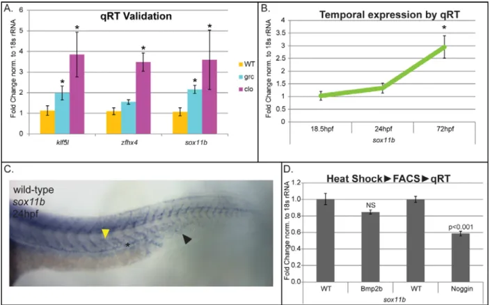
Up-regulation of sox11b in kdrl+ cells in avascular mutant embryos was confirmed by
quantitative RT-PCR (Figure 3A).
During development, sox11b is highly expressed in multiple tissues including
neurons, somites, and retina as previously proposed. In addition, approximately at
24 hpf, sox11b expression was detectable in developing posterior axial vessels in
wild-type embryos (Figure 3B). A similar expression pattern was seen in avascular
27
within endothelial cells, kdrl+ cells were isolated from wild-type embryos and
quantitative RT-PCR was performed. We found that sox11b expression can be
detected as early as 18 hpf, and the level of expression gradually increases until 72
hpf within endothelial cells, consistent with our in situ hybridization result (Figure
3C). Interestingly, the expression of sox11b appears to be induced by Bone
Morphogenetic Protein (Bmp) signaling, as over-expression of Noggin3, an
endogenous antagonist of Bmp signaling, led to a substantially decrease on the level
of sox11b transcript level (Figure 3D). Considering that Bmp signaling functions as
a context-dependent pro-angiogenic cue [18,34], it is possible that Sox11b may
function as one of the effectors in this process.
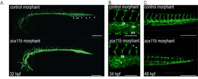
To better assess the function of Sox11b during vascular development, we
attenuated the activity of Sox11b by injecting morpholino (MO) anti-sense
oligonucleotide as previously reported [35]. Embryos injected with sox11b MO
displayed discernible defects in vascular development, compared to control MO
injected embryo (Figure 4A). At 32-34 hpf, the length of intersegmental vessels,
which sprout from the dorsal aorta at this stage [36], was substantially reduced in
sox11b MO injected embryos (Figure 4B). While control MO injected embryos had
an average length of 89.82±1.92μm (N=139 ISVs), sox11b MO injected embryos
intersegmental vessels were significantly shorter, 82.67±2.46μm (N=156 ISVs, N=8
embryos; Figure 4B, 4C and E), indicating that the function of Sox11b is essential for
the morphogenesis of sprouting intersegmental vessels during development. Since
a mammalian ortholog of Sox11b, SOX11 is known to promote transcription of key
28
as well as arrays of Actin binding proteins which modulate cell motility [37], it is
possible down-regulation of Sox11b by MO injection led to a decreased endothelial
proliferation and/or migration.
Since intersegmental vessels at 24 hpf are arterial in nature [36], we
investigated whether sox11b preferentially influences migration of arterial endothelial
cells, by analyzing the effects of sox11b knock-down on sprouting angiogenesis of
caudal vein plexus (CVP). Previously, we reported that the CVP undergoes
morphogenetic changes starting at 30 hpf by forming extensive ventral sprouts [18].
In sox11b MO injected embryos, the number of angiogenic sprouts was drastically
reduced compared to control MO injected embryos at 32 hpf (1.85±0.56 in sox11b
MO injected embryos and 11.4 ±1.0 in control MO injected embryos; Figure 4A,
white arrowheads and 4D). Morphologically, the CVP in sox11b MO injected
embryos failed to undergo proper morphogenesis to generate the dorsal vein and
the ventral vein as in wild-type embryos (Figure 4G), reflecting the attenuated
sprouting angiogenesis in these embryos.
Conclusion
Our results indicate that kdrl+ cells in avascular mutant embryos express a
unique transcriptional profile that allow them to circumvent the initial failure of
endothelial specification, which led to the formation of rudimentary vascular structure
in these embryos. We found that a number of transcription factors were selectively
up-regulated in the kdrl+ cells of avascular mutant embryos, indicating that these
transcription factors may guide an alternative mechanism to generate the endothelial
29
our microarray, sox11b, and found that sox11b plays an important role in early
morphogenesis of the vasculature by mediating sprouting angiogenesis. Taken
together, our data provides a compelling evidence of developmental heterogeneity of
30
FIGURES:
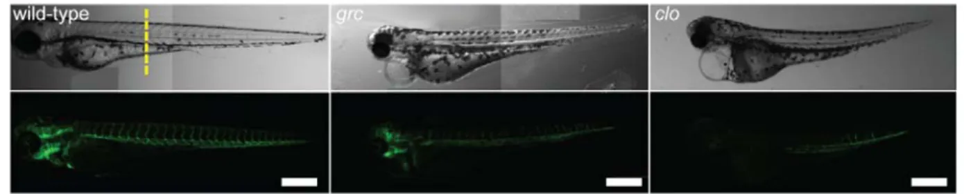
Figure 2.1: Avascular mutant embryos generate endothelial cells at later stages
(a) Gross morphology of 72 hpf wild-type (left), groom of cloche (grc)
(middle), and cloche (clo) embryos in Tg(kdrl:eGFP) background. Both bright-field (top rows) and epifluorescent (bottom rows) images are shown. Scale bar=250μm. (b) Transverse section of 72 hpf embryos taken from the area marked by dashed line in (a). GFP+ endothelial cells are shown in green and nuclei stained with DAPI
31
Figure 2.2: Expression profile of kdrl+ cells isolated from avascular mutant embryos.
(a) Schematic diagram for molecular profiling. (b) Expression profile of known lineage specific markers in microarray (*: q<0.05). (c) Characteristics of genes which are up-regulated in endothelial cells in all three avascular mutant embryos. A total of 32 genes were shown to be up-regulated. (d) Expression profiles of putative transcription factors of which function have not previously implicated in the
32
Figure 2.3: sox11b expression is elevated in kdrl+ cells isolated from avascular mutant embryos.
(a) Quantitative RT-PCR analyses confirmed the up-regulated expression of sox11b in endothelial cells of avascular mutant embryos. Two additional
33
Figure 2.4: sox11b regulates sprouting angiogenesis during development.
(a) Epifluorescent micrographs of control (top) or sox11b (bottom) morpholino (MO) injected embryos. Trunk regions posterior to the end of yolk extension are shown. White arrowheads indicate ventral sprouts. Scale bar=250μm. (b)
34
REFERENCES
1. P. Carmeliet: Angiogenesis in life, disease and medicine, Nature 2005, 438:932-936.
2. L. Pardanaud, D. Luton, M. Prigent, L.M. Bourcheix, M. Catala, F. Dieterlen-Lievre: Two distinct endothelial lineages in ontogeny, one of them related to hemopoiesis, Development 1996, 122:1363-1371.
3. M.J. Ferkowicz, M.C. Yoder: Blood island formation: longstanding observations and modern interpretations, Exp Hematol 2005, 33:
1041-1047.
4. W. Risau, I. Flamme: Vasculogenesis, Annu Rev Cell Dev Biol 1995, 11:
73-91.
5. A. Caprioli, K. Minko, C. Drevon, A. Eichmann, F. Dieterlen-Lievre, T.
Jaffredo: Hemangioblast commitment in the avian allantois: cellular and molecular aspects, Dev Biol 2001, 238:64-78.
6. J. Wilting, B. Brand-Saberi, R. Huang, Q. Zhi, G. Kontges, C.P. Ordahl, B. Christ: Angiogenic potential of the avian somite, Dev Dyn 1995, 202:
165-171.
7. R. Demir, Y. Seval, B. Huppertz: Vasculogenesis and angiogenesis in the early human placenta, Acta Histochem 2007, 109:257-265.
8. T.P. Yamaguchi, D.J. Dumont, R.A. Conlon, M.L. Breitman, J. Rossant: flk-1, an flt-related receptor tyrosine kinase is an early marker for endothelial cell precursors, Development 1993, 118:489-498.
9. D.M. Noden: Embryonic origins and assembly of blood vessels, Am Rev
Respir Dis 1989, 140:1097-1103.
10. D.W. Leung, G. Cachianes, W.J. Kuang, D.V. Goeddel, N. Ferrara: Vascular endothelial growth factor is a secreted angiogenic mitogen, Science
1989, 246:1306-1309.
11. P.J. Keck, S.D. Hauser, G. Krivi, K. Sanzo, T. Warren, J. Feder, D.T. Connolly: Vascular permeability factor, an endothelial cell mitogen related to PDGF, Science 1989, 246:1309-1312.
12. D. Gospodarowicz, J. Cheng, M. Lirette: Bovine brain and pituitary fibroblast growth factors: comparison of their abilities to support the proliferation of human and bovine vascular endothelial cells, J Cell Biol
35
13. J.A. Abraham, A. Mergia, J.L. Whang, A. Tumolo, J. Friedman, K.A. Hjerrild, D. Gospodarowicz, J.C. Fiddes: Nucleotide sequence of a bovine clone encoding the angiogenic protein, basic fibroblast growth factor, Science
1986, 233:545-548.
14. T. Ishikawa, Y. Tamai, A.M. Zorn, H. Yoshida, M.F. Seldin, S. Nishikawa, M.M. Taketo: Mouse Wnt receptor gene Fzd5 is essential for yolk sac and placental angiogenesis, Development 2001, 128:25-33.
15. H. Yamashita, A. Shimizu, M. Kato, H. Nishitoh, H. Ichijo, A. Hanyu, I. Morita, M. Kimura, F. Makishima, K. Miyazono: Growth/differentiation factor-5 induces angiogenesis in vivo, Exp Cell Res 1997, 235:218-226.
16. N. Wernert, M.B. Raes, P. Lassalle, M.P. Dehouck, B. Gosselin, B. Vandenbunder, D. Stehelin: c-ets1 proto-oncogene is a transcription factor expressed in endothelial cells during tumor vascularization and other forms of angiogenesis in humans, Am J Pathol 1992, 140:119-127.
17. J.E. Visvader, Y. Fujiwara, S.H. Orkin: Unsuspected role for the T-cell leukemia protein SCL/tal-1 in vascular development, Genes Dev 1998, 12:473-479.
18. D.M. Wiley, S.W. Jin: Bone Morphogenetic Protein functions as a context-dependent angiogenic cue in vertebrates, Semin Cell Dev Biol
2011, 22:1012-1018.
19. B.L. Martin, D. Kimelman: Canonical Wnt signaling dynamically controls multiple stem cell fate decisions during vertebrate body formation, Dev
Cell 2012, 22:223-232.
20. S.W. Jin, D. Beis, T. Mitchell, J.N. Chen, D.Y. Stainier: Cellular and
molecular analyses of vascular tube and lumen formation in zebrafish,
Development 2005, 132:5199-5209.
21. S.W. Jin, W. Herzog, M.M. Santoro, T.S. Mitchell, J. Frantsve, B. Jungblut, D. Beis, I.C. Scott, L.A. D'Amico, E.A. Ober, H. Verkade, H.A. Field, N.C. Chi, A.M. Wehman, H. Baier, D.Y. Stainier: A transgene-assisted genetic screen identifies essential regulators of vascular development in vertebrate embryos, Dev Biol 2007, 307:29-42.
22. D.Y. Stainier, B.M. Weinstein, H.W. Detrich, 3rd, L.I. Zon, M.C. Fishman:
Cloche, an early acting zebrafish gene, is required by both the
endothelial and hematopoietic lineages, Development 1995, 121:
36
23. M. Westerfield: The Zebrafish Book; A Guide for the Laboratory Use of Zebrafish (Brachydanio rerio) University of Oregon Press, Eugene., 1989.
24. J. Alexander, D.Y. Stainier, D. Yelon: Screening mosaic F1 females for mutations affecting zebrafish heart induction and patterning, Dev Genet
1998, 22:288-299.
25. A. Schroeder, O. Mueller, S. Stocker, R. Salowsky, M. Leiber, M. Gassmann, S. Lightfoot, W. Menzel, M. Granzow, T. Ragg: The RIN: an RNA integrity number for assigning integrity values to RNA measurements, BMC Mol
Biol 2006, 7:3.
26. E.K. Lobenhofer, J.T. Auman, P.E. Blackshear, G.A. Boorman, P.R. Bushel, M.L. Cunningham, J.M. Fostel, K. Gerrish, A.N. Heinloth, R.D. Irwin, D.E. Malarkey, B.A. Merrick, S.O. Sieber, C.J. Tucker, S.M. Ward, R.E. Wilson, P. Hurban, R.W. Tennant, R.S. Paules: Gene expression response in target organ and whole blood varies as a function of target organ injury phenotype, Genome Biol 2008, 9:R100.
27. K.J. Livak, T.D. Schmittgen: Analysis of relative gene expression data using real-time quantitative PCR and the 2(-Delta Delta C(T)) Method,
Methods 2001, 25:402-408.
28. M.B. Veldman, M.A. Bemben, R.C. Thompson, D. Goldman: Gene expression analysis of zebrafish retinal ganglion cells during optic nerve regeneration identifies KLF6a and KLF7a as important regulators of axon regeneration, Dev Biol 2007, 312:596-612.
29. J. Bussmann, N. Lawson, L. Zon, S. Schulte-Merker: Zebrafish VEGF receptors: a guideline to nomenclature, PLoS Genet 2008, 4:e1000064.
30. D.G. Ahn, I. Ruvinsky, A.C. Oates, L.M. Silver, R.K. Ho: tbx20, a new vertebrate T-box gene expressed in the cranial motor neurons and developing cardiovascular structures in zebrafish, Mech Dev 2000, 95:253-258.
31. S.A. Patten, R.K. Sihra, K.S. Dhami, C.A. Coutts, D.W. Ali: Differential expression of PKC isoforms in developing zebrafish, Int J Dev Neurosci
2007, 25:155-164.
32. B. Thisse, S. Pflumio, M. Fürthauer, B. Loppin, V. Heyer, A. Degrave, R. Woehl, A. Lux, T. Steffan, X.Q. Charbonnier, and C. Thisse: Expression of the zebrafish genome during embryogenesis, ZFIN Direct Data
37
33. J. Bowles, G. Schepers, P. Koopman: Phylogeny of the SOX family of developmental transcription factors based on sequence and structural indicators, Dev Biol 2000, 227:239-255.
34. J.D. Kim, H. Kang, B. Larrivee, M.Y. Lee, M. Mettlen, S.L. Schmid, B.L. Roman, Y. Qyang, A. Eichmann, S.W. Jin: Context-dependent
proangiogenic function of bone morphogenetic protein signaling is mediated by disabled homolog 2, Dev Cell 2012, 23:441-448.
35. A. Nasevicius, S.C. Ekker: Effective targeted gene 'knockdown' in zebrafish, Nature Genetics 2000, 26:216-220.
36. S. Isogai, M. Horiguchi, B.M. Weinstein: The vascular anatomy of the developing zebrafish: an atlas of embryonic and early larval
development, Dev Biol 2001, 230:278-301.
37. X. Wang, S. Bjorklund, A.M. Wasik, A. Grandien, P. Andersson, E. Kimby, K. Dahlman-Wright, C. Zhao, B. Christensson, B. Sander: Gene expression profiling and chromatin immunoprecipitation identify DBN1, SETMAR and HIG2 as direct targets of SOX11 in mantle cell lymphoma, PLoS One
CHAPTER 3
A Paired Box Homeodomain Transcription Factor pax9 as a Novel Regulator for the Migration of Endothelial Cells during Vascular Morphogenesis
Summary
Multiple populations of endothelial cells exist during development and into
adulthood. Two avascular zebrafish mutants, clo and grc, which retain small
populations of kdrl+ cells and undergo vascular morphogenesis allow for the study of
endothelium which is able to specify in the absence of clo or etv2. Using clo and grc
as models for endothelial heterogeneity, we performed lineage tracing from shield
stage and found an increase of kdrl- unspecified mesoderm, indicating that much of
the mesoderm fated to become angioblasts fails to specify. Additionally, we
determined that a large portion of early kdrl+ cells in these mutants are specified from
tailbud, as opposed to lateral plate mesoderm. Given the unique developmental
source of endothelium in avascular mutants, we focused on a transcription factor
previously indicated to be up-regulated in avascular mutant kdrl+ cells, pax9. We
found that pax9 is highly enriched in developing zebrafish vasculature and controls
vascular morphogenesis. We also found that pax9 controls scratch closure in
HUVEC cell culture, indicating that its role in vascular morphogenesis is likely
39
Highlights
Avascular mutants undergo endothelial specification at later stages.
Tailbud mesoderm largely contributes to vascular recovery.
pax9 is up-regulated in avascular mutant kdrl+ cells.
Modulation of pax9 controls vascular morphogenesis.
Results and Discussion
In order to define alternative mechanisms of endothelial specification and
organization, we decided to use avascular mutants as a model for endothelial
heterogeneity. We have previously isolated an avascular mutant, groom of
cloches635 (grc635) from a large scale forward genetic screen [1]. At early stages, the
vascular phenotype of grc is comparable to that of cloche (clo), an avascular mutant
that lacks both endothelial and hematopoietic lineages (Fig. 3.1A) [2]. Homozygous
grc mutant embryos completely lack the expression of endothelial specific markers
including kdrl, dab2, and cdh5 at early developmental stages, indicating that the
endothelial lineage failed to be specified or not maintained (Fig. 3.5). However,
approximately at 18 hours post-fertilization (hpf), a small group of kdrl+ cells can be
detected in the posterior region of homozygous grc mutant embryos, which
subsequently expanded. At 72 hpf, a rudimentary vascular structure can be
detected in the majority of homozygous grc mutant embryos (Fig. 3.2A), and a small
fraction (less than 5 percent) of these embryos survive to the adulthood. Similarly,
we found that homozygous clo mutant embryos do possess kdrl+ cells at later stages