Neurobiology of Disease
Maternal Loss of
Ube3a
Impairs Experience-Driven
Dendritic Spine Maintenance in the Developing Visual
Cortex
Hyojin Kim,
1,2* Portia A. Kunz,
1,2,3* Richard Mooney,
4X
Benjamin D. Philpot,
1,2,3and
X
Spencer L. Smith
1,2,31Department of Cell Biology and Physiology,2Neuroscience Center, and3Carolina Institute for Developmental Disabilities, University of North Carolina, Chapel Hill, North Carolina 27599, and4Department of Neurobiology, Duke University School of Medicine, Durham, North Carolina 27710
Dendritic spines are a morphological feature of the majority of excitatory synapses in the mammalian neocortex and are motile structures
with shapes and lifetimes that change throughout development. Proper cortical development and function, including cortical
contribu-tions to learning and memory formation, require appropriate experience-dependent dendritic spine remodeling. Dendritic spine
abnor-malities have been reported for many neurodevelopmental disorders, including Angelman syndrome (AS), which is caused by the loss of
the maternally inherited
UBE3A
allele (encoding ubiquitin protein ligase E3A). Prior studies revealed that UBE3A protein loss leads to
reductions in dendritic spine density and diminished excitatory synaptic transmission. However, the decrease in spine density could
come from either a reduction in spine formation or an increase in spine elimination. Here, we used acute and longitudinal
in vivo
two-photon microscopy to investigate developmental and experience-dependent changes in the numbers, dynamics, and morphology of
layer 5 pyramidal neuron apical dendritic spines in the primary visual cortex of control and AS model mice (
Ube3a
m⫺/p⫹mice). We
found that neurons in AS model mice undergo a greater elimination of dendritic spines than wild-type mice during the end of the first
postnatal month. However, when raised in darkness, spine density and dynamics were indistinguishable between control and AS model
mice, which indicates that decreased spine density in AS model mice reflects impaired experience-driven spine maintenance. Our data
thus demonstrate an experience-dependent anatomical substrate by which the loss of UBE3A reduces dendritic spine density and
disrupts cortical circuitry.
Key words:
two-photon; Angelman syndrome; dendritic spine; E6AP; Ube3a; visual cortex
Introduction
Angelman syndrome (AS) is a neurodevelopmental disorder
caused by loss-of-function of the UBE3A protein (
Kishino et al.,
1997
). The
UBE3A
gene encodes a HECT (homologous to
E6-associated protein C terminus) domain E3 ubiquitin ligase.
Neu-rons in the brain express the maternal, but not paternal, allele of
Received Nov. 23, 2015; revised March 9, 2016; accepted March 24, 2016.
Author contributions: H.K., P.A.K., R.M., B.D.P., and S.L.S. designed research; H.K. and P.A.K. performed research; H.K. analyzed data; H.K., R.M., B.D.P., and S.L.S. wrote the paper.
This work was supported by the Angelman Syndrome Foundation (B.D.P.), the Simons Foundation (Grant SFARI 274426 to B.D.P. and Grant SCGB 325407SS to S.L.S.), the National Institutes of Health (NIH Grants R01NS085093 and R01MH093372 to B.D.P., Grant R01EY024294 to S.L.S., Grant DC002524 to R.M., and Training Grant T32HD040127 to P.A.K.), the Whitehall Foundation (S.L.S.), the Klingenstein Foundation (S.L.S.), and the National Science Foundation (Grant 1450824 to S.L.S.). P.A.K. was funded by Autism Speaks and the Autism Science Foun-dation. The UNC Confocal and Multiphoton Imaging Core is funded by NIH Grants P30NS045892 and U54HD079124.
We thank Vladimir Ghukasyan at the UNC Confocal and Multiphoton Imaging Core for technical assistance and Dr. Kelly Jones for help editing this manuscript.
The authors declare no competing financial interests.
*H.K. and P.A.K. contributed equally to this work and are co-first authors.
Correspondence should be addressed to either Benjamin D. Philpot or Spencer L. Smith, Department of Cell Biology and Physiology, University of North Carolina, 115 Mason Farm Rd., Campus Box 7545, Chapel Hill, NC 27599. E-mail:[email protected]@unc.edu.
DOI:10.1523/JNEUROSCI.4204-15.2016
Copyright © 2016 the authors 0270-6474/16/364888-07$15.00/0
Significance Statement
UBE3A
due to genetic imprinting (
Kishino et al., 1997
;
Landers et
al., 2004
). Therefore, deletions or mutations of the maternal
UBE3A
allele cause the complete loss of UBE3A protein in almost
all central neurons (
Kishino et al., 1997
;
Judson et al., 2014
). AS
phenotypes begin to present during early life (
⬃
1 year) and
are characterized by severe cognitive impairments, seizures,
min-imal speech, hypermotoric behavior, and a short attention span
(
Clayton-Smith and Laan, 2003
).
To help identify dendritic spine deficits contributing to the
anatomical and cognitive impairments in AS, we took advantage
of the visual cortex as a model system to explore the early sensitive
periods during which experience can leave a lasting imprint on
the brain (
Hubel and Wiesel, 1970
;
Mower, 1991
;
Hensch, 2005
;
Levelt and Hu¨bener, 2012
). Such studies have served as a useful
tool for identifying the synaptic mechanisms of learning and
memory and for exposing synaptic deficits that underlie
neuro-logical disorders (
Penzes et al., 2011
;
Cooke and Bear, 2014
).
These studies have shown, for example, that dendritic spines
rap-idly change in number and shape after various sensory and
be-havioral manipulations (
Shepherd et al., 2003
;
Mataga et al.,
2004
;
Hofer et al., 2009
;
Tropea et al., 2010
;
Miquelajauregui et
al., 2015
) and that changes in dendritic spine shape and/or
den-sity are common in neurodevelopmental disorders and their
an-imal models (
Nimchinsky et al., 2001
;
Hutsler and Zhang, 2010
;
Pan et al., 2010
;
Penzes et al., 2011
;
Till et al., 2012
). Although
appropriate changes in dendritic spines support normal learning,
memory, and cognitive function, abnormal dendritic spine
de-velopment may contribute to cortical dysfunction and associated
behavioral abnormalities such as those observed in individuals
with AS.
Reduced dendritic spine density has been one of the most
consistent anatomical observations in both AS patients and AS
model mice. In the only human postmortem anatomical AS study
performed to date, dendritic spine density was shown to be
re-duced in layer 3 (L3) and L5 pyramidal neurons within the visual
cortex (
Jay et al., 1991
). Consistent with those observations, AS
model mice (Ube3a
m⫺/p⫹mice) exhibit reduced dendritic spine
density in L2/3 pyramidal neurons (
Yashiro et al., 2009
) and in
basal dendrites of L5 pyramidal neurons (
Sato and Stryker, 2010
)
in the visual cortex. These changes are correlated with reductions
in excitatory synaptic transmission (
Wallace et al., 2012
),
sug-gesting that reductions in dendritic spines and excitatory synaptic
transmission are causally linked. Reduced spine density has also
been observed on secondary apical dendrites of L3–L5 cortical
pyramidal neurons in AS model mice (
Dindot et al., 2008
). The
loss of UBE3A may cause generalized reductions in dendritic
spine density given that AS model mice also exhibit reduced spine
density in the cerebellum and hippocampus (
Dindot et al., 2008
).
Despite consistent observations of reduced dendritic spine
density in an AS individual and in mouse models, how the loss of
UBE3A affects the experience-driven formation and elimination
of dendritic spines during development remains unclear. To
re-solve this issue, we used longitudinal two-photon microscopy in
wild-type and AS model mice to examine dendritic spine
turn-over in L5 pyramidal neuron tufts, where changes in visual input
are known to alter spine dynamics (
Majewska et al., 2006
;
Tropea
et al., 2010
). We focused on spine maintenance across a critical
period during which visual cortex connectivity is normally
sensi-tive to experience-driven rearrangements, such as the ocular
dominance plasticity that occurs as a consequence of brief
mon-ocular deprivation (
Gordon and Stryker, 1996
). Our studies
re-vealed that AS model mice exhibit a larger proportion of thinner
dendritic protrusions and reduced spine density, the latter of
which resulted from impaired experience-dependent spine
maintenance. Our findings indicate that maternal
Ube3a
plays an
important role in the experience-dependent maturation of
excit-atory synapses in the visual cortex.
Materials and Methods
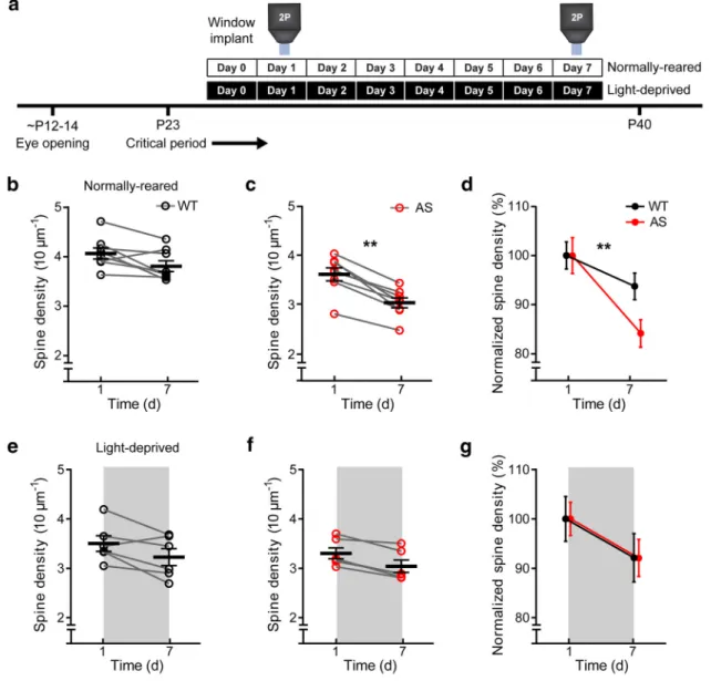
Animals.Mice carrying a Ube3adeletion were bred on a C57BL/6 background (Jiang et al., 1998).Ube3am⫹/p⫺females were crossed withThy1-GFP (line O) males (Feng et al., 2000) to obtain AS model mice (Ube3am⫺/p⫹) and control mice (Ube3am⫹/p⫹) expressing GFP in L5 pyramidal neurons. Male and female mice were used at different ages [postnatal day 14 (P14)–P38] and were raised either on a 12 h light/dark cycle (normally raised; NR) or in constant darkness (light-deprived; LD) starting on the first day of surgery (between P24 and P31) for two-photon imaging. All animal procedures were performed in compliance with animal care guidelines at the University of North Carolina at Chapel Hill.
Cranial window surgery. Mice were anesthetized with ketamine (80 mg/kg) and xylazine (6 mg/kg) and administered ketorolac (10 mg/ kg; Hospira) and cefazolin (22 mg/kg; Sagent Pharmaceuticals) subcuta-neously to reduce edema and local tissue inflammation. A head plate (inner diameter, 5 mm; custom-made, stainless steel) was placed directly on the skull and secured with Vetbond (n-butyl cyanoacrylate; 3M). Mice were positioned in a head-plate holder (custom-made, stainless steel mounting apparatus) for craniotomy. A craniotomy (3– 4 mm diameter) was made over the primary visual cortex and a 3 mm glass coverslip (#1; Warner Instruments) was placed over the pial surface and secured to the skull with Vetbond. Dental acrylic (Ortho Jet; Lang Dental) was then applied across the skull surface and over the edges of the coverslip.
Two-photon imaging.Images were acquired on an upright Zeiss LSM 7MP and a Coherent Vision S titanium–sapphire laser was focused on the specimen by a W-Plan Apochromat 20⫻/1.0 numerical aperture objec-tive. Fluorescence was collected by the same objective, filtered, and re-corded by a GaAsP detector in a BiG module. Image acquisition and processing were controlled through the Zen software package. Dendritic segments of visual cortex were imaged at high resolution (1024⫻1024 pixels, 0.21m/pixel, 1mz-steps). In GFP-O mice, the vast majority of neurons expressing GFP are located in L5 (Feng et al., 2000; also see
Fig. 1b) and, in most cases, the identity of L5 pyramidal neurons was confirmed using low-magnification image stacks collected from the cor-tical surface to the soma (1024⫻1024 pixels, 0.59m/pixel, 4m
z-steps). Therefore, we assumed that the dendritic segments we imaged and analyzed belonged to L5 pyramidal neurons. We acutely or chroni-cally imaged apical tufts of L5 pyramidal neurons (⬃100m below the cortical surface). Blood vessel patterns were used as initial references to guide chronic imaging of the same dendritic segments.
Chronic imaging took place over two imaging sessions; the first session occurred the day after the surgery and the second was performed 7 d after the surgery. Durotomy or bone growth removal was occasionally re-quired for optical clarity the day before the second imaging session (Goldey et al., 2014). The chronic window surgery did not appear to cause abnormal microglial cell activation (Xu et al., 2007). The morphol-ogy and density of Iba1-labeled microglia were not different between the hemispheres ipsilateral and contralateral to the window (quantified the day after surgery; Iba1-positive cells/mm2in contralateral vs ipsilateral to surgery cortex⫽476.0⫾31.39 vs 475.3⫾21.67,p⫽0.979,n⫽3, Student’s pairedttest;Goldey et al., 2014).
previous studies, we observed that spine density was relatively stable between P25 and P38 (seeFig. 1fandHoltmaat et al., 2005;Hofer et al., 2009).
The rates of spine formation and elimination were defined, respec-tively, as the number of spines that appeared and disappeared relative to
the total spine number of the previous imaging session. Spine subtypes (mushroom, stubby, and thin) were quantified using NeuronStudio ( Ro-driguez et al., 2008).
Immunohistochemistry.Mice were anesthetized with sodium pento-barbital (40 mg/kg) before transcardial perfusion with PBS, immediately
followed by 4% paraformaldehyde in PBS, pH 7.4. Brains were postfixed overnight at 4°C before sequential 24 h incubations in PBS with 10%, 20%, and 30% sucrose. Brains were sectioned coronally at 40m (for Iba1 staining) or 100m (for Thy1-GFP visualization) using a freezing sliding microtome (Thermo Scientific). Sections were stored at 4°C in a cryopreservative solution (45% PBS, 30% ethylene glycol, 25% glycerol, by volume). For Iba1 immunostaining, sections were rinsed several times with PBS before blocking with 5% normal goat serum and 0.02% Triton X-100 in PBS for 1 h at room temperature. Sections were then incubated with primary antibody (Wako 019 –19741, rabbit anti-Iba1, 1:600), di-luted in normal goat serum at 4°C overnight, and then rinsed several times with 0.02% Triton X-100 in PBS. Tissues were then incubated with secondary antibody (Life Technologies, Alexa Fluor-568 goat anti-rabbit IgG A11011, 1:500) for 2 h. Images were acquired with a Zeiss LSM 780 confocal microscope.
Statistics. Graphs were generated and statistical analyses were per-formed using GraphPad Prism 6 software. Data are represented as the mean⫾SEM unless otherwise indicated. For statistical analyses, two-tailed Student’s paired and unpairedttests and two-way ANOVA were used as indicated. Statistical significance is represented as follows: *p⬍
0.05, **p⬍0.01, and ****p⬍0.0001.
Results
Decreases in spine density are greater in L5 pyramidal neuron
apical tufts in AS model mice
Chronic imaging reveals a role for
UBE3A in regulating dendritic
spine density
Using longitudinal imaging through a
chronic cranial window (
Holtmaat et al.,
2009
), we tested whether visual
experi-ence differentially modified dendritic
spine dynamics in the absence of maternal
Ube3a. We focused on developmental
changes after P25, when we observed
dif-ferences in spine density between the
ge-notypes in our acute imaging experiments
(
Fig. 1
g). We imaged spines on the apical
dendritic tufts of individual L5 neurons
across a 7 d interval in NR mice (12:12 h
light-dark cycle) and LD mice (24 h
dark-ness;
Fig. 2
a). Spine density did not
change significantly and trended
down-ward across imaging sessions in WT mice
(
p
⫽
0.055, paired
t
test;
Fig. 2
b), but was
significantly reduced over this same
inter-val in AS model mice (p
⫽
0.007, paired
t
test;
Fig. 2
c). A two-way ANOVA revealed
a significant effect of genotype (F
(1,28)⫽
10.2,
p
⫽
0.003), time (F
(1,28)⫽
53,
p
⬍
0.0001), and a genotype–time interaction
(F
(1,28)⫽
10.2,
p
⫽
0.003;
Fig. 2
d).
How-ever, the same chronic imaging approach
in LD mice failed to reveal differences by
genotype (F
(1,20)⫽
0.004,
p
⫽
0.953), but
did suggest that there was a modest but
significant effect of time on spine density
(F
(1,20)⫽
18.10,
p
⫽
0.0004;
Fig. 2
e–g).
These results indicate that visual
experi-ence drives an abnormal reduction in
dendritic spine density in AS model mice
and light deprivation prevents this
geno-typic difference.
AS model mice have impaired experience-driven dendritic
spine maintenance
In visual cortical development, sensory experience influences
dendritic spine turnover, which is determined by the relative
rates of spine formation and elimination (
Holtmaat et al., 2009
).
In many brain regions, synapse formation declines whereas
syn-apse elimination increases during development, resulting in a net
decrease of dendritic spines before the stabilization of spine
den-sity (
Zuo et al., 2005
;
Yang et al., 2009
). In AS model mice, we
observed reduced dendritic spine density during later stages of
the critical period for ocular dominance plasticity, but it is
un-known whether this net spine loss is due to changes in the rates of
spine elimination or formation. To determine how UBE3A
reg-ulates experience-dependent spine dynamics, we quantified
spine formation and elimination over a 7 d window in NR and LD
conditions (
Fig. 3
a). The rates of spine formation were similar
across this 7 d window in WT and AS mice and the rates were not
affected by visual experience (NR: WT vs AS
⫽
14.82
⫾
1.79%
vs 16.43
⫾
0.98%,
p
⫽
0.443; LD: WT vs AS
⫽
14.69
⫾
1.61%
vs 13.48
⫾
0.55%,
p
⫽
0.490; 2-way ANOVA;
Fig. 3
b).
How-ever, under normal rearing conditions, AS mice exhibited
greater dendritic spine elimination across a 7 d period than
WT mice (NR: WT vs AS
⫽
22.73
⫾
1.12% vs 30.23
⫾
2.14%,
p
⫽
0.008; 2-way ANOVA;
Fig. 3
c). However, light deprivation
eliminated the genotype differences (LD: WT vs AS
⫽
23.09
⫾
1.09% vs 24.05
⫾
0.95%,
p
⫽
0.525; 2-way ANOVA;
Fig. 3
c).
These results indicated that the presence of UBE3A helps to
limit spine elimination in response to visual experience.
Prior studies have shown that larger spines, such as
mush-room and stubby spines, are more persistent than smaller spines,
which tend to have shorter lifetimes (
Kasai et al., 2003
;
Holtmaat
et al., 2005
). An increase in the spine elimination rate observed in
AS mice raised the possibility of an abundance of spines with
immature morphology. To address this issue, we categorized
spines into three morphologies (mushroom, stubby, and thin;
Fig. 4
a). Although we observed no effect of genotype or visual
experience on the proportion of mushroom spines, we did find a
much higher proportion of thin spines and a lower proportion of
stubby spines in AS mice than in WT mice (thin: WT vs AS
⫽
17.91
⫾
0.53% vs 31.07
⫾
0.85%,
p
⬍
0.0001; stubby: WT vs
AS
⫽
38.53
⫾
2.25% vs 29.75
⫾
1.11%,
p
⫽
0.004; 2-way
ANOVA;
Fig. 4
b). However, the genotype-dependent changes in
spine types were no longer observed in light-deprived mice (thin:
WT vs AS
⫽
20.17
⫾
0.87% vs 21.07
⫾
1.32%,
p
⫽
0.604; stubby:
WT vs AS
⫽
36.45
⫾
0.77% vs 36.19
⫾
1.44%,
p
⫽
0.878; 2-way
ANOVA;
Fig. 4
b). Together, our data demonstrate that UBE3A
supports the experience-dependent maintenance of mature
spines during visual cortex development.
Figure 3. The absence of maternalUbe3aimpairs experience-driven dendritic spine maintenance.a, Representative two-photon images of dendritic segments taken on day 1 (D1) and D7 in WT and AS mice raised under normal conditions. Yellow arrows indicate spine additions and blue arrows indicate spine eliminations across the two imaging sessions. Scale bar, 5m.b,c, Rate of spine formation (b; rearing condition:F(1, 24)⫽1.22,p⫽0.281; genotype:F(1, 24)⫽0.02,p⫽0.889; interaction:F(1, 24)⫽1.03,
p⫽0.321) and rate of spine elimination (c; rearing condition:F(1, 24)⫽3.85,p⫽0.062; genotype:F(1, 24)⫽7.37,p⫽0.012;
interaction:F(1, 24)⫽4.82,p⫽0.038) between imaging sessions under NR (WT:n⫽8 mice, 1453 spines; AS:n⫽8 mice, 1871
Discussion
We used longitudinal
in vivo
imaging to reveal the abnormal
den-dritic spine dynamics that underlie the reduced spine densities
ob-served in AS model mice (
Yashiro et al., 2009
;
Sato and Stryker,
2010
) and, by extension, in individuals with AS (
Jay et al., 1991
). Our
data demonstrate that, in the apical tufts of L5 pyramidal neurons in
the visual cortex, maternal loss of
Ube3a
impairs the
experience-driven maintenance of dendritic spines during an important
devel-opmental sensitive period. We also observed a greater fraction of
thin spines compared with mushroom or stubby spines in AS model
mice, indicating that the loss of UBE3A disrupts spine maturation.
Thin spines are generally immature, highly dynamic, and have
shorter lifetimes compared with other spine types (
Holtmaat et al.,
2005
). Therefore, these thin spines could be particularly susceptible
to experience-driven spine elimination. This is consistent with our
observation that, when AS model mice and control mice are light
deprived, spine elimination rates are not different between the two
groups. Our data thus support a model in which UBE3A promotes
synapse stabilization during cortical circuit development.
Here, we demonstrated a role for UBE3A in the
experience-dependent maintenance of dendritic spine density in L5
pyrami-dal neurons (
Fig. 2
). Our results are similar to a Golgi-method
study showing that the loss of UBE3A also reduces spine density
in L2/3 pyramidal neurons (
Yashiro et al., 2009
). Therefore, both
L2/3 and L5 pyramidal neurons require UBE3A for normal spine
development and maintenance in the visual cortex, a process that
sensory experience strongly influences (
Hofer et al., 2009
;
Tropea
et al., 2010
). Given that reduced dendritic spine densities have
also been observed in the hippocampus and cerebellum of AS
mice (
Dindot et al., 2008
), it is tempting to speculate that spine
deficits in these circuits might also arise from inappropriate
experience-driven sculpting of excitatory synapses. Such a
possi-bility is consistent with the observations
that the loss of UBE3A impairs excitatory
synaptic plasticity in both the visual
cor-tex (
Yashiro et al., 2009
) and
hippocam-pus (
Jiang et al., 1998
) and that synaptic
plasticity critically regulates the shape,
size, and number of dendritic spines (
Ka-sai et al., 2003
;
Spruston, 2008
).
There-fore, UBE3A likely plays a critical role in
experience-dependent sculpting of
den-dritic spines in many brain regions.
The appropriate experience-driven
ad-dition or elimination of dendritic spines
during sensory experience provides a
physical basis for learning and memory
(
Hofer et al., 2009
;
Caroni et al., 2012
;
Hayashi-Takagi et al., 2015
). Defects in
the stabilization of dendritic spines, which
are the major recipients of excitatory
syn-aptic inputs in the brain, likely underlie
reduced glutamatergic transmission from
the loss of UBE3A (
Wallace et al., 2012
)
and may contribute to the intellectual
dis-ability seen in individuals with AS. Our
finding that maternal loss of
Ube3a
im-pairs
experience-dependent
dendritic
spine plasticity, which is critical for circuit
wiring during development, suggests a
target for pharmacological interventions.
Indeed, Ephexin5, a putative substrate of
UBE3A-mediated degradation, regulates
excitatory synapse development (
Margolis et al., 2010
).
There-fore, this and other relevant UBE3A substrates are viable targets
for correcting dendritic architecture as a treatment for AS.
References
Caroni P, Donato F, Muller D (2012) Structural plasticity upon learning: regulation and functions. Nat Rev Neurosci 13:478 – 490. CrossRef Medline
Clayton-Smith J, Laan L (2003) Angelman syndrome: a review of the clinical and genetic aspects. J Med Genet 40:87–95.CrossRef Medline
Cooke SF, Bear MF (2014) How the mechanisms of long-term synaptic po-tentiation and depression serve experience-dependent plasticity in pri-mary visual cortex. Philos Trans R Soc Lond B Biol Sci 369:20130284.
CrossRef Medline
Dindot SV, Antalffy BA, Bhattacharjee MB, Beaudet AL (2008) The Angel-man syndrome ubiquitin ligase localizes to the synapse and nucleus, and maternal deficiency results in abnormal dendritic spine morphology. Hum Mol Genet 17:111–118.Medline
Feng G, Mellor RH, Bernstein M, Keller-Peck C, Nguyen QT, Wallace M, Nerbonne JM, Lichtman JW, Sanes JR (2000) Imaging neuronal subsets in transgenic mice expressing multiple spectral variants of GFP. Neuron 28:41–51.CrossRef Medline
Goldey GJ, Roumis DK, Glickfeld LL, Kerlin AM, Reid RC, Bonin V, Schafer DP, Andermann ML (2014) Removable cranial windows for long-term imaging in awake mice. Nat Protoc 9:2515–2538.CrossRef Medline
Gordon JA, Stryker MP (1996) Experience-dependent plasticity of binocu-lar responses in the primary visual cortex of the mouse. J Neurosci 16: 3274 –3286.Medline
Hayashi-Takagi A, Yagishita S, Nakamura M, Shirai F, Wu YI, Loshbaugh AL, Kuhlman B, Hahn KM, Kasai H (2015) Labelling and optical erasure of synaptic memory traces in the motor cortex. Nature 525:333–338.
CrossRef Medline
Hensch TK (2005) Critical period plasticity in local cortical circuits. Nat Rev Neurosci 6:877– 888.Medline
Hofer SB, Mrsic-Flogel TD, Bonhoeffer T, Hu¨bener M (2009) Experience
Figure 4. AS mice exhibit more thin, filopodia-like dendritic spines than WT mice.a, Representative classification of dendritic spines in a NR WT and AS mouse. M, Mushroom; S, stubby; T, thin. Scale bar, 5m.b, Percentage of mushroom spines (rearing condition:F(1, 16)⫽2.37,p⫽0.143; genotype:F(1, 16)⫽4.10,p⫽0.060; interaction:F(1, 16)⫽1.67,p⫽0.214), stubby spines
(rearing condition:F(1, 16)⫽2.13,p⫽0.164; genotype:F(1, 16)⫽9.12,p⫽0.008; interaction:F(1, 16)⫽8.11,p⫽0.012), and
thin spines (rearing condition:F(1, 16)⫽17.30,p⫽0.0007; genotype:F(1, 16)⫽56.11,p⬍0.0001; interaction:F(1, 16)⫽43.24,
leaves a lasting structural trace in cortical circuits. Nature 457:313–317.
CrossRef Medline
Holtmaat AJ, Trachtenberg JT, Wilbrecht L, Shepherd GM, Zhang X, Knott GW, Svoboda K (2005) Transient and persistent dendritic spines in the neocortex in vivo. Neuron 45:279 –291.CrossRef Medline
Holtmaat A, Bonhoeffer T, Chow DK, Chuckowree J, De Paola V, Hofer SB, Hu¨bener M, Keck T, Knott G, Lee WC, Mostany R, Mrsic-Flogel TD, Nedivi E, Portera-Cailliau C, Svoboda K, Trachtenberg JT, Wilbrecht L (2009) Long-term, high-resolution imaging in the mouse neocortex through a chronic cranial window. Nat Protoc 4:1128 –1144.CrossRef Medline
Hubel DH, Wiesel TN (1970) The period of susceptibility to the physiolog-ical effects of unilateral eye closure in kittens. J Physiol 206:419 – 436.
CrossRef Medline
Hutsler JJ, Zhang H (2010) Increased dendritic spine densities on cortical projection neurons in autism spectrum disorders. Brain Res 1309:83–94.
CrossRef Medline
Jay V, Becker LE, Chan FW, Perry TL Sr (1991) Puppet-like syndrome of Angelman: a pathologic and neurochemical study. Neurology 41:416 – 422.CrossRef Medline
Jiang YH, Armstrong D, Albrecht U, Atkins CM, Noebels JL, Eichele G, Swe-att JD, Beaudet AL (1998) Mutation of the Angelman ubiquitin ligase in mice causes increased cytoplasmic p53 and deficits of contextual learning and long-term potentiation. Neuron 21:799 – 811.CrossRef Medline
Judson MC, Sosa-Pagan JO, Del Cid WA, Han JE, Philpot BD (2014) Allelic specificity of Ube3a expression in the mouse brain during postnatal de-velopment. J Comp Neurol 522:1874 –1896.CrossRef Medline
Kasai H, Matsuzaki M, Noguchi J, Yasumatsu N, Nakahara H (2003) Structure-stability-function relationships of dendritic spines. Trends Neurosci 26:360 –368.CrossRef Medline
Kishino T, Lalande M, Wagstaff J (1997) UBE3A/E6-AP mutations cause Angelman syndrome. Nat Genet 15:70 –73.CrossRef Medline
Landers M, Bancescu DL, Le Meur E, Rougeulle C, Glatt-Deeley H, Brannan C, Muscatelli F, Lalande M (2004) Regulation of the large (approxi-mately 1000 kb) imprinted murine Ube3a antisense transcript by alterna-tive exons upstream of Snurf/Snrpn. Nucl Acids Res 32:3480 –3492.
CrossRef Medline
Levelt CN, Hu¨bener M (2012) Critical-period plasticity in the visual cortex. Annu Rev Neurosci 35:309 –330.CrossRef Medline
Majewska A, Sur M (2003) Motility of dendritic spines in visual cortex in vivo: changes during the critical period and effects of visual deprivation. Proc Natl Acad Sci U S A 100:16024 –16029.CrossRef Medline
Majewska AK, Newton JR, Sur M (2006) Remodeling of synaptic structure in sensory cortical areas in vivo. J Neurosci 26:3021–3029.CrossRef Medline
Margolis SS, Salogiannis J, Lipton DM, Mandel-Brehm C, Wills ZP, Mardinly AR, Hu L, Greer PL, Bikoff JB, Ho HY, Soskis MJ, Sahin M, Greenberg ME (2010) EphB-mediated degradation of the RhoA GEF Ephexin5 relieves a developmental brake on excitatory synapse formation. Cell 143:442– 455.
CrossRef Medline
Mataga N, Mizuguchi Y, Hensch TK (2004) Experience-dependent pruning of dendritic spines in visual cortex by tissue plasminogen activator. Neu-ron 44:1031–1041.CrossRef Medline
Miquelajauregui A, Kribakaran S, Mostany R, Badaloni A, Consalez GG, Portera-Cailliau C (2015) Layer 4 pyramidal neurons exhibit robust dendritic spine plasticity in vivo after input deprivation. J Neurosci 35: 7287–7294.CrossRef Medline
Mower GD (1991) The effect of dark rearing on the time course of the critical period in cat visual cortex. Brain Res Dev Brain Res 58:151–158.
CrossRef Medline
Nimchinsky EA, Oberlander AM, Svoboda K (2001) Abnormal develop-ment of dendritic spines in FMR1 knock-out mice. J Neurosci 21:5139 – 5146.Medline
Pan F, Aldridge GM, Greenough WT, Gan WB (2010) Dendritic spine in-stability and insensitivity to modulation by sensory experience in a mouse model of fragile X syndrome. Proc Natl Acad Sci U S A 107:17768 –17773.
CrossRef Medline
Penzes P, Cahill ME, Jones KA, VanLeeuwen JE, Woolfrey KM (2011) Den-dritic spine pathology in neuropsychiatric disorders. Nat Neurosci 14: 285–293.CrossRef Medline
Rodriguez A, Ehlenberger DB, Dickstein DL, Hof PR, Wearne SL (2008) Automated three-dimensional detection and shape classification of den-dritic spines from fluorescence microscopy images. PLoS One 3:e1997.
CrossRef Medline
Sato M, Stryker MP (2010) Genomic imprinting of experience-dependent cortical plasticity by the ubiquitin ligase gene Ube3a. Proc Natl Acad Sci U S A 107:5611–5616.CrossRef Medline
Shepherd GM, Pologruto TA, Svoboda K (2003) Circuit analysis of experience-dependent plasticity in the developing rat barrel cortex. Neu-ron 38:277–289.CrossRef Medline
Spruston N (2008) Pyramidal neurons: dendritic structure and synaptic in-tegration. Nat Rev Neurosci 9:206 –221.CrossRef Medline
Till SM, Wijetunge LS, Seidel VG, Harlow E, Wright AK, Bagni C, Contractor A, Gillingwater TH, Kind PC (2012) Altered maturation of the primary somatosensory cortex in a mouse model of fragile X syndrome. Hum Mol Genet 21:2143–2156.CrossRef Medline
Tropea D, Majewska AK, Garcia R, Sur M (2010) Structural dynamics of synapses in vivo correlate with functional changes during experience-dependent plasticity in visual cortex. J Neurosci 30:11086 –11095.
CrossRef Medline
Wallace ML, Burette AC, Weinberg RJ, Philpot BD (2012) Maternal loss of Ube3a produces an excitatory/inhibitory imbalance through neuron type-specific synaptic defects. Neuron 74:793– 800.CrossRef Medline
Xu HT, Pan F, Yang G, Gan WB (2007) Choice of cranial window type for in vivo imaging affects dendritic spine turnover in the cortex. Nat Neurosci 10:549 –551.CrossRef Medline
Yang G, Pan F, Gan WB (2009) Stably maintained dendritic spines are as-sociated with lifelong memories. Nature 462:920 –924.CrossRef Medline
Yashiro K, Riday TT, Condon KH, Roberts AC, Bernardo DR, Prakash R, Weinberg RJ, Ehlers MD, Philpot BD (2009) Ube3a is required for experience-dependent maturation of the neocortex. Nat Neurosci 12: 777–783.CrossRef Medline