http://www.jscdss.com Vol.6 No.4 August 2019: 15-19 Article history:
Accepted 26 May 2019 Published online 26 May 2019
Journal of Soft Computing and Decision
Support Systems
Morphological-Edge Detection Approach for the Human Iris
Segmentation
Neda Ahmadi
1,*1
Department of Computer Engineering, Faculty of Engineering, Shahid Chamran University of Ahvaz, Ahvaz, Iran
* Corresponding author email address:
[email protected]
Abstract
In the new millennium, technology has become more interesting issues and it has salient progress. Therefore, iris recognition systems attract many attention not only because of its huge applications such as security but also due to its importance in our today’s life. Even though, a number of researches have been done in this field; due to the large number of demands from every places like banks, airports, hospitals, market places and so on, it deserves more considerations. In this paper, a new segmentation method is performed in order to segment an exact part of the eyes e.g. iris area. Then, for extracting the top and bottom texture, calculating the texture images, local entropy of grayscale image is utilized. After that, Otsu’s method is applied for globalizing image threshold. Finally, Haar wavelet t ransform is applied for feature extraction step. We use CASIA-Iris V3 database for our experimental results.
Keywords: Iris recognition; Acquisition; Segmentation; Biometrics; Morphological operators
1.
Introduction
In today’s life of human, they will face numerous
advancements of cutting-edge technologies (Ricanek et al.,
2010). Among many of these, individuals need more
reliability and security in all of the facets of their life. So,
biometric traits brought this for facilitating the usage of
almost everything for people (Niinuma et al., 2010). The
process of identification by means of biometric traits uses
the behavioral and physical features of human (Reid et al.,
2013) which it comprises iris (Ahmadi and Akbarizadeh,
2018), vein (Yang et al., 2014), palm (Zhu and Zhang,
2010), face (Park, and Jain, 2010), pupil (Elhoseny et al.,
2018), fingerprint (Cappelli et al., 2015), vessel (Perera et
al., 2015), etc. These features gain lots of popularity
among many scholars and researchers around the world and
they have been utilizing these traits for their researches (De
Marsico et al., 2015). Furthermore, by performing new
machine learning (De Marsico wt al., 2016) and the other
artificial intelligence methods (Alvarez-Betancourt and
Garcia-Silvente, 2016), they have been trying to do their
best and obtain more applicable and reasonable results for
future studies (Raffei et al., 2015).
From the popularity point of view, it is worthwhile to
say that, iris trait has gained more attention among the
researchers and companies (Othman et al., 2016) and it has
appeared as a trustable device in order to distinguish people
(Ahmadi and Akbarizadeh, 2017). The reason which is
behind this is the factors that made the iris distinguishable
among other biometric traits, these features are uniqueness
(Raghavendra and Busch, 2015), reliability (Chen et al.,
2016), constant pattern (Phillips et al., 2007), genetic
independence (Hollingsworth et al., 2011), and so forth.
The common iris recognition framework consists of the
following steps (Ahmadi and Akbarizadeh, 2015): (1)
Image acquisition (Pillai et al., 2011), (2) pre-processing
(Kang, 2010), (3) feature extraction (Ahmadi and Nilashi
2018), and (4) feature matching (Belcher and Du, 2009;
Ahmadi and Akbarizadeh, 2017).
The rest of this paper is summarized as follows. In
Section 2, background and literature review are described.
Section 3 provides our proposed method and results.
Finally, conclusion is presented in Section 4.
2.
Background and literature review
In this section we provide a literature which consists of
the numerous published papers related to iris segmentation,
especially with morphological operator. In (Umer et al.,
2015), they proposed a novel iris recognition system. Based
on their work, the applied Restricted Circular Hough
Transformation (RCHT) approach for iris segmentation;
then, they applied multi-scale morphologic operator for iris
feature extraction step, and finally, the utilized support
vector machine (SVM) and fusion for the classification
step. They tested their study on four well-known iris
databases: (1) UPOL, (2) MMU1, (3) IITD, and (4)
UBIRIS. The author of this paper (Wan et al., 2013),
applied
anisotropic
diffusion
for
non-ideal
iris
segmentation which is circle-based. They also used Laplace
pyramid (LP) in order to diminish the computational
complexity of the both interior and exterior boundaries
localization. Their method was tested on
CASIA-IrisV3-Interval, MMU1, UBIRIS 1.0, and CASIA-IrisV3-Lamp
and according to their experimental results, they got
96.90% segmentation ratio.
Luengo-Oroz et al.
(2010), presented a fast iris
segmentation approach on undetermined noisy iris images
based on mathematical morphology. Furthermore, NICE-I
datasets was used for testing their work.
Jalilian and Uhl
(2017), applied a novel iris recognition approach based on
fully convolutional encoder-decoder networks (FCEDNs).
In the paper presented by Abdullah et al. (2016), they
performed an active contour force for iris segmentation
following by a non-circular normalization for iris
segmented iris. Furthermore, they used
CASIA V4.0,
MMU2, UBIRIS V1, and UBIRIS V2 iris datasets for their
experimental results.
3.
Proposed method and results
For performing this approach, after pre-processing step,
at first, local entropy of grayscale image is utilized. Then,
rough mask is created in order to segment the textures for
the bottom texture and threshold the rescaled image. After
that, for smoothing the edges process and closing all the
open holes in the object, morphologically close image is
employed, and selected a 9-by-9 neighborhood as it chosen
by local entropy of grayscale image. Fig. 1 demonstrates
the block diagram of our proposed method.
3.1 Image Segmentation
The main step which is the most important step in iris
recognition is the image segmentation. Several image
quality measures have been assessed that are comprises
dilation ratio (Fairhurst and Erbilek, 2011) and occlusion
(Pundlik et al., 2008); in addition, in order to make a
conversion of the matrix to grayscale image and create a
texture image, local entropy (Proença, 2014) of grayscale
image is used. Then, rough mask is created in order to
segment the textures for the bottom tissue; after that,
threshold (Jeong et al., 2010) the rescaled image.
Subsequently, for smoothing the edges process and closing
all the open holes in the object, morphologically close
image (Vincent, 1994) is employed and then selected a
9-by-9 neighborhood as it was also chosen by local entropy
of grayscale image. Finally, for extracting the top and
bottom of the texture, and calculating the image tissue,
local entropy of grayscale image is utilized. Furthermore,
Otsu’s method is applied for globalizing image threshold
(see Fig. 2).
3.2 Iris Localization
From the beginning, the grayscale format (Rakshit and
Monro, 2007) is performed in the captured mages. Then,
the holes (the hole is the region which dim pixels
surrounded light pixels in the image) that exist in the gray
level images must be identified. After that, in order to make
an edge map on this gray level image, Canny edge
detection operator (Othman et al., 2019) which is an
effective method is utilized. Subsequently, morphological
operators (de Mira et al., 2015; Ahmadi and Akbarizadeh,
2016) are used to eliminate small objects. Following that,
the
pupil
and
limbic
boundary
are
detected
morphologically. Afterwards, the reflections of the pupil
are removed by creating a mask. Later, the connected
component labeling (Solomatin et al., 2018) on binary
images is carried out. Succeeding that, in order to omit the
noise, the least-squares fit of ellipse to 2D points (Mulleti
and Seelamantula, 2015) is done, and pupil and limbic
boundary are detected. And finally, the iris area is
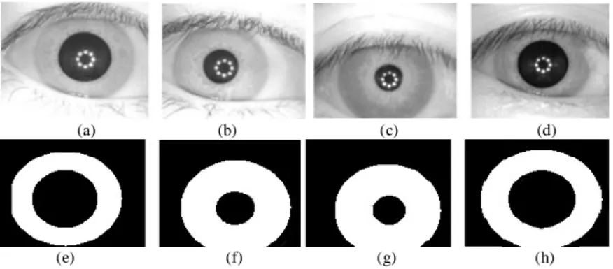
discovered (see Fig. 3 and Fig. 4).
Fig. 1. Block diagram of our proposed method
Iris Image Normalization
Iris Image Segmentation
Iris Image Localization
3.3 Image Normalization
After segmentation process of the iris area and it is
separated from eye image completely, then in order to
calculate the similarity, the iris area is transformed in the
next step for obtaining the constant dimensions. For this
normalization process, Duugman rubber sheet model
(Ahmadi and Nilashi, 2018) is performed as it gives
wonderful result for this process. Fig. 5 shows the
normalized iris image.
3.4
Feature Extraction
In this step, for obtaining the iris features, Haar wavelet
transform is used in order to extract the iris tissue precisely.
This is because of the low computational requirements of
this method and also it has been widely utilized for both
image processing and pattern recognition (Jillela and Ross,
2015; Tapia et al, 2016). It disintegrates the iris data in
some sections, so that it is usable in different resolutions.
(a) (b) (c) (d)
(e) (f) (g) (h)
Fig. 2. (a) Original resized image. (b) ) Local entropy of grayscale. (c) Create rough mask. (d) Morphologically close image. (e, f, g, h) Local entropy of grayscale image, and Otsu's method
(a) (b) (c)
(d) (e) (f)
Fig. 3. Canny edge detection. (b) Remove small objects morphologically. (c) Robust pupil and limbic boundary detection, apply morphological operations on binary images. (d) Pupil boundary detection part mask reflections first. (e) Label connected components on binary image. (f) iris area
(a) (b) (c) (d)
(e) (f) (g) (h)
Fig. 4. Accurate iris localization of CASIA-Iris V3 database: (a, b, c, and d) are the first image from first, second, third, and fourth person, respectively. (e, f, g, and h) are the localized iris detection for image a, b, c, and d, respectively.
4.
Conclusion
In this study according to the fact that segmentation step
is quite prominent for iris recognition framework, we
presented a novel approach towards iris segmentation.
After we obtained the iris image, then we used
pre-processing steps that consists of segmentation, localization,
and normalization steps. In segmentation step, we utilized
local entropy of the grayscale iris image; then, we created a
rough mask; after that we used morphological operators
and Otsu method. In the next step which is localization, we
applied Canny edge detection method. Then, we omitted
the tiny objects morphologically. Afterward, we performed
a detection approach for pupil and limbic boundary and we
applied morphological operators on the binary iris images.
Finally, we utilized connected component labeling method
on the binary iris images. Last but not least, in
normalization step, in order to achieve a normalized iris
image, we employed Duugman rubber sheet model. In the
feature extraction step, we uses Haar wavelet transform
method for feature extraction step and for our experimental
results, we used CASIA-Iris V3 dataset.
In the future study, it is worthwhile to improve iris
segmentation by applying some of the artificial intelligence
methods like Particle Swarm Optimization (PSO)
algorithm, Neuro-Fuzzy method (Nilashi et al., 2019a;
Nilashi et al., 2019b; Nilashi et al., 2015; Yadegaridehkordi
et al., 2018), hybrid Artificial Neural Networks (ANNs),
Decision Trees (Nilashi et al., 2017a; Nilashi et al., 2018;
Nilashi et al., 2017b) and ensemble learning approaches
(Nilashi et al., 2019c). As the segmentation step is one of
the important part in iris recognition system; so, it must
apply very intelligence methods for not only obtaining the
best results in matching step, but also boosting the accuracy
rate.
References
Abdullah, M. A., Dlay, S. S., Woo, W. L., & Chambers, J. A. (2016). Robust iris segmentation method based on a new active contour force with a noncircular normalization. IEEE
transactions on systems, man, and cybernetics:
Systems, 47(12), 3128-3141.
Ahmadi, N., & Akbarizadeh, G. (2015). Iris recognition system based on canny and LoG edge detection methods. Journal of Soft Computing and Decision Support Systems, 2(4), 26-30. Ahmadi, N., & Akbarizadeh, G. (2016). A review of iris
recognition based on biometric technologies. Transylvanian Rev, 24(4), 151-163.
Ahmadi, N., & Akbarizadeh, G. (2017). Hybrid robust iris recognition approach using iris image pre-processing, two-dimensional gabor features and multi-layer perceptron neural network/PSO. Iet Biometrics, 7(2), 153-162.
Ahmadi, N., & Akbarizadeh, G. (2018). Iris tissue recognition based on GLDM feature extraction and hybrid MLPNN-ICA classifier. Neural Computing and Applications, 1-15. Ahmadi, N., & Nilashi, M. (2018). Iris texture recognition based
on multilevel 2-D Haar wavelet decomposition and Hamming distance approach. Journal of Soft Computing and Decision Support Systems, 5(3), 16-20.
Alvarez-Betancourt, Y., & Garcia-Silvente, M. (2016). A keypoints-based feature extraction method for iris
recognition under variable image quality
conditions. Knowledge-Based Systems, 92, 169-182. Belcher, C., & Du, Y. (2009). Region-based SIFT approach to iris
recognition. Optics and Lasers in Engineering, 47(1), 139-147.
Cappelli, R., Ferrara, M., & Maltoni, D. (2015). Large-scale fingerprint identification on GPU. Information Sciences, 306, 1-20.
Chen, J., Shen, F., Chen, D. Z., & Flynn, P. J. (2016). Iris recognition based on human-interpretable features. IEEE Transactions on Information Forensics and Security, 11(7), 1476-1485.
De Marsico, M., Nappi, M., Riccio, D., & Wechsler, H. (2015). Mobile iris challenge evaluation (MICHE)-I, biometric iris dataset and protocols. Pattern Recognition Letters, 57, 17-23. De Marsico, M., Petrosino, A., & Ricciardi, S. (2016). Iris recognition through machine learning techniques: A survey. Pattern Recognition Letters, 82, 106-115.
de Mira, J., Neto, H. V., Neves, E. B., & Schneider, F. K. (2015). Biometric-oriented iris identification based on mathematical morphology. Journal of Signal Processing Systems, 80(2), 181-195.
Elhoseny, M., Elkhateb, A., Sahlol, A., & Hassanien, A. E. (2018). Multimodal biometric personal identification and verification. In Advances in Soft Computing and Machine Learning in Image Processing (pp. 249-276). Springer, Cham.
Fairhurst, M., & Erbilek, M. (2011). Analysis of physical ageing effects in iris biometrics. IET Computer Vision, 5(6), 358-366.
Hollingsworth, K., Bowyer, K. W., Lagree, S., Fenker, S. P., & Flynn, P. J. (2011). Genetically identical irises have texture similarity that is not detected by iris biometrics. Computer Vision and Image Understanding, 115(11), 1493-1502. Jalilian, E., & Uhl, A. (2017). Iris segmentation using fully
convolutional encoder–decoder networks. In Deep Learning for Biometrics (pp. 133-155). Springer, Cham.
Jeong, D. S., Hwang, J. W., Kang, B. J., Park, K. R., Won, C. S., Park, D. K., & Kim, J. (2010). A new iris segmentation method for non-ideal iris images. Image and vision computing, 28(2), 254-260.
Jillela, R. R., & Ross, A. (2015). Segmenting iris images in the
visible spectrum with applications in mobile
biometrics. Pattern Recognition Letters, 57, 4-16.
Kang, J. S. (2010). Mobile iris recognition systems: An emerging biometric technology. Procedia Computer Science, 1(1), 475-484.
Luengo-Oroz, M. A., Faure, E., & Angulo, J. (2010). Robust iris
segmentation on uncalibrated noisy images using
mathematical morphology. Image and Vision
Computing, 28(2), 278-284.
Mulleti, S., & Seelamantula, C. S. (2015). Ellipse fitting using the finite rate of innovation sampling principle. IEEE Transactions on Image Processing, 25(3), 1451-1464. Niinuma, K., Park, U., & Jain, A. K. (2010). Soft biometric traits
for continuous user authentication. IEEE Transactions on information forensics and security, 5(4), 771-780.
Nilashi, M., Ahani, A., Esfahani, M.D., Yadegaridehkordi, E., Samad, S., Ibrahim, O., Sharef, N.M. and Akbari, E., (2019b). Preference learning for eco-friendly hotels recommendation: A multi-criteria collaborative filtering approach. Journal of Cleaner Production, 215, 767-783. Nilashi, M., Ahmadi, H., Shahmoradi, L., Ibrahim, O., & Akbari,
Nilashi, M., bin Ibrahim, O., Ahmadi, H., & Shahmoradi, L. (2017a). An analytical method for diseases prediction using machine learning techniques. Computers & Chemical Engineering, 106, 212-223.
Nilashi, M., bin Ibrahim, O., Ithnin, N., & Sarmin, N. H. (2015). A multi-criteria collaborative filtering recommender system for the tourism domain using Expectation Maximization (EM) and PCA–ANFIS. Electronic Commerce Research and Applications, 14(6), 542-562.
Nilashi, M., Ibrahim, O., Ahmadi, H., & Shahmoradi, L. (2017b). A knowledge-based system for breast cancer classification using fuzzy logic method. Telematics and Informatics, 34(4), 133-144.
Nilashi, M., Ibrahim, O., Samad, S., Ahmadi, H., Shahmoradi, L., & Akbari, E. (2019a). An analytical method for measuring the Parkinson’s disease progression: A case on a Parkinson’s telemonitoring dataset. Measurement, 136, 545-557. Nilashi, M., Ibrahim, O., Yadegaridehkordi, E., Samad, S.,
Akbari, E., & Alizadeh, A. (2018). Travelers decision making using online review in social network sites: A case on TripAdvisor. Journal of computational science, 28, 168-179.
Othman, N., Dorizzi, B., & Garcia-Salicetti, S. (2016). OSIRIS: An open source iris recognition software. Pattern Recognition Letters, 82, 124-131.
Othman, Z., Abdullah, A., Kasmin, F., & Ahmad, S. S. S. (2019). Effect of Supervised Region of Interest Against Edge Detection Method for Iris Localisation. In Intelligent and Interactive Computing (pp. 249-263). Springer, Singapore. Park, U., & Jain, A. K. (2010). Face matching and retrieval using
soft biometrics. IEEE Transactions on Information Forensics and Security, 5(3), 406-415.
Perera, L. P., Oliveira, P., & Soares, C. G. (2015). System identification of vessel steering with unstructured uncertainties by persistent excitation maneuvers. IEEE Journal of Oceanic Engineering, 41(3), 515-528.
Phillips, P. J., Bowyer, K. W., & Flynn, P. J. (2007). Comments on the CASIA version 1.0 iris data set. IEEE Transactions on Pattern Analysis and Machine Intelligence, 29(10), 1869-1870.
Pillai, J. K., Patel, V. M., Chellappa, R., & Ratha, N. K. (2011). Secure and robust iris recognition using random projections and sparse representations. IEEE transactions on pattern analysis and machine intelligence, 33(9), 1877-1893. Proença, H. (2014). Iris recognition: What is beyond bit
fragility?. IEEE Transactions on Information Forensics and Security, 10(2), 321-332.
Pundlik, S. J., Woodard, D. L., & Birchfield, S. T. (2008, June). Non-ideal iris segmentation using graph cuts. In 2008 IEEE Computer Society Conference on Computer Vision and Pattern Recognition Workshops (pp. 1-6). IEEE.
Raffei, A. F. M., Asmuni, H., Hassan, R., & Othman, R. M. (2015). A low lighting or contrast ratio visible iris recognition using iso-contrast limited adaptive histogram equalization. Knowledge-Based Systems, 74, 40-48. Raghavendra, R., & Busch, C. (2015). Robust scheme for iris
presentation attack detection using multiscale binarized statistical image features. IEEE Transactions on Information Forensics and Security, 10(4), 703-715.
Rakshit, S., & Monro, D. M. (2007). An evaluation of image sampling and compression for human iris recognition. IEEE Transactions on Information Forensics and Security, 2(3), 605-612.
Reid, D. A., Nixon, M. S., & Stevenage, S. V. (2013). Soft biometrics; human identification using comparative
descriptions. IEEE Transactions on Pattern Analysis and Machine Intelligence, 36(6), 1216-1228.
Ricanek, K., Savvides, M., Woodard, D. L., & Dozier, G. (2010).
Unconstrained biometric identification: Emerging
technologies. Computer, 43(2), 56-62.
Solomatin, I. A., Matveev, I. A., & Novik, V. P. (2018). Locating the visible part of the iris with a texture classifier with a support set. Automation and Remote Control, 79(3), 492-505.
Tapia, J. E., Perez, C. A., & Bowyer, K. W. (2016). Gender classification from the same iris code used for recognition. IEEE Transactions on Information Forensics and Security, 11(8), 1760-1770.
Umer, S., Dhara, B. C., & Chanda, B. (2015). Iris recognition using multiscale morphologic features. Pattern Recognition Letters, 65, 67-74.
Vincent, L. (1994). Morphological area openings and closings for grey-scale images. In Shape in Picture (pp. 197-208). Springer, Berlin, Heidelberg.
Wan, H. L., Li, Z. C., Qiao, J. P., & Li, B. S. (2013). Non-ideal iris segmentation using anisotropic diffusion. IET Image Processing, 7(2), 111-120.
Yadegaridehkordi, E., Hourmand, M., Nilashi, M., Shuib, L., Ahani, A., & Ibrahim, O. (2018). Influence of big data adoption on manufacturing companies' performance: An
integrated DEMATEL-ANFIS approach. Technological
Forecasting and Social Change, 137, 199-210.
Yang, L., Yang, G., Yin, Y., & Xi, X. (2014). Exploring soft
biometric trait with finger vein
recognition. Neurocomputing, 135, 218-228.