Journal of Global Pharma Technology
Available Online at:
www.jgpt.co.in
RESEARCH ARTICLE
The Effect of Extra Virgin Olive Oil to Decrease HSP-90, TNF-Α
and ET-1, in Pre-Eclampsia Rat Model
Bambang Rahardjo
1*,Wenny Rahmawati
2,Alfima Rahasti
3,Dwi Norma
Retnaningrum
4,Hidayat Sujuti
5,Noorhamdani AS
6,Tri Yudani Mardining
Raras
71. Dept. Obstetri Ginecology, Faculty of Medicine, Brawijaya University/ Saiful Anwar General Hospital.
2. Magister of Midwifery, Faculty of Medicine, Brawijaya University.
3. Magister of Midwifery, Faculty of Medicine, Brawijaya University.
4. Magister of Midwifery, Faculty of Medicine, Brawijaya University.
5. Dept. Biochemistry And Biomolecular, Faculty of Medicine, Brawijaya University, Malang, Indonesia.
6. Dept. Microbiology, Faculty of Medicine, Brawijaya University, Malang, Indonesia.
7. Dept. Biochemistry and Biomolecular, Faculty of Medicine, Brawijaya University, Malang, Indonesia.
*Corresponding Author:Bambang Rahardjo
Abstract
Background: Preeclampsia characterised by new-onset hypertension with systolic blood pressure ≥ 140 mmHg or diastolic blood pressure ≥ 90 mmHg, measured on two occasions at least four hours apart, and proteinuria of > 0.3 g per 24 hours or ≥ 1+ proteinuria, detected by urine dipstick after 20 weeks of pregnancy, or in the absence of proteinuria. Objectived: This study aimed to determine effect of EVOO as a strong exogenous antioxidant to decrease TNF-α, HSP-90 and ET-1 level in preeclampsia rat model. Method: This research consisted of five groups; negative control, positive control (preeclampsia rat model), doses 1, 2, and 3 that were preeclampsia rats given EVOO in 3 different doses (0.5 mL/day, 1 mL/day and 2 mL/day respectively). After sacrificed, placentas were collected to determine HSP-90, TNF-α and ET-1 level. Result: Result of this study showed that there was a reduction of HSP90 (p=0, 00), TNF-α (p=0.011), ET-1 (p=0.02). Conclusion: Administration EVOO decreased HSP-90, TNF-α and ET-1 Level in preeclampsia rat model
Keywords: Preeclampsia, EVOO, HSP-90, TNF-α, ET-1, Antioxidant, Oxidative stress.
Introduction
Preeclampsia affects 3-14% of all pregnancies world-wide and can have a significant impact on health for both mother and fetus. For the fetus, preeclampsia can result in IUGR. For the mother complications of preeclampsia include renal failure, HELLP syndrome (haemolysis, elevated liver enzymes, and thrombocytopenia), seizures, stroke or death [1]. Approximately 72,000 pregnant women die every year because of eclampsia and severe preeclampsia. That amounts to nearly 200 women every day. The risk that a woman in a developing country will die of preeclampsia or eclampsia is about 300 times that of a woman in a developed country [2]. Preeclampsia characterised by new-onset
oedema, or cerebral or visual problems [3]. Medical treatment depends on the severity of preeclampsia, and relies on antihypertensive medications and magnesium sulfate. Medical treatment does not alter the course of the disease, but aims at preventing the occurrence of intracranial hemorrhages and seizures [4]. Delivery of the placenta remains the only known treatment for this clinical disease, suggesting that the placenta is the principal contributor to the pathogenesis of preeclampsia [5]. The decision of terminating pregnancy and perform delivery is based on
gestational age, maternal and fetal
conditions, and severity of preeclampsia [4]. One factors in the pathophysiology of preeclampsia is deficient conversion of the uterine spiral arteries. The placenta is supplied by maternal spiral arteries, which
undergo major modifications during
pregnancy to accommodate the increase in uterine blood flow.
Abnormal conversion of the maternal spiral arteries supplying the placenta and with subsequent placental malperfusion result in placental oxidative stress and release of a complex mix of factors, including pro-inflammatory cytokines, apoptotic debris and angiogenic regulators into the maternal circulation [6].
The condition of preeclampsia will cause vascular hypoxia. Hypoxic conditions in the
vascular stimulate increased hypoxia
inducible factor (HIF). HIF is a
transcriptional activator that regulates cell death or survival and has the ability to mediate adaptive responses to changes in cellular oxygen tension. HIF-1 is very sensitive to hypoxic stimulus. Furthermore, an increase in HIF-1 and heat shock
conditions in cells will stimulate
transcription of heat shock factor (HSF). HSF-1 plays a central role in controlling the heat stress response (HSR).
Increased HSF-1 is very important for transcription of heat shock protein (HSP) during hypoxic and reoxygenated events [7,8, 9]. HSP is a protein that has an effect on the transcription process so as to produce proinflammatory cytokines such as IL1β and TNF-α. Stimulation of proinflammatory cytokines (IL-1β and TNF-α) in endothelial blood vessels will release monocyte adhesion molecules, one of which is VCAM-1. VCAM-1 in monocytes will release the MMP-9 enzyme
which can damage type 4 and vascular collagen. Vascular damage causes endothelial dysfunction which is part of the maternal inflammatory reaction, causing ischemic arterial spirals which will have an impact on decreased placental perfusion and the
appearance of clinical symptoms of
preeclampsia [9].One of HSP or also known as stress protein which is dominantly induced in cells is heat shock protein 90 (HSP90). In a non-stress state, the amount of HSP90 is abundant in cells, which is around 1-2% of the total protein and can be found in the cytosol, nucleus and endoplasmic reticulum of cells. HSP90 functions as an anti-apoptosis and has the function of chaperone which is maintenance of cell survival after various pathological conditions such as the process of endothelial dysfunction in preeclampsia [10, 11, 12].Recent studies have shown that HSP90 as a stress protein plays an important role in the regulation of a number of inflammatory signaling pathways.
HSP90 can also be a stimulating response to systemic inflammatory immunity that endangers pregnancy. In the case of preeclampsia, an increase in HSP90 levels is a marker of hypoxia, oxidative stress and systemic inflammation [9]. The inflammatory response involves activation of various transcription factors.
One that plays an important role in the inflammatory response is Nuclear Factor Kappa Beta (NF-kβ). NF-kβ is regulatior of the expreesion of inflammatory genes, and triggers the production of cytokin that effect inflamation such as tumor necrosis factor-α (TNF-α) [13]. The increase in TNF-α is proven to occur in the condition of preeclampsia, therefore it can be used as a marker of the incidence of preeclampsia. Exposure to inflammatory cytokines, will also interfere with the work of endothelial cells [14].
Oxidative stress, inflammation, genetics and immunological disorders are real processes
that underlie the pathogenesis of
preeclampsia. This condition can cause ischemia or hypoxia in the placenta. Hypoxia Inducible Factor 1a (HIF-1a), Flt-1 (sFlt-1), angiotensin II type receptor autoantibodies (AT1-AA), and placental growth factor PlGF) are factors known to be involved in preeclampsia. Increased these factors cause
bioavailability of Nitric Oxide (NO) and increase Reactive Oxygen Species (ROS) and Endothelin (ET-1), which cause changes in kidney function, increase Total Peripheral Resistance (TPR), and eventually become hypertensive [15]. Endothelin (ET)-1 is another factor that is found to be elevated in
pre-eclampsia compared with normal
pregnancy. In plasma from healthy pregnant women, the concentration of ET-1 ranges from 5 to 10 pg/ml, whereas the concentration is 20-50 pg/ml in the presence of pre-eclampsia [16].
There are several factors that can stimulate the production of ET-1 such as cytokines, thrombin, stress and also hypoxic conditions [17].Increased plasma concentrations of ET-1, cause vasoconstriction of the uterine
arteries and contribute to systemic
hypertension [18].One alternative that might
be used in additional therapy or support for preeclampsia is the provision of Extra virgin olive oil or commonly called EVOO. Extra virgin olive oil is one type of oil that comes from the first juice of olives [19].
The antioxidants contained in EVOO have the role of delaying the oxidation process. In this case, the main antioxidant that inhibits the oxidation process at EVOO is OP (Olive Phenols), which acts as a chain breaker by donating hydrogen radicals to alkylperoxyl radicals produced by lipid oxidation and the formation of stable derivatives during reaction [20]. With the increase in antioxidants is expected to reduce endhotel dysfunction and reduce levels of HSP90, TNF-α and ET-1.
Materials and Methods
Animal Model
This research was in vivo laboratory research. The design was a Post Test Only Control Group design.This research was use
experimental animals rats (Rattus
norvegicus) wistar strain. This study used 20 pregnant rat and divided into 5 groups. The negative control group was normal pregnant rats; the positive control was pregnant pre-eclampsia rats (pre-pre-eclampsia rat model); and the treatment group 1, 2, and 3 were
preeclampsia rat given Extra Virgin Olive Oil
(EVOO) in three different doses (0,5 mL/day, 1 mL/day and 2 mL/day) respectively [21]. The next day after mating was assumed to be the day 1 of pregnancy.
The sacrifice of rat did in 19 day of
pregnancy. The sample used in the study was
placenta and plasma. The research was
carried out in the Laboratory of Biosains
Universitas Brawijaya, Laboratory of
Physiology and Laboratory of Biomolecular
Biochemistry, Faculty of Medicine
Universitas Brawijaya Malang.
Preeclampsia Induction and EVOO Administration
The material used for induction of preeclampsia was using NOS inhibitors, L-NAME (C7H15N5O4 • HCl) from Sigma-Aldrich (Merck KGaA, Darmstadt, Germany) [22]. Pregnant rat were randomly placed in 5 groups consisting of negative control group, positive control group and three treatment groups. Each group contains 4 pregnant rats. Intraperitoneal injection of L-NAME with a dose of 125 mg L-NAME / kilogram of body weight was given to rats with 13-19 days of gestation [23]. Preeclampsia rat model can be
made by injection of L-NAME
intraperitoneally used dose of 125
mg/Kilogram of body weigt was injected to rats with 13 days of gestation until 19 days of gestation [19].
Clinical and Sample Examination
Measurement of HSP90 level was performed with ELISA KIT HSP90 (No catalog E-EL-0042).ELISA kit with pre-coated plates rat TNF-α No. E-EL-R0019. ELISA KIT ET-1 level was performed with ET-1 ELISA KIT (No catalog E-EL-R0167). Data was analyzed statistically with ANOVA.
HSP90 Activity Measurement
Measurement of HSP90 levels was carried out using the protocol found in the Elabscience kit with the catalog number E-EL-0042.Sample used blood of rats. Prepare
wells for blanks, standards and
Enter 90 µl Substrate Reagents in all wells. Protect Substrate Reagents from light.Cover with adhesive strips then incubate for 15 minutes at 370 C.Put a 50 µl stop solution on all wells.Determine optical density (O.D) using a 450 nm ELISA reader.
TNF-α Activity Measurement
Measurement of TNF-α levels will be carried out using the ELISA method with No catalog: E-EL-R00197 produced by Elabscience. Sample used blood of rat. All reagents are placed at room temperature before use. Continue to work on standard and double or triple samples use one standard curve for each essay. Add standard solutions to the first two columns: each concentration of solution is added duplicates. Fill each well with 100µL.. Incubation for 90 minutes at 37ºC. Discard the sanple without washing. Add 100 µL of Biotinylated immediately to each well. Incubated for 1 hour at 37ºC. Pour the soution from each well.
Add 350µL buffer, turn it on for 1-2 minutes then discard the solution to each well. Cover with a plate sealer. Incubate for 30 minutes at 37ºC in dark place. Aspiration or pour the solution from each well. Repeat washing five times like step 4. Add 90 µL of reagent substrate to each hole. Close the new plate sealer. Incubate for 15 minutes at 37º C in a dark place. Add 50 µL from the stop solution to each wel. Read absorbance at 450 nm within 30 minutes with ELISA reader.
ET-1 Activity Measurement
Measurement of endothelin-1 levels will be carried out using the ELISA method with No
catalog: E-EL-R0167 produced by
Elabscience. The placenta was smoothed using PBS liquid in a ratio (tissue weight (grams): BS (mL) = 1: 9). Homogenation was
done by centrifugation for 5 minutes to get the supernatant. Then a standard solution was added to each well (100 μL in each well) and incubated for 90 minutes at 37 ° C. Remove the liquid in each well without washing. Immediately add 100 μL of Biotinylated Detection Ab working to each well. Cover using a Plate Sealer. Incubate for 1 hour at 37 ° C.
Wash each well, repeats the process twice with a total of three washing times. Wash by filling each well with Wash Buffer (350μl) using a multi-channel pipette, manifold dispenser, or auto washer, and leave it for 2 minutes, making sure the liquid in each well is completely empty. After the final washer, clean the remaining washes. Turn over well and clean with a clean paper towel. Then add 100μl HRP-avidin to each well.
Cover the microtiter plate with a new adhesive strip. Incubate for 30 minutes at 37 ° C. Repeat the aspiration / washing process five times. Add 90μl of TMB substrate to each well. Incubate for 15 minutes at 37 °C. Protect from light. Add 50μl Stop Solution to each well. Optical density was read on a micro pet reader with a wavelength of 450nm.
Data Analysis
Data analysis using SPSS 25.00.Test used to
observe group mean different was One Way Anova Test.
Result and Discussions
Effect of EVOO on HSP-90 Level Preeclampsia Rat Model
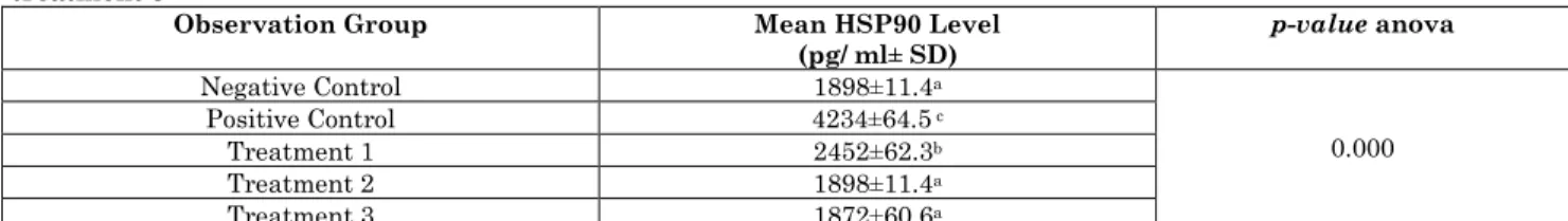
Table 1 shows that the preeclampsia rat that were no treated with EVOO had a higher HSp90 levels compared to the preeclampsia rat model treated with EVOO.
Table 1: Comparison of mean level of HSP-90 of negative control, positive control, treatment 1, treatment 2 and treatment 3
Observation Group Mean HSP90 Level (pg/ ml± SD)
p-value anova
Negative Control 1898±11.4a
0.000
Positive Control 4234±64.5 c
Treatment 1 2452±62.3b
Treatment 2 1898±11.4a
Treatment 3 1872±60.6a
Note: Negative control is a normal pregnant rat. Positive control is a pregnant rat model of preeclampsia. Treatment 1 was a preeclampsia model rat that was given EVOO 0.5 cc. Treatment 2 was a preeclampsia model rat that was given EVOO 1 cc. Treatment 3 was a preeclampsia model rat that was given EVOO 2 cc
Our study confirmed that HSP90 levels significantly different between rat didn’t give EVOO and rat administrated with EVOO (p
doses. EVOO 0.5 cc/day did not appear to significantly decrease HSP90 levelin the preeclampsia rat model. The data showed that the levels of HSP90 in the treated group at 1 and 2 cc/day dose differ significantly from the mean in the normal pregnant rat group.
This means that the optimum dose of EVOO to reduce HSP90 levels in the preeclampsia rat model was 1 and 2 cc/day. Extra Virgin Olive Oil contains flavonoids which have a role in scavenging free radicals. Scavenging activity in flavonoids begins with the administration of hydrogen or electron groups to free radicals which then produce flavonoid radical molecules and stable molecules (RH). Flavonoid radicals have lower reactivity than free radicals.
Then flavonoid radicals bind with other radicals to become non-reactive compounds. Flavonoids contained in EVOO will inhibit lipid peroxidation in vitro in the early stages with its role as a scavenger of superoxide anions and hydroxyl radicals.
This radical reaction chain will then be broken by donating hydrogen atoms to peroxyl radicals so that flavonoid radicals are formed which will react with free radicals to break the ROS chain [20, 24].
Effect of EVOO on TNF-α Level Preeclampsia Rat Model
Table 2 shows that the preeclampsia rat that were no treated with EVOO had a higher TNF-α levels compared to the preeclampsia rat model treated with EVOO.
Table 2: Comparison of mean level of TNF-α of negative control, positive control, treatment 1, treatment 2 and treatment 3
Observation Group Mean ET-1 Level (pg/ ml± SD)
p-value anova
Negative Control
99.9±10.9b
0.011 Positive Control
312.6±171.6a
Treatment 1
143.5±29.3b
Treatment 2
167.0±52.3b
Treatment 3
92.1±2.9b
Note: Negative control is a normal pregnant rat. Positive control is a pregnant rat model of preeclampsia. Treatment 1 was a preeclampsia model rat that was given EVOO 0.5 cc. Treatment 2 was a preeclampsia model rat that was given EVOO 1 cc. Treatment 3 was a preeclampsia model rat that was given EVOO 2 cc.
Our study confirmed that TNF-α levels significantly different between rat didn’t give EVOO and rat administrated with EVOO (p < 0.05). A significant decrease in TNF-α levels was observed in the preeclampsia rat administered EVOO at 05cc/day, 1 cc/day and 2 cc/day doses. The decrease in TNF- α levels was in line with the increase in EVOO dose given, although the three types of EVOO doses were not statistically significant. In general it can be said that there is a decrease
in serumreatm TNF-α levels by
administrering EVOO at various doses.
So EVOO administration oh trese three doses can reduce TNF-α levels in preeclamsia pregnant mice. EVOO dose which is considered the fastest able to reduce TNF-α level is dose 1 by administering 0.5 mL/hr, because with smaller administration EVOO has been able to reduce TNF-α levels in dose 1 group with mean values close to TNF-α
levels in the group negative control. However, because there was no significant difference in the mean TNF-α levels between the three treatment dose.
This situation means that the three EVOO dose given have the same ability in terms of reducing TNF-α levels. Giving extra virgin olive oil (EVOO) to wistar rats with
preeclamsia models can reduce
proinflammatory cytokines in cases of preeclamsia because EVOO has polyphenol content that is antiinflamatory and shows the ability to inhibit inflammatory cytokines such as Tα, interleukin-6, and factor NF-kB transcription [25].
mg/day) can reduce the expression of several inflammatory genes including NF-kB and COX-2 [27].
Effect of EVOO on ET-1 Level Preeclampsia Rat Model
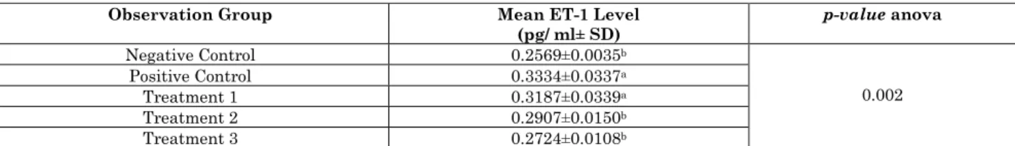
Table 3 shows that the preeclampsia rat that were no treated with EVOO had a higher ET-1 levels compared to the preeclampsia rat model treated with EVOO.
Table 3 Comparison of mean level of ET-1 of negative control, positive control, treatment 1, treatment 2 and treatment 3
Observation Group Mean ET-1 Level
(pg/ ml± SD) p-value anova
Negative Control 0.2569±0.0035b
0.002
Positive Control 0.3334±0.0337a
Treatment 1 0.3187±0.0339a
Treatment 2 0.2907±0.0150b
Treatment 3 0.2724±0.0108b
Note: Negative control is a normal pregnant rat. Positive control is a pregnant rat model of preeclampsia. Treatment 1 was a preeclampsia model rat that was given EVOO 0.5 cc. Treatment 2 was a preeclampsia model rat that was given EVOO 1 cc. Treatment 3 was a preeclampsia model rat that was given EVOO 2 cc
Our study confirmed that ET-1 levels significantly different between rat didn’t give EVOO and rat administrated with EVOO (p < 0.05). A significant decrease in ET-1 levels was observed in the preeclampsia rat administered EVOO at 1 cc/day and 2 cc/day doses. EVOO 0.5 cc/day did not appear to significantly decrease ET-1 levelin the preeclampsia rat model. The data showed that the levels of ET-1 in the treated group at 2 cc/day dose did not differ significantly from the mean in the normal pregnant rat group. This means that the optimum dose of EVOO to reduce ET-1 levels in the preeclampsia rat model was 2 cc/day. This is in line with research conducted by Rahma (2017) antioxidants in black cumin can reduce the expression of ET-1 in mice preeclampsia models.
A study said that flavonoids found in chocolate can reduce levels of ET-1 in endothelial cells induced by plasma from patients with preeclampsia. These flavonoids reduce ET-1 by inactivating ECE (endothelin converting enzyme) together with the modulation of NO bioavailability production. Oxidative stress, inflammation cause an increase in factors such as Hypoxia Inducible Factor 1a (HIF-1a), sFlt-1, angiotensin II type receptor autoantibodies (AT1-AA), cause
endothelial dysfunction, reduce the
bioavailability of Nitric Oxide (NO) and increase endothelin (ET-1).
At the systemic level, an increase in ET-1 can cause vasoconstriction and remodeling of blood vessels to become more resistant which causes changes in kidney function, increases Total Peripheral Resistance (TPR), and eventually becomes hypertension [15].The mechanism of decreasing ET-1 by EVOO through the potential of EVOO as an antioxidant. Antioxidants prevent damage to organs by free radicals by preventing the formation of ROS. Antioxidants also suppress the production of NF-kB and NF-kB itself inhibits the occurrence of endothelial dysfunction in preeclampsia by reducing the production of AT1-AA, by decreasing the production of AT-1AA resulting in decreased markers of endothelial dysfunction, such as decreasing ET-1.
Antioxidants will suppress the formation of peroxynitrit (ONOO-) which will further reduce the occurrence of endothelial dysfunction which is characterized by a decrease in ET-1 which in turn will cause smooth muscle relaxation that will reduce blood pressure [28].
Conclusion
Administration EVOO decreased HSP-90
level, TNF-α level and ET-1 Level in
preeclampsia rat model.
References
1. Finger Irminger-Finger, Nicole Jastrow,
Irion Oliver (2008) Preeclampsia: A danger growing in disguise. Int J Biochem Cell Biol., 40(10):1979-83.
2. Sanjay Gupte, Girija Wagh (2014)
Preeclampsia-Eclampsia. J. Obstet.
Gynecol. India, 64(1):4-13.
Pre-eclampsia: its pathogenesis and
pathophysiolgy. Cardiovasc J. Afr.
[Internet]. 27(2):71-8. Available from: http://cvja.co.za/onlinejournal/vol27/vol27_i ssue2/#17/z
4. Lambert G, Brichant JF, Hartstein G,
Bonhomme V, Dewandre PY (2014)
Preeclampsia: An update. Acta
Anaesthesiol. Belg., 65(4):137-49.
5. Sircar M, Thadhani R, Karumanchi SA
(2015) Pathogenesis of preeclampsia. Curr Opin Nephrol. Hypertens, 24(2):131-8.
6. Jain A (2012) Endothelin-1: A key
pathological factor in pre-eclampsia?
Reprod Biomed Online [Internet].
25(5):443-9. Available from:
http://dx.doi.org/10.1016/j.rbmo.2012.07.01 4
7. Baird NA, Turnbull DW, Johnson EA
(2006) Induction of the Heat Shock
Pathway during Hypoxia Requires
Regulation of Heat Shock Factor by, 281(50):38675-81.
8. Agarwal A, Aponte-mellado A, Premkumar
BJ, Shaman A, Gupta S (2012) The effects
of oxidative stress on female reproduction :
a review. Reprod Biol Endocrinol
[Internet]. 10(1):1. Available from:
Reproductive Biology and Endocrinology
9. Padmini E, Uthra V, Lavanya S (2012)
Effect of HSP70 and 90 in Modulation of JNK, ERK Expression in Preeclamptic Placental Endothelial Cell. Cell Biochem Biophys. 64(3):187-95.
10.E P, BV G (2008) Placental heat shock
protein 70 overexpression confers
resistance against oxidative stress in
preeclampsia. Turkish J Med Sci
[Internet]. 38(1):27-34. Available from: http://www.embase.com/search/results?sub action=viewrecord&from=export&id=L351 346796%5Cnhttp://journals.tubitak.gov.tr/ medical/issues/sag-08-38-1/sag-38-1-5-
0705-8.pdf%5Cnhttp://sfx.library.uu.nl/utrecht?s id=EMBASE&issn=13000144&id=doi:&ati tle=Placental+heat+s
11.Li J, Soroka J, Buchner J (2012) The
Hsp90 chaperone machinery:
Conformational dynamics and regulation by co-chaperones. Biochim Biophys Acta - Mol Cell Res [Internet]. 1823(3):624-35.
Available from:
http://dx.doi.org/10.1016/j.bbamcr.2011.09.
003
12.Surai F P (2016) Antioxidant Systems in
Poultry Biology: Superoxide Dismutase. J. Anim. Res Nutr., 01: 01.
13.Laresgoiti-Servitje E, Gomez-Lopez N
(2012) The Pathophysiology of
Preeclampsia Involves Altered Levels of Angiogenic Factors Promoted by Hypoxia and Autoantibody-Mediated Mechanisms1. Biol. Reprod., 87(2):1-7.
14.Rahardjo B, Widjajanto E, Sujuti H,
Keman K (2014) Different levels of IL-1α, IL-6, TNF-α, NF-κB and PPAR-γ in monocyte cultures exposed by plasma
preeclampsia and normotensive
pregnancy. Pregnancy Hypertens
[Internet]. 4(3):187-93. Available from: http://dx.doi.org/10.1016/j.preghy.2014.03. 001
15.Rahma H, Indrawan IWA, Nooryanto M,
Rahajeng, Keman K (2017) Effect of a black cumin (Nigella sativa) ethanol extract on placental angiotensin II type 1-receptor autoantibody (AT1-AA) serum levels and endothelin-1 (ET-1) expression in a preeclampsia mouse model. J Taibah Univ Med Sci [Internet]. 12(6): 528-33.
Available from:
https://doi.org/10.1016/j.jtumed.2017.06.00 2
16.Fiore G, Florio P, Micheli L, Nencini C,
Rossi M, Cerretani D, et al (2005) Endothelin-1 triggers placental oxidative stress pathways: Putative role in preeclampsia. J. Clin Endocrinol Metab., 90(7):4205-10.
17.Kohan DE, Rossi NF, Inscho EW, Pollock
DM (2011) Regulation of blood pressure and salt homeostasis by endothelin. Physiol Rev., 91(1):1-77.
18.Dechanet Clotilde, Aurélie F,
Barbero-Camps E, Dechaud H, Virsolvy A, Richard
S (2011) Endothelin-Dependent
Vasoconstriction in Human Uterine Artery: Application to Preeclampsia. PLoS One, 6(1):e16540.
19.Vitoratos N, Hassiakos D, Lavazzo C
(2008) Molecular mechanisms of
preeclampsia. Microvasc Res, 75(1):1-8.
20.Servili Maurizio, Sordini Beatrice, Esposto
Sonia, Urbani Stefania, Veneziani
Gianluca, Maio Ilona Di, et al (2013)
Biological Activities of Phenolic
Antioxidants, 3(1):1-23.
21.Cicerale S, Lucas L, Keast R (2010)
Biological activities of phenolic compounds present in virgin olive oil. Int. J. Mol. Sci., 11(2):458-79.
22.Amaral TAS, Ognibene DT, Carvalho
LCRM, Rocha APM, Costa CA, Moura RS, et al (2018) Differential responses of mesenteric arterial bed to vasoactive
substances in L-NAME-induced
preeclampsia: Role of oxidative stress and
endothelial dysfunction. Clin Exp
Hypertens [Internet]. 40(2):126-35.
Available from:
https://doi.org/10.1080/10641963.2017.133 9073
23.Meilina (2017) Extra virgin olive oil
menurunkan kadar mda
(Malondialdehyde) pada tikus (Rattus norvegicus) jantan galur wistar yang dipapar asap rokok. Intisari Sains Medis. 8(2):97-101.
24.Salles AMR, Galvao TF, Silva MT, Motta
LCD, Pereira MG (2012) Antioxidants for preventing preeclampsia: A systematic review. Sci. World J., 2012.
25.Camargo A, Ruano J, Fernandez JM,
Parnell LD, Jimenez A, Santos-Gonzalez M, et al (2010) Gene expression changes in mononuclear cells in patients with metabolic syndrome after acute intake of
phenol-rich virgin olive oil. BMC
Genomics, 11: 1.
26.Impellizzeri D, Esposito E, Mazzon E,
Paterniti I, Di Paola R, Bramanti P, et al (2011) The effects of oleuropein aglycone, an olive oil compound, in a mouse model of carrageenan-induced pleurisy. Clin Nutr [Internet]. 30(4):533-40. Available from: http://dx.doi.org/10.1016/j.clnu.2011.02.004
27.Cicerale S, Lucas LJ, Keast RSJ (2012)
Antimicrobial, antioxidant and anti-inflammatory phenolic activities in extra virgin olive oil. Curr Opin Biotechnol [Internet]. 23(2):129-35. Available from: http://dx.doi.org/10.1016/j.copbio.2011.09.0 06
28.Leong X-F, Rais Mustafa M, Jaarin K
![語料庫導向之方位短句於固定框架的共現概念統計分析 (A Corpus-driven Pattern Analysis in Locative Phrases: A Statistical Comparison of Co-appearing Concepts in Fixed Frames) [In Chinese]](data:image/gif;base64,R0lGODlhAQABAIAAAP///wAAACH5BAEAAAAALAAAAAABAAEAAAICRAEAOw==)