THE DROSOPHILA MELANOGASTER PROTEIN SUPPRESSOR OF SABLE [SU(S)] NEGATIVELY REGULATES TRANSPOSON-‐CONTAINING TRANSCRIPTS BY A MECHANISM THAT
INVOLVES DIRECTLY BINDING TO ITS RNA TARGET
Lonna Finnic Mollison
A dissertation submitted to the faculty of The University of North Carolina at Chapel Hill in partial fulfillment of the requirements for the degree of Doctor of Philosophy in the
Curriculum of Genetics and Molecular Biology
Chapel Hill
2016
Approved by: Lillie Searles
Robert Duronio
William Marzluff
Mark Peifer
©2016
Lonna Finnic Mollison ALL RIGHTS RESERVED
ABSTRACT
Lonna Finnic Mollison: The Drosophila melanogaster Protein Suppressor of sable [Su(s)] Negatively Regulates Transposon-‐Containing Transcripts by a Mechanism that Involves
Directly Binding to its RNA Target (Under the direction of Lillie Searles)
RNA quality control systems operate at various stages of gene expression to prevent
aberrant RNAs from accumulating. The nuclear pathways that lead to the identification
and elimination of defective pre-‐mRNAs are incompletely understood, especially in
multicellular organisms. The Suppressor of sable (Su(s)) protein of D. melanogaster plays a role in this process. Su(s) is a nuclear RNA-‐binding protein that negatively regulates the
accumulation of RNA from genes that contain transposon insertions in the 5’ transcribed
region. Previous studies have shown that the Su(s)-‐regulatory pathway induces premature
transcription termination and degradation of the resulting RNAs. Here, I present in vitro and in vivo evidence that Su(s) recognizes specific sequences in one of its biological targets. I found that a U-‐rich element is efficiently bound by Su(s) and a G-‐rich element appears to
be a weaker binding site. The results of reporter gene analysis confirmed that the U-‐rich
and G-‐rich elements are relevant regulatory sequences. However, a GUA-‐rich element that
contributes significantly to this regulation is not a Su(s) binding site. These results indicate
that this regulation depends on the direct binding of Su(s) and, possibly, one or more other
proteins to the RNA.
To my sister Tina and my Father
ACKNOWLEDGEMENTS
I would like to thank Dr. Lillie Searles for allowing me to join her lab and for her
continued commitment to my success. Lillie has been a patient, kind, and understanding
mentor with a unique ability to bring out the best in me while challenging me to become
more cautious and well reasoned in my scientific viewpoint. I have grown immensely from
her mentorship and guidance.
I owe so much gratitude and appreciation to my family. First and foremost I want to
thank, my parents, who lovingly raised eight children. They were the first example of the
type of person I should strive to be. They instilled in me the attributes that are required to
persevere through the rigors of the PhD journey and for this I am forever grateful.
Although my father has passed, my mother continues to find the strength to encourage her
adult children. She has been my biggest champion, supporter and friend, I love you
Mommy. To my seven siblings, who have always protected their youngest sister, I thank
you for your love and support. My sister Shona, who encouraged me to go back to school
after I had gone through a layoff, thank you for being my best friend. To my older sisters
Eva, Reva and Kleva, I admire your strength to raise children and pursue your own career
and I thank you for your love and support over the years. To my only brother Joel, I
appreciate your sense of humor and your kind nature. To my sister Tina, I miss you every
day but you inspired me to reach for my goals. Finally I want to thank my nephew Trenton
and my friends, for providing balance, fun, and a voice of reason.
PREFACE
The work presented in Chapter II determines that the RNA binding activity of Su(s)
contributes to its regulatory activity. While I performed the majority of the experimental
procedures used to investigate the Su(s)/αβ RNA interaction in vitro and in vivo, a research specialist in the Searles lab, Paul Brewer-‐Jensen, made significant contributions to the
reporter gene analysis. I generated and tested the reporter constructs for Region 2 of αβ RNA, which contains invader1 sequences. These constructs determined that Su(s) binding sites are important for regulation of Region 2. However, Paul performed repeat
experiments to produce publication-‐quality Northern blot images. Additionally, all
reporter constructs for Region 1, which contain Dm88 sequences, were generated and
tested by Paul. Therefore, Paul’s efforts truly helped to create a more comprehensive
TABLE OF CONTENTS
LIST OF TABLES……….. ix
LIST OF FIGURES...x
LIST OF ABBREVIATIONS……… xi
CHAPTER I: INTRODUCTION... 1
Transcription by Pol II ...1
Co-transcriptional RNA Modifications ...2
Cotranscriptional Nuclear RNA Quality Control ...6
Cotranscriptional Nuclear RNA Quality Control of Pervasive Transcription ...7
The Drosophila melanogaster Protein Suppressor of Sable Negatively Regulates Aberrant Transcripts... 10
Targets Identified by PCIF Analysis ... 11
The Su(s) Protein ... 13
Su(s) is an RNA Binding Protein... 13
Su(s) has Two Distinct RNA Binding Domains ... 14
The ARMs and the ZFs Contribute to Su(s) Mediated Regulation in vivo... 15
Identification of Components of the Su(s) Regulatory Pathway ... 16
Su(s) Interacts with Wdr82 to Regulate Hsp70-αβ Transcripts ... 17
CHAPTER II: THE INHIBITION OF ABERRANT RNA ACCUMULATION BY THE SUPPRESSOR OF SABLE (SU(S)) PATHWAY DEPENDS ON THE RNA-BINDING ACTIVITY OF SU(S) AND MULTIPLE, DISTINCT REGULATORY ELEMENTS ...19
Introduction... 19
Generation and purification of MBP-‐Su(s) fusion proteins……….. 23
RNA binding experiments………. 23
Analysis of Su(s)-‐RNA interactions in cultured cells……… 24
Generation and Analysis of Reporter Gene Constructs……….. 25
Results... 26
Su(s) Binds αβ RNA in vitro………. 26
Su(s) Binds αβ RNAs in vivo………. 31
Sequence Analysis of UV-‐crosslinked cDNAs Reveal Potential Sites of Contact Between Su(s) and the 209nt Region of αβ Transcripts………..…….. 33
Loss of In Vivo Binding Sites Reduces Binding Affinity to αβ Transcripts In Vitro……… 34
U-‐rich and G-‐rich Elements in Invader1 Contribute to Regulation by Endogenous Su(s)……… 36
A GUA-‐rich Element in the Dm88 LTR Mediates Regulation by Su(s) but is Not a Su(s) Binding Site………. 39
Discussion... 41
CHAPTER III: DISCUSSION AND FUTURE DIRECTIONS...47
Further Analysis of the ARMs... 47
Further Analysis of the CCCH-ZFs ... 49
Possible Secondary Structure within αβ RNAs... 50
Analysis of the Dm88 Region 1... 52
Identification of Global Targets of Su(s)... 53
The Su(s)-Wdr82 Regulatory Pathway ... 54
APPENDIX 1...56
REFERENCES ...59
LIST OF TABLES
Table A1-1-‐ Primers used to make substrates for in vitro RNA binding assays………..……... 56
Table A1-2-‐ Primers used for RIP, CLIP, and PAR-‐CLIP analysis..………..……….... 56
Table A1-3-‐ Synthetic oligomers used to generate the invader1 reporter constructs...….... 57
LIST OF FIGURES
Figure 1- Schematic map of the structure and transcripts of Hsp70-‐αβ
Elements.………... 21
Figure 2- MBP-‐Su(s) fusion proteins used for in vitro RNA binding assays.………... 27
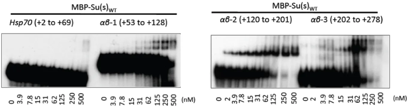
Figure 3- Nitrocellulose filter binding assays demonstrate the variable
affinity of MBP-‐Su(s) for various αβ RNA fragments……….... 28
Figure 4- EMSAs reveal that MBP-‐Su(s)WT and the αβ RNA fragments form
distinct complexes……….……… 29
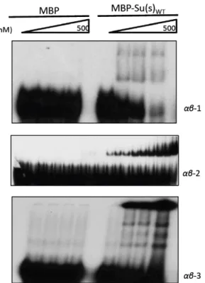
Figure 5-‐ MBP does not form stable complexes with αβ RNA……….……….. 30
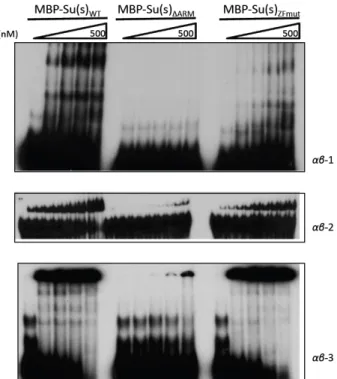
Figure 6-‐ The ARMs mediate stable interaction with αβ RNA...……….….. 31
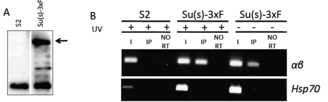
Figure 7-‐ Su(s)-‐3xF binds to αβ transcripts in cultured Drosophila cells……….. 32
Figure 8- Sequence analysis of UV-‐crosslinked cDNAs……….…………. 34
Figure 9-‐ Deletion of the U-‐rich putative Su(s)-‐3xF crosslinking sites affects
the affinity of MBP-‐Su(s)WT for αβ RNA in vitro……….. 35
Figure 10-‐ Deletion of the G-‐rich putative Su(s)-‐3xF crosslinking sites
affects the affinity of MBP-‐Su(s)WT for αβ RNA in vitro………... 36
Figure 11-‐ Deletion of Su(s) binding sites affects regulation by
endogenous Su(s) in cultured cells……….. 38
Figure 12- Nucleotide substitution mutations in the Dm88 LTR disrupt
Su(s)-‐mediated regulation.………... 40
Figure 13- Nucleotide substitution mutations in the Dm88 LTR do not
affect the RNA binding of MBP-‐Su(s)WT in vitro………...….. 41
Figure 14-‐ Putative model of the Su(s)-‐Wdr82 mediated regulation αβ transcripts……….. 45
Figure 15-‐ RNAfold analysis of the secondary structure of Region 2 of αβ RNA………….….. 51
LIST OF ABBREVIATIONS
αβ alpha/beta
APT Associated with Pta1
ARMs Arginine Rich Motifs
CBC Cap-‐Binding Complex
CCCH Cysteine-‐Cysteine-‐Cysteine-‐Histidine
cDNA complimentary DNA
CE Capping Enzyme
CFI Cleavage Factor I
CLIP Crosslinking-‐Immunoprecipitation
CPF Cleavage and Polyadenylation Factor
CPSF Cleavage and Polyadenylation Specificity Factor
CPM Counts Per Minute
CstF Cleavage Stimulation Factor
CTD C-‐Terminal Domain
CUTS Cryptic Unstable Transcripts
DNA Deoxyribonucleic acid
EJC Exon-‐Junction Complex
EMSA Electrophoretic Mobility Shift Assay
eRNAs enhancer RNAs
HIV Human Immunodeficiency Virus
kDA kilodaltons
lRNAs long RNAs
m7G 7-‐methylguanosine cap
mRNA messenger RNA
miRNA microRNAs
ncRNAs non-‐coding RNAs
NEXT Nuclear-‐Exosome Targeting Complex
NNS Nrd1-‐Nab3-‐Sen1
NRQC Nuclear RNA Quality Control
NXF1 Nuclear Export Factor
PAP Poly (A) polymerase
PAS Poly (A) Signal
PASR Promoter Associated small RNAs
PCIF Polytene Chromosome Immunoflourescence
Pol II RNA polymerase II
Poly(A) Polyadenylic acid
Poly (G) Polyguanylic acid
Poly (U) Polyuridylic acid
PROMPTS Promoter Upstream Transcripts
RBPs RNA Binding Proteins
RIP RNA Immunoprecipitation
RNA Ribonucleic acid
RNMT RNA guanine-‐7 methyltransferase
RRE Rev Response Element
RRM RNA Recognition Motif
sRNAs small RNAs
SELEX Systematic Evolution of Ligand by Exponential Enrichment
Ser-‐5P Serine-‐5 Phosphorylation
Ser-‐2P Serine-‐2 Phosphorylation
snRNAs small nuclear RNAs
snRNPs small nuclear Ribonucleoproteins
snoRNAs small nucleolar RNAs
Su(s) Suppressor of sable
TASRs Termination Associated small RNAs
TSS Transcription Start Site
UTR Untranslated Region
WT Wild Type
CHAPTER I: INTRODUCTION
Introduction
The life cycle of a messenger RNA (mRNA) molecule begins in the nucleus where the
pre-‐mRNA is transcribed by RNA polymerase II (Pol II) and co-‐transcriptionally processed.
After being exported to the cytoplasm the mRNA undergoes multiple rounds of translation
and is ultimately degraded. Each step in gene expression depends on the proper
association of RNA binding proteins (RBPs) that facilitate these processes (1). RBPs act to
modify, protect, and survey the RNA quality throughout the life cycle, thus ensuring the
informational integrity of the transcriptome.
Transcription by Pol II
Pol II transcribes protein-‐coding genes to generate mRNAs as well as a subset of
non-‐coding RNAs (ncRNAs), including small nucleolar RNAs (snoRNAs), small nuclear
RNAs (snRNAs) and micro RNAs (miRNAs). Pol II is unique among polymerases in that the
largest subunit contains a conserved, C-‐terminal domain (CTD) that consists of a hepta-‐
peptide repeat (Tyr-‐Ser-‐Pro-‐Thr-‐Ser-‐Pro-‐Ser) (2). The CTD exists as an extended structure
that undergoes extensive post-‐translational modifications that are essential for the proper
progression through the phases of transcription and for the recruitment of protein factors
involved in RNA processing events. The post-‐translational modification of the CTD that has
Pol II is recruited to the promoters of genes when the CTD is in an
unphosphorylated state. Hyper-‐phosphorylation of the CTD at Serine 5 (Ser-‐5P) of the
heptad repeat occurs during initiation and is thought to facilitate release of Pol II from the
promoter (4). In multicellular organisms, the early elongation Pol II complex often pauses
a short distance downstream of the transcription start site (TSS). Release of Pol II from this
paused state is facilitated by phosphorylation of Ser-‐2 (Ser-‐2P). As Pol II escapes into the
body of the gene Ser-‐5P is removed and Ser-‐2P increases. Thus, phosphorylation levels
peak for Ser-‐5 at the 5’ region of genes and at the 3’ region for Ser-‐2, with intermediate
levels of both Ser-‐5P and Ser2P within the body of the gene (4). Therefore this CTD code is
informative of the phase of transcription of Pol II and the protein factors that may be
associated with the CTD and the transcription machinery during those phases.
Co-transcriptional RNA Modifications
As mentioned above, the phosphorylation state of the CTD orchestrates the
dynamic, co-‐transcriptional association and dissociation of proteins required to process the
nascent pre-‐mRNA molecule to generate mRNA. During pre-‐mRNA maturation, the 5’ end
is capped, introns are removed and exon sequences are ligated together and the 3’ end is
generated by cleavage of the RNA followed by the addition of a protective poly adenosine
(poly(A)) tail (5).
The process of modifying a nascent RNA molecule begins when the first 20-‐30
nucleotides have been synthesized and the growing RNA emerges from the Pol II exit
channel. At this stage the CTD is hyperphosphorylated at Ser-‐5, which facilitates
recruitment and activation of the capping enzyme (CE) and the RNA (guanine-‐7)
(m7G) cap that serves to protect the RNA from the actions of 5’ to 3’ exonucleases (6). The
completed cap structure is cotranscriptionally bound by the cap binding complex (CBC)
which aids in protecting the RNA and in the transition to productive elongation by
interactions with the CTD (7).
The process of splicing, which removes introns and fuses exons together, is
performed by the large multi-‐subunit ribonucleoprotein (RNP) complex called the
spliceosome. The spliceosome is comprised of five subunits called small nuclear RNPs
(snRNPs) and many protein cofactors. The RNA components (snRNAs) are non-‐coding and
non-‐polyadenylated (8). snRNAs assemble with seven Sm proteins in the cytoplasm to
form the various snRNP molecules. Upon reentry into the nucleus, the holo-‐spliceosome is
formed by the consecutive association of snRNPs with active areas of transcription
coordinated by interactions with the CTD (9). In particular both U1snRNP and a
component of the U2 associated factors (U2AF) are believed to directly associate with the
CTD (8,9). Splicing proceeds in a stepwise manner. U1snRNP recognizes the 5’ splice site
and CBC and SR proteins stabilize this interaction (8). U2snRNP then binds to the 3’ splice
site that is further defined by U2AF and SR1. Recognition of the adenosine-‐branch point by
U2snRNP and interaction with U1snRNP define the intron and recruits U4, U5 and
U6snRNP to complete the spliceosome (8). Splicing then proceeds in a two-‐step reaction
with the initial cleavage at the 5’splice site followed by cleavage of the 3’ splice site and
fusion of the exons (8).
Two models have been proposed to explain how splicing might be coupled to
transcription, and there is experimental support for both models (9). In the recruitment
machinery and associated factors thereby facilitating splicing. The observation that intron-‐
containing RNAs transcribed by a bacteriophage RNA polymerase are not efficiently spliced
is consistent with this model and indicates that the CTD of Pol II is necessary to recruit
splicing components (10). Also, antibodies directed against the CTD inhibit splicing by
preventing the association of splicing components (9,10). The kinetic model states that the
rate of transcription affects splicing efficiency. This is best demonstrated by alternative
splicing in which certain exons are skipped or included in the mRNA. A fast Pol II
elongation rate favors skipping an alternate exon, whereas a slower rate favors the
inclusion of the alternate exon (11).
The final step in RNA processing involves generating a 3’ end by an endonucleolytic
cleavage event and the addition of a poly (A) tail. Furthermore, RNA Pol II must terminate,
which requires the release of Pol II from the DNA template. The proper formation of a 3’
end involves several multi-‐protein complexes that cooperate to recognize pre-‐mRNA cis-‐ elements that serve to position the cleavage machinery (12). Positioning of the cleavage
machinery involves a tripartite mechanism: first the recognition of the conserved
(AAUAAA) poly(A) signal (PAS) located 10 to 30 nts upstream of the cleavage site (cleavage
usually follows a CA dinucleotide). Secondly two downstream elements (GU-‐rich and U-‐
rich) are found about 30 nt downstream of the cleavage site. Third, UGUA sequences are
found 40 to 100 nts upstream of the cleavage site for several genes (13). The cleavage and
polyadenylation specificity factor (CPSF) complex recognizes the PAS and a subunit of CPSF
(CPSF-‐73) performs the endonucleolytic cleavage. The downstream elements are
recognized by a component of the cleavage stimulation factor (CstF) and the upstream
by poly(A) polymerase (PAP) and its processivity is enhanced by both PABNI and CPSF,
which causes the rapid addition of 200 to 300 adenosines (12). Polyadenylation is coupled
to transcription as Pol II transcribes the PAS before the 3’ end processing steps can occur.
Interestingly, transcription of the PAS enhances processing of the terminal intron, and
processing the terminal intron facilitates cleavage and poly (A) via a direct interaction of
U1 and U2snRNP and CPSF (14).
Following cleavage and poly (A) synthesis, Pol II continues to transcribe, and
termination, the release of Pol II from the DNA template, can occur proximal to the cleavage
site or several kilobases downstream (15). There are two prevailing ideas about how
termination occurs. The allosteric model suggests that conformational changes occur
following the recognition of the PAS that reduces the stability of the elongation complex.
Secondly, the torpedo model postulates that the 3’ cleavage event exposes an unprotected
5’ end of the downstream RNA; this allows entry for the 5’ to 3’ exonuclease Xrn2/Rat1 to
degrade the RNA and catch up to Pol II, causing its release (16,17). However, it is likely
that both models contribute to efficient termination. For instance, the CTD-‐binding
cleavage factor protein, Pcf11, was shown to disassociate Pol II from the DNA template by
bridging the CTD to the RNA (3). Yet, Xrn2/Rat1 has been shown to be required for
efficient termination in yeast and human cells. Both Pcf11 and the Rat1 associated protein,
Rtt103 interact with the Ser-‐2P CTD (3). Thus, allosteric rearrangements of the Pol II CTD
and associated factors and degradation of the RNA generated following cleavage are both
important in termination. Thus, the CTD is integral in the seamless coupling of every step
in transcription and RNA processing.
Cotranscriptional Nuclear RNA Quality Control
Superimposed on the complex yet elegant coupling of transcription and RNA processing, is the need to survey the growing transcript via cotranscriptional quality
control mechanisms. Nuclear RNA quality control (NRQC) mechanisms ensure rapid
degradation of transcripts that are not properly processed, contain aberrant sequences, or
improperly integrated into an RNP complex. This prevents the accumulation of aberrant
transcripts which are potentially harmful to the cell (18). To deal with aberrant transcripts
most NRQC components act at the site of transcription, while the RNA is still associated
with the DNA template. NRQC employs three different methods to address faulty
transcripts: degradation, nuclear retention, or down-‐regulation of the expressed gene (18).
The 5’ m7G cap is the first RNA processing step that subject to surveillance by NRQC
mechanisms. RNA that is uncapped or improperly capped will fail to recruit CBC and this
renders the transcript susceptible to the 5’-‐3’ exonuclease activity of XRN2/Rat1 (7).
Additionally, yeast Rat1 protein associates with the decapping enzymes Dox1 and Rai1 that
can facilitate the removal of methylated or unmethylated caps and the hydrolysis of
uncapped triphosphate ends to monophosphate, respectively, thereby providing a suitable
substrate for Rat1 (19). Together these enzymes enforce a NRQC checkpoint that ensures
the 5’ ends of transcripts are properly capped or rapidly degraded.
Improper splicing potentially introduces errors in the sequence and structure of the
mRNA, and thus surveillance of this step of RNA processing is also important. Splicing
defects can cause the transcript to be retained at the site of transcription. The association
of proteins required for splicing, such as SR proteins, with the nascent transcript is
Mex69p (20). Also, in mammals, the cotranscriptionally deposited exon-‐junction complex
(EJC) mediates the recruitment of the nuclear export factor, NXF1, to the mRNA, suggesting
that proteins involved in RNA processing also exert NRQC as the lack of their proper
association impedes downstream activities including nuclear export (21). Improperly
spliced transcripts also undergo degradation by XRN2/Rat1 and the 3’-‐5’ exonuclease, the
nuclear exosome (22). The nuclear exosome is comprised of a nine-‐subunit core that lacks
catalytic activity. The catalytic activity of the exosome is provided by the association of two
RNases, RRP6, a 3’-‐5’ exonuclease, and RRP44/DIS3, which has both endonuclease and 3’-‐5’
exonuclease activity (23). In cells deficient in either XRN2 or the exosome, aberrantly-‐
spliced transcripts accumulate, indicating these nucleases are fundamental components of
NRQC (22).
Processing of the 3’ end of pre-‐mRNA is another step in which NRQC mechanisms
are required. One example is that failure to splice the terminal intron, which requires the
exon-‐defining complex and CPSF, causes retention of the transcript at the gene, as only fully
spliced transcripts with a poly(A) tail are released by the cleavage and polyadenylation
machinery (24). Furthermore, in yeast, mutation of the poly(A) polymerase caused rapid
degradation of mRNA in an RRP6 dependent manner (25). These findings demonstrate
that the canonical 3’ processing factors impose NRQC by tethering the transcript to the
gene until a proper poly(A) tail is formed or signal for the rapid degradation via a
mechanism involving the nuclear exosome component RRP6.
Cotranscriptional Nuclear RNA Quality Control of Pervasive Transcription
transcriptome led to the discovery that transcription is pervasive, meaning the majority of
the genome, from yeast to humans, is transcribed at some level (26). Several different
classes of pervasive non-‐coding transcripts have been identified, and these have arbitrarily
been placed into two categories based on their size; small RNAs (sRNAs) are 20 nts to 200
nts in length and long RNAs (lRNAs) range from 200 nts to over 1000 nts (27). Many of the
small RNAs cluster around promoters (promoter associated sRNAs (PASR)) or around the
3’ end (termination-‐associated sRNAs (TASRs)) of genes. Interestingly, PASRs are found in
both the same and divergent orientation to the promoter from which they originate and
their abundance is directly proportional to the strength of the promoter. In yeast, mutation
of catalytically active components of the exosome or its cofactor, the TRAMP complex,
facilitated the identification of RNAs that are highly unstable and undetectable in WT cells.
These RNAs were named cryptic unstable transcripts (CUTs) (28). CUTs, at 200 nts to 600
nts, are larger than PASRs, and are predominantly antisense to the promoter region with
heterogenous 3’ ends. The 5’ ends of CUTs originate upstream of the TSS from nucleosome-‐
depleted regions that can broadly define promoter regions. This indicates that promoters
are inherently bidirectional (29). Like yeast, human cells produce unstable promoter
upstream transcripts (PROMPTS) that also map to nucleosome-‐depleted regions.
PROMPTS are transcribed from both stands but are generally antisense to the downstream
promoter (30). Another type of long non-‐coding RNA are enhancer RNAs (eRNAs), which
are generated from the transcription of enhancer elements (31). Similar to PROMPTS,
eRNAs are bidirectional yet enriched for antisense transcripts (32) and rapidly degraded
The identification of protein factors involved in down regulation of pervasive
transcripts has been most extensively examined in yeast. The Nrd1-‐Nab3-‐Sen1 (NNS)
complex regulates CUTs. Both Nrd1 and Nab3 bind to short tetramer sequences in the
nascent RNA to direct termination (33). The NNS pathway interacts with the Ser-‐5P form
of the CTD and thus this termination pathway is limited to the region proximal to the TSS
(31). Therefore a factor in the fate of CUTs is the presence of cis-‐termination sequences relative to the TSS. Sites bound by NNS are enriched in the TSS-‐proximal region in
antisense transcripts and yet they are depleted from protein-‐coding mRNA transcripts
(31). Thus the direct association of Nrd1 with the Ser-‐5P CTD of Pol II and the association
of the NNS complex with the exosome, couples the transcription of CUTs to their
termination and exosome mediated rapid degradation (34).
In humans, the position of transcript termination sequences in the promoter-‐
proximal region is also involved in the regulation of antisense transcripts such as PROMPTs
and eRNAs. The presence of a canonical PAS (AAUAAA) in the 5’ region of the transcript
invokes early termination coupled to rapid degradation. The CBC appears to interact with
the nuclear-‐exosome-‐targeting (NEXT) complex to facilitate exosome-‐mediated
degradation (35). Similar to NNS binding sites, cryptic PAS are enriched in antisense
transcripts whereas protein-‐coding transcripts are depleted of cryptic PAS (36).
Additionally, binding by U1 snRNP protects the nascent RNA by preventing the recognition
of PAS that could otherwise induce early termination and thus aids in determining
promoter directionality (37). Recently a role for the WD40 repeat protein, Wdr82, in
Other proteins may also be involved in the regulation of pervasive and aberrant transcripts
in metazoans.
The Drosophila melanogaster Protein Suppressor of Sable Negatively Regulates Aberrant Transcripts
The D. melanogaster protein Suppressor of sable (Su(s)) is a 140 kDa RNA binding protein that functions in NRQC. Su(s) was initially identified as a recessive sex-‐linked locus
in which mutations suppressed spontaneous mutant alleles at a second site. Particularly,
mutations at su(s) suppress spontaneous mutations associated with the sable, purple, speck
and vermilion genes. It was determined that the spontaneous mutations at vermilion, purple and speck were caused by insertions of the 412 retrotransposon (39). Further analysis of the suppressible 412 induced mutations at the vermilion (v1, v2, vk) locus revealed that 412 inserted within the 5’ UTR in the opposite orientation to the vermilion gene (40) and that 412 sequences are removed from the primary transcript through the use of cryptic splice sites located in the long terminal repeats (LTRs) (41,42). In a WT su(s)
background mature mutant vermilion mRNAs fail to accumulate, yet in a mutant su(s) background mature vermilion mRNAs accumulate and are similar in size as the WT
vermilion despite having a 7.5kb 412 insertion (40). Intriguingly, the full 412 sequence was not required for su(s) mediated regulation, as a single, 480-‐bp LTR from 412 was sufficient (43). In a su(s) mutant background the single LTR also resulted in higher accumulation of
vermilion transcripts that both retained or spliced out the single LTR (43).
background. In a mutant su(s) background two transcripts accumulate, one containing the P-‐element insertion and one in which the P-‐element is removed from the primary
transcript via recognition of cryptic donor splice sites in the 39bp-‐inverted repeat of the P-‐
element and acceptor sites within the yellow transcript (44). These findings led to the hypothesis that the su(s) locus may encode a protein that is involved in preventing the recognition of cryptic splice sites, thus causing destabilization of the insertion containing
transcripts (44). However, analysis of the molecular genetic interactions between su(s) and
the suppressible alleles provided limited information about the role of the Su(s) protein
product. Thus, additional targets of this regulation needed to be identified. Furthermore,
molecular characterization of the su(s) gene and resultant protein were critical.
Targets Identified by PCIF Analysis
Polytene chromosome immunofluorescence (PCIF) studies demonstrated that Su(s)
protein localized to distinct chromosomal regions (45,46). Further analysis revealed that
Su(s) colocalized with the Ser5-‐P form of Pol II, present during initiation and the early
elongation phase, but not the Ser2-‐P form of Pol II, which is present during active
elongation, and the 3’ region of genes (47). A strong signal for Su(s) was observed at the 3C
locus of the X-‐chromosome. This region contains salivary specific intronless genes (ng 1-4,
Sgs4, and Pig1) that lie within the introns of a larger gene. The normal expression pattern of these genes is known with ng genes being expressed earlier in third instar larvae. When ng mRNA levels decline, Sgs4 is rapidly induced, with no overlap in the expression.
However, in a mutant su(s) background, ng mRNA levels remained elevated while Sgs4 was expressed (47). Also, Sgs4 mRNA levels accumulated much earlier, while ng mRNA levels
when compared to WT su(s) background. This data suggest that Su(s) plays a role in maintaining the normal temporal expression pattern for these developmental genes and
the rapid induction of Sgs4 following the decline of ng RNA levels (47).
PCIF analysis of the distribution of Su(s) during heat-‐shock revealed that Su(s)
protein localized to 87C. There are four heat-‐shock genes at the 87C locus. Between two of
the genes there is a 38 kb repetitive insert of the remnants of the retrotransposon
elements, invader1 and Dm88. These elements were named alpha/beta (αβ) elements. A
subset of these elements are fused to a duplication of the HSP70 promoter (gamma element), and these hybrid retrotransposons are referred to as alpha-gamma elements (47). Under mild heat-‐shock conditions, noncoding, polyadenylated transcripts of various
lengths arise from the αβ RNA region (48). Northern analysis revealed that αβ transcripts, similar to the vermilion transcripts, accumulate at significantly higher levels in a mutant
su(s) background indicating that Su(s) also regulates these aberrant transposon-‐containing transcripts. When Su(s) is active Hsp70-‐αβ elements generate short, unstable transcripts with heterogeneous 3’ ends. Conversely, when Su(s) is inactive, longer, stable transcripts
are produced that terminate following a canonical PAS (49). Unlike the antisense 412 insertion in vermilion, purple and speck, the αβ elements are in a sense orientation with
respect to the Hsp70 promoter (47). Analysis of the sequences required for the Su(s)-‐ mediated negative regulation of αβ transcripts revealed that sequences spanning nucleotides +1 to +278 are sufficient for regulation, and Hsp70 promoter and 5’ UTR sequences (+1 to +69) do not contribute to regulation (47).
The Su(s) Protein
The su(s) locus is located between the cytological regions of 1B10 – C1 of the X-‐ chromosome (50). A fortunate P-‐element-‐induced mutation in a mutant vermilion strain resulted in a revertant to a WT vermilion phenotype. The su(s) locus was the only known
suppressor of vermilion, and thus the P-‐element insertion into su(s) facilitated cloning of the su(s) gene via transposon-‐tagging, leading to the identification of a 5kb transcript derived from this gene (50). Subsequent temporal analysis revealed that the su(s) mRNA is produced throughout the life cycle of the fly and the resultant 150 kDa protein localized to
the nucleus (51). Initial examination of the protein sequence revealed that Su(s) contained
a highly charged region with 28.6% sequence identity to the arginine-‐serine region of the
Drosophila U1 70K protein that is involved in splicing and a putative C-‐terminal region
with low similarity to a RNA recognition motif (RRM) (51). However, more extensive
analysis of this putative RRM showed that this region did not fit the criteria for a true RRM
and the arginine rich region of Su(s) lacked the arginine-‐serine repeats present in U1 70K.
Thus, while Su(s) contained regions that were loosely reminiscent of proteins involved in
RNA processing, the function of Su(s) had yet to be determined (52).
Su(s) is an RNA Binding Protein
To examine the biochemical functions of Su(s), our lab generated recombinant full
length and truncated Su(s) protein. Full length Su(s) bound to ftz RNA with a high affinity and this binding could be out-‐competed by excess poly(U) or poly(G) (52). Furthermore,
systematic evolution of ligand by exponential enrichment, SELEX, experiments identified
an enriched consensus sequence, UCAGUAGUCU, which was present in many of the high
identified that were enriched for GU-‐dinucleotides (52). To delineate the region of Su(s)
responsible for the RNA binding activity the 1325 amino acids that comprise the full length
Su(s) were divided into four sections and high affinity binding was localized the N-‐terminal
434 amino acids (52). Immunohistochemical analysis of the endogenous Su(s) distribution
pattern in embryos and the nuclei of salivary glands demonstrated that Su(s) localized to
the nucleoplasm and to multiple locations on polytene chromosomes. Interestingly, in some
regions, Su(s) co-‐localized with U1 70K, suggesting Su(s) is found at a subset of areas
undergoing active transcription where pre-‐mRNAs are being processed (52).
Su(s) has Two Distinct RNA Binding Domains
High affinity RNA binding displayed by Su(s) had been localized to the N-‐terminal region (52). Sequence alignment analysis determined that this region contained two CCCH
zinc-‐finger motifs (ZFs) (53). The CCCH-‐ZF motif is found in other RNA interacting proteins
such as the mRNA destabilizing protein, TTP (Tis11) (54,55), the 3’ end pre-‐mRNA
processing factor subunit CPSF-‐30/CLIPPER (56), and the splicing factor subunit U2AF-‐35
(57). TTP binds to AU-‐rich (UUAUUUAUU) elements in the 3’ UTR to destabilize the
targeted mRNA (54) while U2AF-‐35 binds to the AG-‐dinucleotide at the 3’SS (57).
Furthermore, in vitro, endonuclease activity was localized to the five CCCH-‐ZFs of
Drosophila CPSF-‐30/CLIPPER which were required to bind and cleave RNA hairpins (58); however, the biological relevance of this activity has not been established. In addition to
their RNA binding activity, the CCCH-‐ZFs of TTP also serve as a protein-‐protein interaction
domain and facilitate the association with the nuclear-‐pore protein Nup214 (59). Based its
CCCH-‐ZF motifs, Su(s) has been placed in an orthologous group with CPSF-‐30 (Yth1 in
Additional in vitro RNA binding analysis of various fragments of the N-‐terminal region of Su(s) localized high affinity binding to two arginine-‐rich motifs (ARMs) (45).
ARMs have been most studied in proteins derived from viruses such as the HIV Rev and Tat
proteins and the N-‐proteins of bacteriophages. The ARM of Rev is essential for binding to
the highly structured Rev Response Element (RRE) located in the intron of the Env gene, and this allows incompletely spliced viral transcripts to exit the nucleus for translation
(60). Tat is a trans-‐activator of transcription initiated in the LTR of HIV provirus. Tat binds
to the trans-‐activator response (TAR) element, also a structured RNA hairpin, in the 5’ UTR, to increase the rate of transcription (61). However, ARMs also recognize unstructured
RNAs. For instance, the CPSF component Fip1, binds to U-‐rich sequences via an ARM to aid
in enhancing the activity of PAP (62). Interestingly, some of the SELEX consensus RNAs
bound by Su(s) were predicted to form hairpin structures, whereas SELEX RNAs that
lacked the consensus were less likely to form structures (45). Furthermore, ARM1 of Su(s)
was found to preferentially bind to RNAs containing the consensus, while ARM2 bound
non-‐consensus RNAs with a higher affinity. Loss of both ARMs eliminated binding to SELEX
RNAs, even though the CCCH-‐ZFs were present, demonstrating that, in vitro, the ZFs do not contribute to RNA binding (45).
The ARMs and the ZFs Contribute to Su(s) Mediated Regulation in vivo
The ARMs had been identified as the domain that confers high affinity binding in vitro and thus our lab wanted to determine if the loss of this RNA binding domain would affect Su(s) mediated regulation in vivo. Initial experiments using WT or ARM deletion su(s) cDNA transgenes that were ectopically expressed at high levels demonstrated that
expressed su(s) transgenes in a su(s) null background revealed that loss of ARM1, but not ARM2, eliminated localization to polytene chromosomes under non-‐heat-‐shock conditions
but did not affect nuclear localization (45). Interestingly, deletion of both ARMs increased
mutant Su(s) protein and mRNA levels, suggesting that the ARMs are involved in regulating
expression from these transgenes (45). Analysis of the effect of deleting of the ARMs or the
ZFs on regulation of the suppressible full 412-‐ insertion-‐containing vermilion transcript, showed that regulation was lost only when ARM1 and ARM2 were deleted together while
destabilizing ZF mutations minimally affected Su(s) mediated regulation (46). However
loss of the ARMs or mutation of the ZFs greatly inhibited the ability of Su(s) to autoregulate
su(s) genomic transgenes, expressed under the control of the endogenous promoter (46). Furthermore, loss of the ARMs or the ZFs reduced or eliminated, respectively, the ability of
Su(s) to down-‐regulate αβ transcripts. The enhanced regulatory role of the ZFs with the αβ
transcripts rather than the ARMs might be expected as mutation of the ZFs resulted in loss
of localization to the 87C heat-‐shock locus (47). These studies suggest that different
targets of Su(s) may interact with different domains by promoting various modes of
binding, and, potentially different protein complexes may form in a target-‐dependent
manner (46).
Identification of Components of the Su(s) Regulatory Pathway
Our lab determined that the nuclear exosome is a component of the Su(s) regulatory
pathway. Northern analysis demonstrated that degradation of αβ transcripts depends on the nuclear exosome, as loss of the enzymatically active components partially restores αβ RNA accumulation (47).
Su(s) Interacts with Wdr82 to Regulate Hsp70-αβ Transcripts
Recently our lab reported that Su(s) interacts with Wdr82 and both of these
proteins are required for the negative regulation of Hsp70-αβ transcripts (49). Wdr82 is a 35-‐kDa protein, containing seven WD40 repeats which typically serve as a β-‐propeller like
platform to which proteins can bind. Wdr82 is conserved from yeast to humans. The yeast
homologue, Swd2, has been shown to function in two distinct complexes; the
SET1/COMPASS complex, responsible for mediating histone 3 lysine 4 (H3K4)
trimethylation, and the APT complex, part of the larger holo-‐enzyme, cleavage and
polyadenylation factor (CPF) complex, which processes the 3’ end of RNA Pol II transcripts
(63). 3’ end polyadenylation processing by CPF does not seem to require APT(64).
However, loss of Swd2 in yeast leads to improper transcription termination of some genes,
particularly snoRNAs, which are not polyadenylated. Also, loss of Swd2 results in reduced
global H3-‐K4 methylation. In human cells, Wdr82 interacts with the C-‐terminal domain
(CTD) of RNA Pol II, which then allows recruitment of Setd1A to the promoter of genes by
an interaction between Wdr82 and the RNA recognition motif (RRM) of Setd1A (65).
Intriguingly, in humans cells, Wdr82 interacts with the putative human ortholog of Su(s),
ZC3H4 (C19orf7, KIAA1099) (49,66). Perhaps the Su(s)-‐Wdr82 regulatory pathway is
conserved in higher organisms, however this will require further investigation.
We demonstrated that the Su(s)-‐Wdr82 pathway regulates Hsp70-αβ transcripts via a mechanism that involves induced promoter-‐proximal termination and non-‐canonical
polyadenylation (49). Furthermore, Su(s)-‐Wdr82 regulation depends on cis-‐sequence elements within the LTRs of Dm88 and invader1 that are proximal to the Hsp70 promoter
5’ region of some actively transcribed genes, explaining why Su(s) colocalized with Ser-‐5P
Pol II (47), and thus restricting the Su(s) regulation to the promoter proximal region. Su(s)
may then bind to RNA sequences in the promoter-‐proximal region to induce premature
termination, similar to NNS pathway and the regulation of CUTS (31). However we had not
previously determined if Su(s) interacts directly with Hsp70-αβ RNA transcripts nor had we defined biologically relevant binding sites. In the findings presented here, I demonstrate that Su(s) binds αβ transcripts in vitro and in living S2 cells. I also define sequences that
are bound by Su(s) and determine that Su(s) binding sites contribute to regulation of αβ RNAs.
CHAPTER II: THE INHIBITION OF ABERRANT RNA ACCUMULATION BY THE SUPPRESSOR OF SABLE (SU(S)) PATHWAY DEPENDS ON THE RNA-BINDING ACTIVITY
OF SU(S) AND MULTIPLE, DISTINCT REGULATORY ELEMENTS Introduction
The maturation of eukaryotic pre-‐mRNAs involves several processing reactions
(capping, splicing, and polyadenylation) that are intricately coupled to RNA polymerase II
(Pol II) transcription. When each of these reactions is efficient, the resulting mRNAs and
associated-‐proteins (mRNPs) are exported to the cytoplasm for translation. However, if
any step in this process is impaired, e.g., because of mutations that affect key regulatory
sequences, the aberrant transcripts are detected and eliminated by one of several RNA
quality control (RQC) systems (18,67). Nuclear RQC (NRQC) often involves co-‐
transcriptional degradation of defective pre-‐mRNAs by the 5’ → 3’exonuclease XRN2/Rat1
(XRN2) or the nuclear exosome, a multi-‐subunit complex that includes 3’ → 5’
exonucleases. XRN2 has been implicated in handling improperly capped pre-‐mRNAs and
stalled Pol II complexes, whereas nuclear exosome components degrade nascent
transcripts with exposed 3’ ends. The latter situation occurs when transcription elongation
or 3’-‐processing is impaired (68,69). Although considerable progress has been made in
elucidating NRQC mechanisms, much remains to be learned, especially in multicellular
organisms, about how defective pre-‐mRNAs are identified and the regulatory proteins that
The Su(s) regulatory pathway in D. melanogaster functions in NRQC. Su(s) downregulates RNAs from transcription units with aberrant sequences in the 5’-‐
transcribed region by a mechanism that involves early transcription termination and
degradation of the RNAs by nuclear exosome components (47,49). Our lab recently
identified the WD40 domain protein Wdr82 (yeast Swd2) as a component of this
regulatory pathway (49). Wdr82 is known to function in two different promoter-‐proximal
gene regulatory processes, i.e., regulation of histone H3 lysine 4 methylation at active genes
and transcription termination at yeast genes that produce short, noncoding RNAs (64,65).
Recently, an additional role for mammalian Wdr82 in promoting early transcription
termination of intergenic transcripts and enhancer RNAs has been reported (38).
Several transcription units that are subject to regulation by the Drosophila Su(s)
pathway have transposon sequences inserted a short distance downstream of the
promoters of protein-‐coding genes (39). The best-‐studied example of this occurs within a
genomic region that contains a cluster of Hsp70 protein-‐coding genes and noncoding Hsp70-αβ elements. These elements consist of remnants of two different retrotransposons (Dm88 and invader1) inserted downstream of Hsp70 promoter/5’ UTR sequences (Figure 1). During a mild heat shock induction, Su(s)-‐Wdr82 act to inhibit αβ RNA accumulation,
but does not affect Hsp70 mRNAs (47). Under these conditions, Hsp70-αβ elements produce short (~100 to 400 nt), unstable RNAs with heterogeneous 3’ ends (Figure 1).
Although the details of this regulatory process have not been fully sorted out, it appears
that Su(s)-‐Wd82 induces transcription termination in the promoter-‐proximal region of
Hsp70-αβ and the unstable RNAs are degraded by nuclear exosome components (47,49).