THE KINEMATIC AND ELECTROMYOGRAPHIC RESPONSE TO OPTICAL FLOW BALANCE PERTURBATIONS IN WALKING: VISUOMOTOR ADAPTATION AND THE
ACUTE EFFECTS OF AGE AND FALLS HISTORY
Jessica Dorothy Thompson
A thesis submitted to the faculty at the University of North Carolina at Chapel Hill in partial fulfillment of the requirements for the degree of Master of Science in the Biomedical
Engineering Department in the College of Arts and Sciences.
Chapel Hill 2017
ii © 2017
iii ABSTRACT
Jessica Dorothy Thompson: The Kinematic and Electromyographic Response to Optical Flow Perturbations in Walking: Visuomotor Adaptation and the Acute Effects of Age and Falls
History
(Under the direction of Jason R. Franz).
Visual perturbations can be used to study balance control and balance deficits due to their ability to elicit corrective motor responses. Advanced age increases the reliance on visual feedback for motor planning and execution. Because older adults are more susceptible to these perturbations than young adults, they may represent a promising diagnostic tool for age-associated falls risk.
iv
ACKNOWLEDGMENTS
v
TABLE OF CONTENTS
LIST OF TABLES ... vii
LIST OF FIGURES ... viii
CHAPTER 1: Introduction ... 1
CHAPTER 2: Do Kinematic Metrics of Walking Balance Adapt to Perturbed Optical Flow? ... 4
2.1 Introduction ... 4
2.2 Methods... 6
2.2.1 Experimental Procedures and Measurements ... 7
2.2.2 Data Analysis ... 8
2.2.3 Statistical Analysis ... 10
2.3. Results ... 11
2.3.1 Initial response to perturbed optical flow ... 11
2.3.2 Propensity for visuomotor adaptation ... 16
2.3.3 Presence of perturbation aftereffects ... 16
vi
2.5 Conclusions ... 21
CHAPTER 3: Age and falls history effects on antagonist leg muscle coactivation during walking with optical flow perturbations ... 23
3.1 Introduction ... 23
3.2 Methods... 25
3.3 Results ... 29
3.4 Discussion ... 31
3.5 Conclusion ... 33
CHAPTER 4: Other Significant Contributions... 35
4.1 The Neuromuscular Origins of Kinematic Variability during Perturbed Walking ... 35
4.2 Neuroimaging of human balance control: A systematic review ... 37
vii
LIST OF TABLES
viii
LIST OF FIGURES Figure 1 - Experimental figure and mediolateral sacrum motion and lateral step placement during normal walking and walking with
the largest amplitude visual perturbation for a representative subject ... 9 Figure 2 - Mean perturbation-induced effects on step width, step length,
step width variability, and step length variability for each perturbation amplitude ... 12 Figure 3 - Group-average spectrum of ML sacrum motion and mean peak
mediolateral sacrum motion at each perturbation frequency as a function
of perturbation amplitude and time compared to normal, unperturbed walking ... 13 Figure 4 - Normalized EMG linear envelopes for normal and perturbed
walking. MH/VL coactivation on the top panel, MG/TA coactivation in
the middle and SOL/TA on the bottom. Shaded area indicates stance phase. ... 28 Figure 5 - Mean (S.E.) antagonist leg muscle coactivation versus perturbation
1
CHAPTER 1: Introduction
With the average age of the population of the United States increasing, falls are an unrelenting concern. About one third of adults over the age of 65 has a fall annually and of these falls, 25-30% lead to moderate to severe injury. Not only do these falls frequently result in a devastating personal loss of independent mobility, but it is projected that falls will result in 67.7 billion dollars in medical expenses in the year 2020. The cause of these falls is likely multifactorial, arising from a series of internal (e.g., sensorimotor decline) and external (e.g., environmental) factors. Despite efforts from funding agencies such as the National Institute of Health, falls rate has remained unabated for the past twenty years. Thus, there is an immediate need for new, innovative approaches to diagnose those at risk of falling and also rehabilitating those who have already fallen.
2
D. G. Thelen, 2015; Jeka, Allison, & Kiemel, 2010; Lord & Webster, 1990; Yeh, Cluff, & Balasubramaniam, 2014). Neural changes associated with aging decrease the selectivity of cells in the visual cortex and could underline the degradation of visual function with age (Schmolesky, Wang, Pu, & Leventhal, 2000).
These sensorimotor changes may explain why older adults have an incredible susceptibility to optical flow perturbations (i.e., augmented optical flow giving the visual perception of imbalance). For example, my lab has demonstrated that, while step width and length variabilities are statistically indistinguishable during normal walking, mediolateral visual perturbations elicited changes that could then differentiate between the two groups (Francis et al., 2015). Even further, we also found that such perturbations were able to disrupt control of lateral step placement and decrease local dynamic stability more in older adults than younger adults – metrics that were both comparable during normal walking (J. R. Franz et al., 2015). Because optical flow perturbations have consistently been able to expose age-related differences in balance control that are not otherwise apparent during normal walking, they are intriguing tool to study age-related balance impairment with translational potential in the diagnosis and rehabilitation of falls risk.
3
during walking over the course of prolonged exposure to optical flow perturbations of different amplitudes.
4
CHAPTER 2: Do Kinematic Metrics of Walking Balance Adapt to Perturbed Optical Flow?1
2.1 Introduction
Walking balance control depends on integrating reliable sensory feedback and planning and executing appropriate motor responses (O'Connor & Kuo, 2009). Accordingly, sensory perturbations are increasingly used to study balance control mechanisms in walking. Visual (i.e., optical flow) perturbations in particular can elicit strong and acute motor responses to regulate balance from step to step (O'Connor & Kuo, 2009; Terry et al., 2012). Moreover, these acute motor responses are remarkably more intense in subjects with sensorimotor deficits, such as those due to advancing age (J. R. Franz et al., 2015). These results forecast the promising potential of optical flow perturbations applied during walking in the diagnosis of people at risk of falls. However, some evidence from the postural control of standing suggests that subjects may adapt to such perturbations, effectively adjusting their sensitivity to visual feedback over time (Jeka et al., 2010). Although highly relevant to their translational potential, it remains unclear whether walking balance exhibits this time-dependent behavior, which we refer to as visuomotor adaptation.
Multisensory reweighting, the central process that determines the relative priority placed on somatosensory, visual, and vestibular feedback, is considered an essential component of balance control (Horak, Shupert, & Mirka, 1989; Jeka et al., 2010; Oie, Kiemel, & Jeka, 2002).
1 This chapter previously appeared in the journal of Human movement Science. The original citation is as follows:
5
However, to the best of our knowledge, this adaptive sensorimotor process has been exclusively studied in the context of postural sway during standing. For example, Jeka et al. (2010) used anterior-posterior (AP) optical flow perturbations to reveal that the relative priority placed on visual feedback in regulating standing balance is reduced when perturbation amplitudes are larger, and that this dynamic response is tuned over prolonged durations. Indeed, depending on environmental conditions, one would expect sensory feedback modalities deemed more reliable to be those prioritized in balance control. Although optical flow perturbations have been used in studies of walking, these studies have not been designed to investigate the propensity for visuomotor adaptation, with trial durations limited to between 30 s and 3 min and time-averaged outcome measures generally reported.
6
2015; Latt, Menz, Fung, & Lord, 2008) . Specifically, human walking exhibits naturally emerging entrainment to frequencies directly present in ML optical flow perturbations, and the strength of this entrainment can be interpreted to signify one’s sensitivity to visual stimuli However, time-dependent changes in foot placement variability and ML CoM motion following exposure to perturbations in walking have yet to be investigated. A return of these metrics of walking balance control toward values seen during normal, unperturbed walking despite ongoing perturbations may allude to the occurrence of visuomotor adaptation.
The purpose of this study was to investigate the propensity for visuomotor adaptation in walking balance control using prolonged exposure to optical flow perturbations of different amplitudes. We used a virtual reality environment to apply continuous ML optical flow perturbations during treadmill walking and recorded the time course of effects on measures of balance control. We first hypothesized that subjects would exhibit visuomotor adaptation, such that the effect of perturbations on walking balance would decrease with walking duration. We also hypothesized that this adaptation would scale with perturbation amplitude, with larger perturbations exhibiting more persistent effects on walking balance.
2.2 Methods
7 2.2.1 Experimental Procedures and Measurements
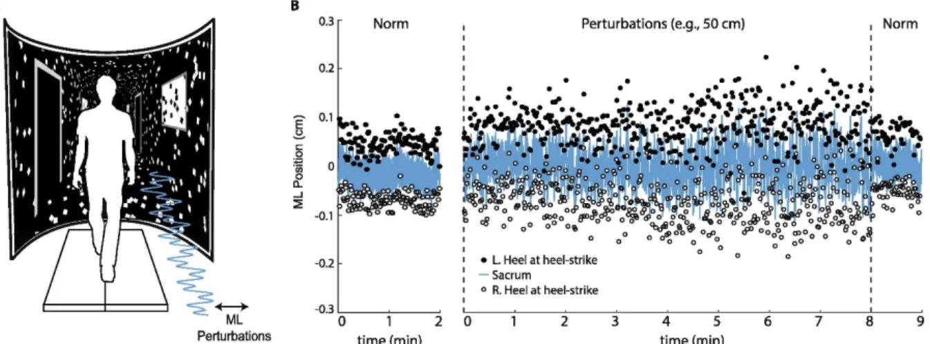
Subjects first walked down a 10 m walkway at their self-selected comfortable speed. We calculated subjects’ preferred overground walking speed as the average of two times taken to traverse the middle 4 m of the walkway (1.38 ± 0.13 m/s). Subjects then completed all treadmill walking trials on a force-sensing treadmill (Bertec, Inc., Columbus, OH) at their preferred overground speed while watching a custom, speed-matched virtual hallway (Fig. 1). The virtual hallway was rear-projected onto a semicircular curved screen that surrounded the treadmill and measured 2.75 m high and 2.25 m wide. Prior to completing the treadmill trials, we instructed subjects simply to “walk while looking down the hallway” so that subjects could naturally respond to the projected visual stimuli.
8
placed on subjects’ right and left heels and sacrum, the latter used as a surrogate for their center of mass (Yang & Pai, 2014).
2.2.2 Data Analysis
9
Fig. 1 (A) Subjects walked on a treadmill while watching a speed-matched, immersive virtual hallway with and without continuous mediolateral optical flow perturbations of
different amplitudes. (B) Mediolateral (ML) sacrum motion and lateral step placement during normal walking (“Norm”) and walking with the largest amplitude visual perturbation for a representative subject. Perturbation trials consisted of 8 min of prolonged exposure followed by 1 min of normal, unperturbed walking. L.heel and R.heel refer to the mediolateral
10
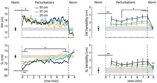
over the trial durations, rather than step to step variations, we used a moving average with a window of 30 steps to remove short term fluctuations (Dingwell & Cusumano, 2010). Using the original time series, we calculated step width and length variabilities (SWV and SLV, respectively) as the standard deviation of SW and SL over steps occurring in 60 s bins.
Finally, we used spectral analysis to quantify each subject’s dynamic response to optical flow perturbations as the intensity of mediolateral sacrum motion (i.e., ML sway intensity) at each of the three frequencies comprising the perturbations (Loughlin & Redfern, 2001). Specifically, algorithms implemented in Matlab computed the fast Fourier transform (FFT) of the ML sacrum marker trajectories, partitioned into 60 s bins. From these 60 s bins, we extracted peaks values of sway intensity occurring at each perturbation frequency. This analysis provided three amplitudes of ML sway (i.e., one each occurring at 0.125, 0.250 and 0.442 Hz) that we quantified for each 60s bin comprising the protocol.
2.2.3 Statistical Analysis
11
perturbation amplitude (i.e., 20, 35, and 50 cm; Hypothesis 2) on each outcome measure. When a significant main effect or interaction was found, we performed post-hoc pairwise comparisons using Tukey’s Honest Significant Difference for multiple comparisons. Finally, in an exploratory test for the presence of perturbation aftereffects, a one-way rmANOVA compared the final minute of unperturbed walking after removal of each perturbation to normal walking at the beginning of the session. We defined significance for all comparisons using an alpha level of 0.05.
2.3. Results
2.3.1 Initial response to perturbed optical flow
12
13
Fig. 3 (A) Group-average (standard error) spectrum of ML sacrum motion reveals distinct peaks of signal intensity at, and thus naturally emerging entrainment to, each of the three perturbation frequencies 0.125, 0.250 and 0.442 Hz). (B) Mean (standard error) peak mediolateral (ML) sacrum motion at each perturbation frequency as a function of
14
15
Table 1 Repeated measures ANOVA results (Perturbation trial, Minutes 1-8). Bold values indicate those reaching statistical significance (i.e., p<0.05).
Amplitude Time Amplitude x Time
F statistic p-Value Partial η2 F statistic p-Value Partial η2 F statistic p-Value Partial η2 Step
Width
F(2, 18) = 0.126 0.883 0.014 F(1, 9) = 4.30 0.153 0.213 F(2, 18) = 0.684 0.517 0.071
Step Length
F(2, 18) = 5.240 0.016 0.368 F(1, 9) = 9.555 0.013 0.515 F(2, 18) = 0.274 0.764 0.03
SWV F(2, 18) = 8.353 0.003 0.481 F(7, 63) = 3.375 0.004 0.273 F(14, 126) = 0.679 0.791 0.07
SLV F(2, 18) = 7.432 0.004 0.452 F(7, 63) = 1.100 0.374 0.109 F(14, 126) = 0.763 0.706 0.078
Sway (0.125 Hz)
F(2, 18) = 6.325 0.008 0.413 F(7, 63) = 0.667 0.699 0.069 F(14, 126) = 0.446 0.956 0.047
Sway (0.250 Hz)
F(2, 18) = 5.953 0.01 0.398 F(7, 63) = 3.989 0.001 0.307 F(14, 126) = 1.425 0.151 0.137
Sway (0.442 Hz)
F(2, 18) = 4.553 0.025 0.336 F(7, 63) = 2.881 0.011 0.243 F(14, 126) = 0.658 0.81 0.068
SWV : step width variability; SLV: step length variability
16 2.3.2 Propensity for visuomotor adaptation
As perturbations continued, SL exhibited a significant time dependence (Table 1), tending toward values seen during normal, unperturbed walking (Fig. 2). Post-hoc comparisons during the final minute revealed that the largest amplitude perturbation (i.e., 50 cm) elicited persistent, though small, reductions in step length compared to 20 cm (p=0.01) and 35 cm (p=0.05). SW did not exhibit a significant main effect of time (p=0.15) (Table 1). However, SW values at the end of the perturbation trials did not differ significantly from unperturbed walking (p=0.10). Neither SWV, SLV, nor the intensity of ML sacral motion at the individual perturbation frequencies decreased from perturbation beginning to end. Rather, SWV exhibited a more complex time-dependent response (p<0.01) (Table 1), with significant increases beyond the first three minutes as perturbations continued (Fig. 2). We also observed a significant time-dependent increase in the intensity of ML sacrum motion at the two fastest perturbation frequencies (0.25 Hz: p<0.01; 0.442 Hz: p=0.01) (Table 1). Pairwise comparisons revealed that this effect was driven by further increases in ML sacral motion after the third minute of walking with perturbations (Fig. 2).
2.3.3 Presence of perturbation aftereffects
17 2.4 Discussion
We investigated the prevalence of visuomotor adaption in walking balance control using the time-dependent response of gait kinematics to visual (i.e., optical flow) perturbations of different amplitudes. Consistent with prior studies, we found that optical flow perturbations elicited an immediate increase in mediolateral sacrum motion and shorter, wider, and more variable steps compared to normal walking - effects that generally scaled with perturbation amplitude (J.R. Franz, C.A. Francis, M.S. Allen, S.M. O'Connor, & D.G. Thelen, 2015; S.M. O'Connor et al., 2012; Terry et al., 2012). We first hypothesized that walking balance control would adapt to prolonged optical flow perturbations, evidenced by a return of these outcome measures to values seen during normal, unperturbed walking. In partial support of this hypothesis, step length and width exhibited a time-dependent return toward unperturbed values. However, step width variability and step length variability did not decrease from perturbation beginning to end, nor did mediolateral sacrum motion. Rather, these outcome measures exhibited a more complex response to prolonged perturbations, with significant increases as perturbations continued beyond the third minute. Thus, our findings demonstrate that while walking balance control exhibits persistent susceptibility to perturbed optical flow, time-dependent changes in gait kinematics do emerge with prolonged exposure, but not in the manner hypothesized. As we elaborate more below, these time-dependent changes may provide insight into strategies used to accommodate perturbations during walking which may also be highly relevant to their translational potential.
18
lateral margins of stability and shorter steps both better position the body’s CoM within those margins and are more resistant to unexpected slips or trips (Espy, Yang, & Pai, 2010). Thus, this general anticipatory balance control strategy adopted at the onset of perturbations was a logical response from subjects experiencing a novel stimuli designed to elicit a perceived and unpredictable loss of balance. However, as the perturbations progressed, step width and step length tended toward values seen during normal, unperturbed walking. We interpret these changes to suggest that as subjects grew accustomed to the perturbations, they progressively abandoned, or at least deprioritized, the general anticipatory balance control strategy.
19
changes in movement direction while walking. When the prompted change in movement direction was unanticipated, subjects adopted wider steps and increased their margins of stability, described by the authors as general anticipatory adjustments. Finally, we also hypothesized that adaptation to perturbations would scale with perturbation amplitude, with larger perturbations exhibiting more persistent effects on walking balance. Step length exhibited the hypothesized return to values seen during normal walking as perturbations progressed. Here, in partial support of our second hypothesis, we found that the largest amplitude perturbation elicited persistent reductions in step length. Compared to subjects’ response to smaller amplitude perturbations, this suggests that our most challenging condition may have prompted a more persistent dependence on the general anticipatory strategy.
20
movement patterns to minimize metabolic energy expenditure during walking (Selinger, O'Connor, Wong, & Donelan, 2015)
There are alternative, or perhaps complementary explanations for the time-dependent changes in subjects’ response to optical flow perturbations. In studying the dynamics of visual reweighting during standing, Jeka et al. (2010) noticed that some subjects’ response to larger amplitude anterior-posterior optical flow perturbations actually increased over time. These complex dynamics are similar to the time-dependent increases in CoM motion evident at the end of our walking trials. Jeka et al. interpreted their findings to suggest that subjects’ initial downweighting of visual feedback at perturbation onset went too far, resulting in small increases in the relative priority placed on vision as perturbations continued. It is possible that a similar process of multisensory fine-tuning could have governed our subjects’ time-dependent response to optical flow perturbations during walking and/or governed the modest aftereffects in step length variability that persisted after perturbation cessation.
21
walking. However, we acknowledge that alternative approaches exist (e.g., time-dependent filtering). Second, although unlikely, we cannot exclude the possibility that subjects learned to anticipate the continuous perturbations used in this study. Although the perturbations were continuous, they prescribed a complex combination of three sinusoids of different frequencies that would be challenging for subjects to predict. Nevertheless, time-dependent adaptation to these perturbations, even if driven in part by prediction, are important to consider; similar combinations of sinusoidal perturbations are frequently used to study walking balance control (Dingwell & Cusumano, 2010; J.R. Franz et al., 2015; S.M. O'Connor et al., 2012). Finally, we used a series of kinematic outcome measures derived from a reduced lower extremity marker set that may not fully describe the dynamics of walking balance control. Other highly relevant metrics of walking balance control include margins of stability, whole-body angular momentum, and dynamic stability, and the time-dependent nature of these metrics during perturbed walking has yet to be carefully examined (Dingwell & Cusumano, 2010; Neptune & Mcgowan, 2016). In addition, to investigate the naturally-emergent response to perturbations, we did not instruct subjects to keep one foot on each treadmill belt. Thus, we could not resolve the individual leg ground reaction forces necessary to compute metrics of balance control based on kinetic measurements.
2.5 Conclusions
22
23
CHAPTER 3: Age and falls history effects on antagonist leg muscle coactivation during walking with optical flow perturbations
3.1 Introduction
Currently about a third of the population over the age of 65 has a fall annually and of these falls, 25-30% lead to moderate to severe injury. The implications are not only devastating for the individual and can result in a loss of independence, but also results in an enormous financial burden. The physiological mechanisms underlying age-related falls risk are likely multifactorial but may include reduced sensory acuity and slowed neuromuscular performance (Richardson & Ashton-Miller, 1996). While numerous studies and clinical trials have attempted to decrease the prevalence and severity of falls, falls rates have been resistant to change (Gardner, Robertson, & Campbel, 2000; Kraemer, Dewane, Bobula, Nelson, & Heiderscheit, 2009; Rubenstein et al., 2000). Recent research has begun to use sensory perturbations to study balance control mechanisms in walking due to their ability to elicit corrective motor responses from step to step (O'Connor & Kuo, 2009; Terry et al., 2012). These perturbations have been able to produce differences in balance control between young and old adults that are not evident using conventional balance and mobility testing.
24
(J.R. Franz et al., 2015). Presumably governed by an increased reliance on visual feedback for motor planning and execution, optical flow perturbations elicit more profound changes of step width and length variability, control of lateral step placement and local dynamic stability in older adults than in young adults when compared to normal walking (Francis et al., 2015; J. R. Franz et al., 2015). Moreover, some evidence suggests that the dependence on vision for balance control is even more pronounced in older adults with a history of falls (Lord & Webster, 1990). While multiple studies have demonstrated kinematic differences in older adults in response to optical flow perturbations, the neuromuscular mechanisms governing these differences have yet to be explored. Electromyography (EMG) recordings of muscle activities may elucidate age and falls history effects on the neuromuscular control mechanisms involved in walking balance control and the response to optical flow perturbations.
25
adults, in comparison to young adults, showed increased antagonist lower leg muscle coactivation during level and downhill walking (2013). However, although downhill walking is correlated with greater movement variability that is seen during compromised balance, they found that antagonist coactivation increased similarly for both young and old adults when walking downhill compared to normal walking (Hunter, Hendrix, & Dean, 2010). Thus, based on the prevailing evidence and purported mechanism, we suspected that older adults, especially those with a falls history, may disproportionately rely on the coactivation of antagonist leg muscle pairs when walking in the presence of balance perturbations.
The purpose of this study was to investigate the effects of age and falls history on antagonist leg muscle coactivation during walking with and without optical flow perturbations of different amplitudes. We used a virtual reality environment to apply continuous mediolateral optical flow perturbations during treadmill walking while recording Electromyographic (EMG) activities of antagonist upper and lower leg muscle pairs. We first hypothesized that, compared to young adults, aging and falls history would increase antagonist muscle coactivation during walking. We also hypothesized that these anticipated differences from age and falls history would increase in the presence of optical flow perturbations
3.2 Methods
Eleven healthy young adults (6 female, mean±sd, age: 24.8±4.8 years, height: 1.72±0.01 m, mass: 67.2±8.8 kg), eleven healthy older adults (6 female, age: 75.3±5.4 years, height:
1.75±0.01 m, mass: 73.4±16.1) and eleven older adults with a history of falls (7 female, age:
26
considered to have a falls history if they had fallen one or more times in the past year. For this
study, falls counted towards the self-reported total were defined according to the Kellogg
International Work Group Definition. Subjects also completed a health questionnaire prior to
participating which we used to exclude subjects based on: BMI≥30, sedentary lifestyle, orthopedic
or neurological condition, or taking medication that causes dizziness. The experimental protocol
was approved and conducted in accordance with the University of North Carolina Internal Review
Board, and subjects provided written informed consent prior to participating.
We first recorded all subjects’ preferred overground walking speed (Young: 1.29 ±0.18
m/s, Nonfallers: 1.19±0.20 m/s, Fallers: 1.03±0.22 m/s) using two photocells (Brower Timing,
Draper, UT) as the time taken to walk the middle 2 m of a 10 m walking. The remainder of the
testing was conducted on a dual-belt, force-measuring treadmill (Bertec Cor., Columbus, OH)
which was surrounded by a semi-circular curved screen measuring 2.24 m high and 2.83 m wide.
Subjects first walked on the treadmill at their preferred walking speed for 5 minutes in order to
acclimate to the treadmill. The subjects were then secured into an overhead harness, which was
worn for each walking trial. Next, subjects completed four 2-minute walking trials, in randomized
order, while watching a speed-matched, virtual hallway rear projected onto the screen. The
walking trials consisted of normal, unperturbed walking and continuous mediolateral optical flow
perturbations at amplitudes of 20, 35, and 50 cm. Each perturbation was comprised of the sum of
three sinusoids, such that the full amplitude was applied at 0.250 Hz and half that amplitude was
applied at 0.125 Hz and 0.442 Hz. The perturbations were consistent with visual feedback
associated with head movement, meaning that the hallway’s end moved very little compared to
27
Young adults walked at 1.25 m/s and both groups of older adults walked at their preferred walking
speed.
28
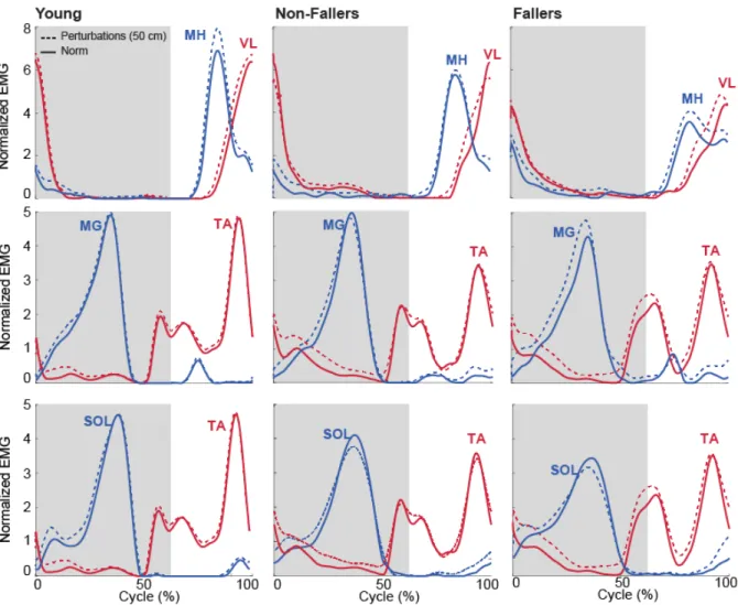
Figure 4. Normalized EMG linear envelopes for normal and perturbed walking. MH/VL coactivation on the top panel, MG/TA coactivation in the middle and SOL/TA on the bottom. Shaded area indicates stance phase.
29
CI(EMG1, EMG2) = 2 ×� ∫min(EMG1, EMG2)
∫min(EMG1, EMG2) + ∫max(EMG1, EMG2)�× 100
Finally, the EMG activity was averaged within two gait cycle phases: stance (0 – 60%) and swing (60-100%) (Perry & Burnfield, 2010).
A two-way repeated measures analysis of variance (ANOVA) tested for main effects of perturbation amplitude and muscle coactivation for each subject group during stance and leg swing phases. Finally, exploratory independent t-tests compared fallers and non-fallers walking with and without perturbations. For all analyses, we defined significance using an alpha level of 0.05.
3.3 Results
Age, independent of falls history, significantly increased stance phase lower leg antagonist muscle coactivation during walking compared to young adults. During stance, the MG/TA pair increased by an average of 138 % and SOL/TA increased by an average of 120% for older adults in comparison to young adults (p’s<0.05). Compared to walking normally, optical flow perturbations had no significant effect on leg muscle coactivation in young adults. In contrast, perturbations significantly increased TA/SOL antagonist coactivation in older adults, by on average up to 58% during stance (Fig 5C).
30
31
Finally, we found no significant difference between older fallers and non-fallers for any measure of antagonist coactivation during unperturbed walking. However, perturbations revealed up to 109% more stance phase VL/MH coactivation in older fallers compared to non-fallers (Fig. 5A).
3.4 Discussion
We investigated differences in antagonist leg muscle coactivation during walking between healthy young, older non-fallers, and older adults with a history of falls responding to balance perturbations. We first hypothesized that aging and falls history would increase antagonist muscle coactivation during walking. In partial support of this hypothesis, lower leg muscle coactivation during walking did increase with age, independent of falls history. Also in support of our hypotheses, we found that between-group differences in antagonist leg muscle coactivation were larger in the presence of optical flow perturbations, with more pervasive effects in older adults with a history of falls.
32
When exposed to optical flow perturbations, young adults showed no significant difference in antagonist muscle coactivation in comparison to normal walking. In contrast, aging effects on muscle coactivation were even more pronounced during perturbed walking than during normal walking. While it has been highlighted in multiple studies that aging increases the amount of antagonist leg muscle coactivation, we found significant increases in antagonist coactivation that were completely unique to older nonfallers but only evident during perturbed walking. Thus, in accordance with our second hypothesis, perturbations were able to bring about even more pronounced balance corrections and were able to create between-group differences that are not otherwise apparent during normal, unperturbed walking. In further support, we also found that upper leg coactivation only increased in subjects with a history of falls when walking with optical flow perturbations. Ochi et al. also found differing upper leg muscle activation in fallers during the swing phase of a step recovery task (2014). They suggested that the increase in activation could have contributed to the slower step velocity they also observed. A correlation between higher upper leg muscle coactivation and slower stepping speed could suggest that fallers are less able to appropriately respond to a trip or unexpected loss of balance. Another explanation could be that older adults who have fallen previously have fear of falling again which has been linked to increased antagonist coactivation (Nagai et al., 2012).
33
agonist muscles and the coactivation of agonist muscles, older adults nearly completely activate the agonist and thus require a much higher level of concurrent antagonist muscle activity. Thus, perhaps older adults respond to the balance challenge of optical flow perturbations by increased antagonist muscle coactivation to stiffen their joints and stabilize their step to step corrections needed to respond appropriately to perturbations. This strategy may be even more prominent in older adults with a history of falls due to some sort of compensatory measure for an underlying issue such as muscle strength.
There are a few important limitations to our study. First, we used a complex combination of sinusoids that would be challenging for subjects to anticipate. However unlikely, we cannot exclude the possibility that subjects learned to predict the perturbation movement. By using continuous, mediolateral optical flow perturbations, it is possible that these muscular adjustments may not be observed when responding to more discrete instances of imbalance. Additionally, older adult subjects may have less treadmill walking experience than young adults which could have led to increased anxiety and thus more muscle tensing. Lastly, subjects were confined to a set walking speed on the treadmill and thus had a limited ability to respond to perturbations by altering their speed.
3.5 Conclusion
34
35
CHAPTER 4: Other Significant Contributions
4.1 The Neuromuscular Origins of Kinematic Variability during Perturbed Walking2
We investigated the neuromuscular contributions to kinematic variability and thus step to step adjustments in posture and foot placement across a range of walking speeds in response to optical flow perturbations of different amplitudes applied in a custom virtual environment. We found that perturbations significantly increased step width, decreased step length, and elicited larger trunk sway compared to normal walking. However, perturbation-induced effects on the corresponding variabilities of these measurements were much more profound. Consistent with our hypotheses, we found that: (1) perturbations increased EMG activity of the gluteus medius and postural control muscles during leg swing, and increased the coactivation of antagonistic leg muscles during limb loading in early stance, and (2) changes in the magnitude of step to step adjustments in postural sway and lateral foot placement positively correlated with those of postural control and gluteus medius muscle activities, respectively, in response to perturbations. However, (3) interactions between walking speed and susceptibility to perturbations, when present, were more complex than anticipated. Our study provides important mechanistic neuromuscular insight
2
36
37
4.2 Neuroimaging of human balance control: A systematic review3
This review examined 83 articles using neuroimaging modalities to investigate the neural correlates underlying static and dynamic human balance control, with aims to support future mobile neuroimaging research in the balance control domain. Furthermore, this review analyzed the mobility of the neuroimaging hardware and research paradigms as well as the analytical methodology to identify and remove movement artifact in the acquired brain signal. We found that the majority of static balance control tasks utilized mechanical perturbations to invoke feet-in-place responses (27 out of 38 studies), while cognitive dual-task conditions were commonly used to challenge balance in dynamic balance control tasks (20 out of 32 studies). While frequency analysis and event related potential characteristics supported enhanced brain activation during static balance control, that in dynamic balance control studies was supported by spatial and frequency analysis. Twenty-three of the 50 studies utilizing EEG utilized independent component analysis to remove movement artifacts from the acquired brain signals. Lastly, only eight studies used truly mobile neuroimaging hardware systems. This review provides evidence to support an increase in brain activation in balance control tasks, regardless of mechanical, cognitive, or sensory challenges. Furthermore, the current body of literature demonstrates the use of advanced signal processing methodologies to analyze brain activity during movement. However, the static nature of neuroimaging hardware and conventional balance control paradigms prevent full mobility and limit our knowledge of neural mechanisms underlying balance control.
3
38
CHAPTER 5: Conclusion
Through my master’s thesis research, I have sought to improve our scientific understanding of walking balance control mechanisms and deficits thereof due to aging and falls history. In a novel approach to examining balance control, I used a custom virtual reality environment to apply continuous mediolateral optical flow perturbations during treadmill walking. I first examined the effects of prolonged exposure to optical flow perturbations on gait kinematics in young adults. While there is some evidence that subjects may adapt to optical flow perturbations through research on postural control during standing, the use of such perturbations during walking has generally been limited to short trial durations. My findings suggest that gait kinematics do indeed change over time and allude to shifts in the neuromechanical strategies used to govern walking balance control. At their onset, perturbations elicited shorter, wider steps indicative of a more cautious, general anticipatory balance control strategy that scaled with perturbation amplitude. As perturbations continued, subjects abandoned, or at least deprioritized, this anticipatory strategy in favor of using a more reactive, task-specific strategy of step to step adjustments. These responses were also amplitude dependent; only the largest, and presumably most challenging, perturbation elicited persistent effects on step length lasting the entire trial. I now propose that prolonged exposure to optical flow perturbations may have clinical utility to reinforce reactive, task-specific balance control through training.
39
foot placement during walking. EMG recordings of muscle activity provides crucial insight into the neuromuscular control mechanisms involved in walking balance control. I found that lower leg antagonist leg muscle coactivation during walking increased with age, independent of falls history; this muscle coactivation was even larger in the presence of optical flow perturbations. I found that increased antagonist muscle coactivation effects were more pervasive in older adults with a history of falls than in healthy older adults. Additionally, antagonist upper leg muscle coactivation was elevated during large perturbations in older adults with a history of falls which suggests an increased susceptibility to optical flow perturbations.
40
REFERENCES
Bauby, C. E., & Kuo, A. D. (2000). Active control of lateral balance in human walking. Journal of Biomechanics, 33, 1433-1440.
Bentley, T. A., & Haslam, R. A. (1998). Slip, trip and fall accidents occuring during the delivery of mail. Ergonomics, 41(12), 1859-1872.
Bertram, J. E., & Ruina, A. (2001). Multiple walking speed-frequency relations are predicted by constrained optimization. Journal of Theoretical Biology, 209(4), 445-453.
Bruijn, S. M., Impe, A. V., Duysens, J., & Swinnen, S. P. (2012). Split-belt walking: adaptation differences between young and older adults. Journal of Neurophysiology, 108(4), 1149-1157.
Collins, S. H., & Kuo, A. D. (2013). Two independent contributions to step variability during overground human walking. Plos One, 8(8).
Cram, J. R., & Kasman, G. S. (1998). Introduction to surface electromyography. Gaithersburg: Aspen Publishers.
Dingwell, J. B., & Cusumano, J. P. (2010). Re-Interpreting Detrended Fluctuation Analyses of Stride-To-Stride Variability in Human Walking. Gait and Posture, 32(3), 348-353. Donelan, J. M., Shipman, D. W., Kram, R., & Kuo, A. D. (2004). Mechanical and metabolic
requirements for active lateral stabilization in human walking. Journal of Biomechanics, 37, 827-835.
Eikema, D. J., Hatzitaki, V., Konstantakos, V., & Papaxanthis, C. (2013). Elderly adults delay proprioceptive reweighting during the anticipation of collision avoidance when standing.
Neuroscience, 234, 22-30.
Espy, D. D., Yang, F., & Pai, Y.-C. (2010). Control of center of mas motion state through cuing and decoupling of spontaneous gait parameters in level walking. Journal of
Biomechanics, 43(13), 2548-2553.
Falcon, K., & Winter, D. A. (1985). Quantitative assessment of co-contraction at the ankle joint in walking. Electromyography and Clinical Neurophysiology, 25(2-3), 135-149.
Finley, J. M., Dhaher, Y. Y., & Perreault, E. J. (2012). Contributions of feed-forward and feedback strategies at the human ankle during control of unstable loads. Experimental Brain Research, 217(1), 53-66.
41
Franz, J. R., Francis, C. A., Allen, M. S., O'Connor, S. M., & Thelen, D. G. (2015). Advanced age brings a greater reliance on visual feedback to maintain balance during walking.
Human Movement Science, 40, 381-392. doi:10.1016/j.humov.2015.01.012
Franz, J. R., Francis, C. A., Allen, M. S., & Thelen, D. G. (2016). Visuomotor Entrainment and the Frequency-Dependent Response of Walking Balance to Perturbation. IEEE Trans Neural Syst Rehabil Eng. doi:10.1109/TNSRE.2016.2603340
Franz, J. R., & Kram, R. (2013). How does age affect leg muscle activity/coactivity during uphill and downhill walking? Gait and Posture, 37(3).
Fujiki, S., Aoi, S., Funato, T., Tomita, N., Senda, K., & Tsuchiya, K. (2015). Adaptation mechanism of interlimb coordination in human split-belt treadmill walking through learning of foot contact timing: a robotics study. Journal of the Royal Society Interface, 12(110), 20150542.
Gardner, M. M., Robertson, M. C., & Campbel, A. J. (2000). Exercise in preventing falls and fall related injuries in older people: a review of randomised controlled trials. British Journal of Sports Medicine, 34(1), 7-17.
Goodworth, A., Perrone, K., Pillsbury, M., & Yargeau, M. (2015). Effects of visual focus and gait speed onw alking balance in the frontal plane. Hum Mov Sci, 42, 15-26.
Horak, F. B., Shupert, C., & Mirka, A. (1989). Components of postural dyscontrol in the elderly: A review. Neurobiology of Aging, 10, 727-738.
Hortobagyi, T., & deVita, P. (2006). Mechanisms responsible for the age-associated increase in coactivation of antagonist muscles. Exercise and Sport Science Reviews, 34(1), 29-35. Hortobagyi, T., Finch, A., Solnik, S., Rider, P., & DeVita, P. (2011). Association between
muscle activation and metabolic cost of walking in young and old adults. Journals of Gerontology Series A Biological Sciences and Medical Sciences, 66, 541-547. Hortobagyi, T., Stanislaw, S., Gruber, A., Rider, P., Steinweg, K., Helseth, J., & DeVita, P.
(2009). Interaction between age and gait velocity in the amplitude and timing of antagonist muscle coactivation. Gait and Posture, 29(4), 558-564.
Hunter, L. C., Hendrix, E. C., & Dean, J. C. (2010). The cost of walking downhill: is the preferred gait energetically optimal? Journal of Biomechanics, 43, 1910-1915.
42
Kraemer, K., Dewane, J., Bobula, S., Nelson, T., & Heiderscheit, B. (2009). The effect of a modified OTAGO exercise program on gait speed and strength in community dwelling older adults. Wisconsin Physical Therapy Association.
Kuo, A. D. (2001). A simple model of bipedal walking predicts the preferred speed-step length relationship. Journal of Biomechanical Engineering, 123, 364-269.
Latt, M. D., Menz, H. B., Fung, V. S., & Lord, S. R. (2008). Acceleration patterns of the head and pelvis during gait in older people with parkinson's disease: A comparison of fallers and nonfallers. The Journals of Gerontology Series: Medical Science, 64A(6), 700-706. Lord, S. R., & Webster, I. W. (1990). Visual field dependence in elderly fallers and non-fallers.
The International Journal of Aging and Human Development, 31, 267-277.
Loughlin, P. J., & Redfern, M. S. (2001). Spectral characteristics of visually induced posutral sway in healthy elderly and healthy young subjects. IEEE Trans Neural Syst Rehabil Eng, 9(1), 24-30.
Maki, B. E. (1997). Gait changes in older adults: Predictors of falls or indicators of fear. Journal of the American Geriatrics Society, 45(3), 313-320.
Malone, L. A., & Bastian, A. J. (2010). Thinking About Walking: Effects of Conscious Correction Versus Distraction on Locomotor Adaptation. Journal of Neurophysiology, 103(4), 1954-1962.
Mian, O. S., Thom, J. M., Ardigo, L. P., Narici, M. V., & Minetti, A. E. (2006). Metabolic cost, mechanical work, and efficiancy during walking in young and older men. Acta
Physiologica, 186, 127-139.
Nagai, K., Yamada, M., Uemura, K., Tanaka, B., Mori, S., Yamada, Y., . . . Tsuboyama, T. (2012). Effects of fear of falling on muscular coactivtion during walking. Aging Clinical and Experimental Research, 24(2).
Neptune, R. R., & Mcgowan, C. P. (2016). Muscle contribution of frontal plane angular momentum during walking. Journal of Biomechanics, 49(13), 2975-2981.
Noel, M., Fortin, K., & Bouyer, L. J. (2009). Using an electrohydraulic ankle foot orthosis to study modifications in feedforward control during locomotor adaptation to force fields applied in stance. Journal of NeuroEngineering and Rehabilitation, 6(1), 16.
O'Connor, S. M., & Kuo, A. D. (2009). Direction-Dependent Control of Balance During Walking and Standing. Journal of Neurophysiology, 102(3), 1411-1419.
doi:10.1152/jn.00131.2009
43
Ochi, A., Shinya, Y., Abe, T., Yamada, K., Hiroshige, T., & Ichihashi, N. (2014). Differences in muscle activation patterns during step recovery in elderly women with and without a history of falls. Aging Clinical and Experimental Research, 26(2), 213-220.
Oie, K. S., Kiemel, T., & Jeka, J. J. (2002). Multi-sensory fusion: Simultaneous re-weighting of vision and touch for the control of human posture. Cognitive Brain Research, 14, 164-176.
Perry, J., & Burnfield, J. M. (2010). Gait Analysis: Normal and Pathological Function (2 ed.). Thorofar, NJ: Slack Incorporated.
Peterson, D. S., & Martin, P. E. (2010). Effects of age and walking speed on coactivation and cost of walking in healthy adults. Gait and Posture, 31, 355-359.
Redfern, M. S., Cham, R. G.-P., K., Gronqvist, R., Hirvonen, M., Lanshammar, H., Marpet, M., Powers, C. (2001). Biomechanics of slips. Ergonomics, 44(13), 1138-1166.
Richardson, J. K., & Ashton-Miller, J. A. (1996). Peripheral neuropathy: an often-overlooked cause of falls in the elderly. Postgraduate Medicine, 99(6).
Rubenstein, L. Z., Josephson, K. R., Trueblood, P. R., Loy, S., Harker, J. O., Pietruszka, F. M., & Robbins, A. S. (2000). Effects of a group exercise program on strength, mobility, and falls among fall-prone elderly men. The Journals of Gerontology Series A: Biological Science and Medical Science, 55(6), M317-M321.
Schmolesky, M. T., Wang, Y., Pu, M., & Leventhal, A. G. (2000). Degradation of stimulus selectivity of visual cortical cells in senescent rhesus monkeys. Nat Neurosci, 3, 384-390. Selinger, J. C., O'Connor, S. M., Wong, J. D., & Donelan, J. M. (2015). Humans can
continuously optimize energetic cost during walking. Current Biology, 25(18), 2452-2456.
Terry, K., Sinitski, E. H., Dingwell, J. B., & Wilken, J. M. (2012). Amplitude effects of medio-lateral mechanical and visual perturbations on gait. Journal of Biomechanics, 45, 1979-1986.
Wezenberg, D., de Haan, A., van Bennekom, C. A. M., & Houdijk, H. (2011). Mind your step: Metabolic energy cost while walking an enforced gait pattern. Gait and Posture, 33(4), 544-549.
44
Yang, F., & Pai, Y. C. (2014). Can Sacral Marker Approximate Center of Mass During Gait and Slip-Fall Recovery Among Community-Dwelling Older Adults? Journal of
Biomechanics, 47(16), 3807-3812.
Yeh, T. T., Cluff, T., & Balasubramaniam, R. (2014). Visual reliance for balance control in older adults persists when visual information is disrupted by artificial feedback delays. Plos One, 9, e91554.