Role of Glycated Proteins in the
Diagnosis and Management of
Diabetes: Research Gaps and
Future Directions
Diabetes Care 2016;39:1299–1306|DOI: 10.2337/dc15-2727
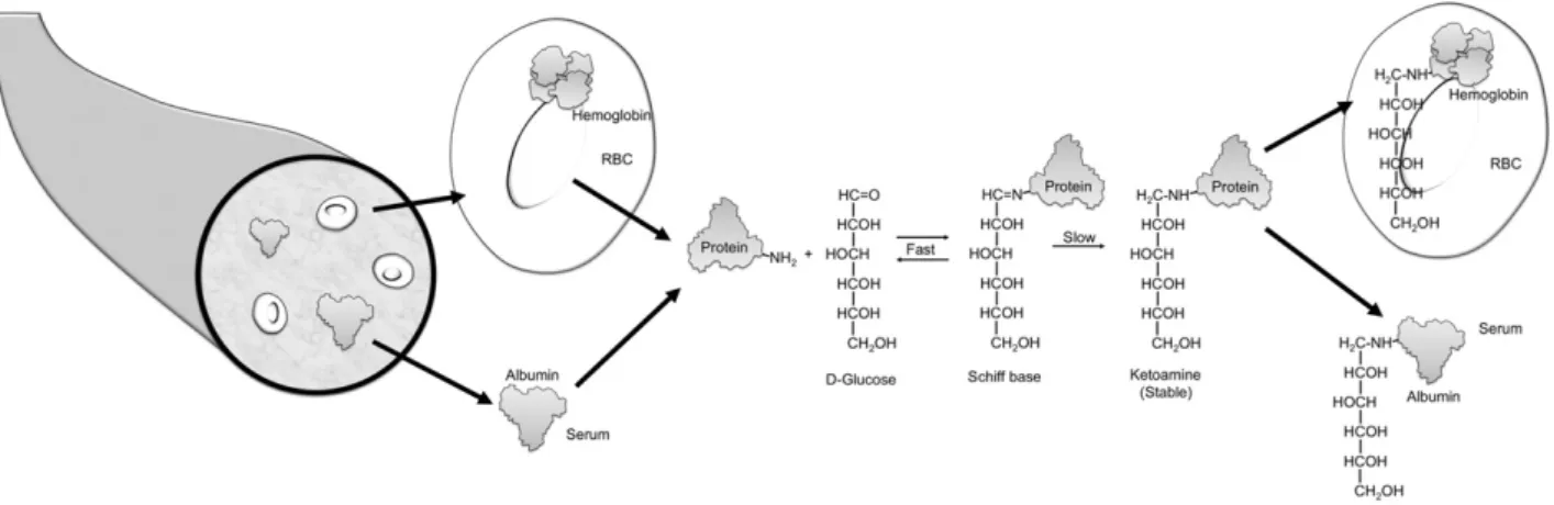
Blood oligosaccharides are attached to many proteins after translation, forming glycoproteins. Glycosylation refers to an enzyme-mediated modification that alters protein function, for example, their life span or their interactions with other pro-teins (1). By contrast, glycation refers to a monosaccharide (usually glucose) attach-ing nonenzymatically to the amino group of a protein. Glycated hemoglobin is formed by the condensation of glucose with select amino acid residues, commonly lysine, in hemoglobin to form an unstable Schiff base (aldimine, pre-HbA1c) (Fig. 1).
The Schiff base may dissociate or may undergo an Amadori rearrangement to form a stable ketoamine.
Glycated hemoglobin, particularly HbA1c, has for decades been widely
incorpo-rated into the management (and, more recently, the diagnosis) of patients with diabetes. An important attribute is that glycation occurs continuously over the lifetime of the protein, so the concentration of the glycated protein reflects the average blood glucose value over a period of time. This contrasts with the measure-ment of blood glucose, which reveals the glucose concentration at the instant blood is sampled and which is acutely altered by multiple factors such as hormones, ill-ness, food ingestion, and exercise (2). While HbA1cis by far the most extensively
useddand studieddglycated protein (2–4), other glycated proteins that have been evaluated in clinical studies include fructosamine, glycated albumin, and advanced glycation end products (AGEs).
HEMOGLOBIN A1c
HbA1cis glycated hemoglobin in which glucose is attached to the N-terminal valine
residue of eachb-chain of hemoglobin A (HbA). Glucose can also be attached at other amino acids, predominantly lysine, in either thea- orb-chain of hemoglobin (5). However, modern methods that measure HbA1cdo not report these other
glycated hemoglobin species. The extent of hemoglobin glycation is influenced by the concentration of glucose in the blood. Since the life span of erythrocytes is;120 days, HbA1creflects the average glucose concentration over the preceding
8–12 weeks (3).
HbA1chas been recommended by the American Diabetes Association since 1988
for routine monitoring of patients with diabetes (6). Although the association of chronic hyperglycemia with the risk of chronic complications of diabetes was sus-pected for many years, landmark trials such as the Diabetes Control and Complica-tions Trial (DCCT) in type 1 diabetes (7) and the UK Prospective Diabetes Study (UKPDS) in type 2 diabetes (8) and their follow-up studies (9,10) confirmed that lowering mean glucose, as measured by HbA1c, significantly reduced the onset and
progression of complications. This led to the development of treatment goals for HbA1cand the use of HbA1cas a performance measure. The increasing use of HbA1c
in patient management is evident from the increase in the number of clinical lab-oratories that are enrolled in proficiency testing surveys conducted by the College of American Pathologists (Fig. 2). Note the large (more than threefold) increase in participants during the 4 years after the publication of the DCCT results in 1993.
HbA1cwas recently included as a diagnostic criterion for diabetes by the
Ameri-can Diabetes Association (11), European Association for the Study of Diabetes,
1
Department of Laboratory Medicine, National Institutes of Health, Bethesda, MD
2Department of Medicine, University of North
Carolina, Chapel Hill, NC
Corresponding author: David B. Sacks, sacksdb@ mail.nih.gov.
Received 16 December 2015 and accepted 13 April 2016.
© 2016 by the American Diabetes Association. Readers may use this article as long as the work is properly cited, the use is educational and not for profit, and the work is not altered.
See accompanying articles, pp. 1458 and 1462.
Kerry J. Welsh,1M. Sue Kirkman,2and David B. Sacks1
PER
S
PEC
TI
VES
IN
CAR
International Diabetes Federation, and World Health Organization (12). This rec-ommendation was motivated by improve-ments in the measurement of HbA1cand
by the certain advantages of its measure-ment over that of glucose, such as the convenience of not requiring the patient
to fast and the reduced intraindividual variability compared with fasting or glu-cose measurements after loading (11).
HbA1ccan be measured by
immunoas-says, high-performance liquid chromatog-raphy (HPLC) (the two most commonly used methods in the U.S. and many other
developed countries), affinity chromatog-raphy, capillary electrophoresis, and en-zymatic assays (13). Standardization of methods by the NGSP (formerly called the National Glycohemoglobin Standard-ization Program) (14,15) and the Interna-tional Federation of Clinical Chemistry and Laboratory Medicine (16) has yielded highly consistent HbA1cresults for a blood
sample, regardless of the method used (provided the method is certified by NGSP).
Interference
There are numerous published reports of conditions that change HbA1c
inde-pendent of glucose (reviewed in refs. 17 and 18). Based on the nature of the interference, these can be conve-niently divided into two groups: condi-tions that influence interpretation (i.e., change HbA1c concentration in ways
unrelated to changes in glucose) and conditions that interfere with HbA1c
measurement (i.e., analytic interfer-ences) (Table 1).
Factors That Influence HbA1cInterpretation
Physiological Factors. HbA1c
concentra-tions increase by;0.1% per decade af-ter 30 years of age (19). It is not known whether this gradual increase reflects an effect of age on the relationship of mean glycemia to HbA1cor merely the
higher prevalence of prediabetes and diabetes with aging (a true increase in mean glycemia). There is contention sur-rounding the influence of race on HbA1c
concentrations. Herman (20) posits that African Americans have higher HbA1c
for any given level of mean glycemia,
Figure 1—Formation of glycated protein. A reversible interaction between a primary amino group (depicted as NH2) of a protein and the carbonyl
group ofD-glucose yields a labile intermediate, called a Schiff base. This can undergo a slow and spontaneous Amadori rearrangement to form a
stable ketoamine. HbA1cis formed if glucose attaches to the N-terminal valine of theb-chain of hemoglobin. If the glucose attaches to proteins in the
plasma, fructosamine or glycated albumin results. RBC, red blood cell.
Figure 2—Progressive increase in HbA1ctesting over time. The number of clinical laboratories
whereas Selvin (21) argues that the in-creased mean HbA1c is a reflection of
truly higher mean glycemia in African Americans.
Chronic Renal Failure.Chronic renal fail-ure (CRF) is a common complication of diabetes, and diabetes is the leading cause of end-stage renal disease (22). Red blood cell survival is reduced in CRF, decreasing HbA1c. In addition, many
pa-tients with CRF are treated with erythro-poietin to stimulate erythropoiesis. The subsequent increase in the number of young erythrocytes further reduces the HbA1c. Therefore the HbA1c
concentra-tion in patients with diabetes and with CRF may not accurately indicate glyce-mic control.
Iron-Deficiency Anemia. Iron deficiency and iron-deficiency anemia occur fre-quently. Some studies, generally with small sample sizes, have reported in-creased HbA1c in individuals with iron
deficiency. Two recent systematic views reached opposite conclusions re-garding the effects of iron deficiency on HbA1c. Thefirst, a meta-analysis and
sys-tematic review, concluded that there was no statistically significant difference in HbA1cmeasured by HPLC in the
pres-ence of iron deficiency or iron-deficiency anemia (23). By contrast, another assess-ment determined that iron deficiency, with or without anemia, increased HbA1c
(24). This discrepancy is likely due to the differences in the studies selected and the method of analysis. Several studies included in both meta-analyses of HbA1c
in iron deficiency were limited by their small sample sizes and the heterogeneity
of the methods. Two large investigations of the National Health and Nutrition Ex-amination Survey (NHANES) data have been conducted. Kim et al. (25) evalu-ated 6,666 female NHANES participants without diabetes from 1999 to 2006 and concluded that iron deficiency was asso-ciated with an increase in HbA1c from
,5.5% to 5.5–6.0%; however, this asso-ciation was not apparent at higher HbA1c
concentrations. A second investigation of NHANES data from 1999 to 2002 in-cluded 8,296 patients with and without diabetes and found an adjusted increase in HbA1cfrom 5.46 to 5.56% in the
pres-ence of iron deficiency (26). Thus, while HbA1cseems to increase slightly with
iron deficiency, the clinical significance of thisfinding remains to be determined. We agree with Ford et al. (26) that cau-tion should be exercised in diagnosing prediabetes and diabetes when HbA1c
is near the decision threshold in patients with iron deficiency.
Erythrocyte Life Span.A change in eryth-rocyte survival alters HbA1c. For example,
assume HbA1cis 7.0% (53 mmol/mol),
with a normal erythrocyte life span of 120 days. If the red blood cell life span is 10 days shorter or longer, the corre-sponding HbA1cvalues would be 6.4%
(46 mmol/mol) and 7.6% (60 mmol/mol), respectively. HbA1cdoes not accurately
reflect average blood glucose concentra-tion if erythrocyte survival is significantly altered, as in, for example, hemolytic anemia or severeb-thalassemia. Since measurement of red blood cell life span is extremely difficult, one cannot easily solve this problem by, for example, applying a correction factor for erythro-cyte age.
Variable Glycation.Intraindividual vari-ability of HbA1cis very low.
Neverthe-less, interindividual variation occurs and has been ascribed by some to dif-ferences in glycation rates (27,28). This postulate is contentious (29,30) because the data validating significantly different rates of glycation are minimal and no mechanism for differences in this non-enzymatic process has been documented. Moreover, a recent analysis, although in-direct, reveals that even the rate of glyca-tion of hemoglobin variants S, C, D, E, J, and G is not significantly different from that of HbA (31), undermining the pre-mise of variable rates of glycation of HbA. There has been speculation that the rate of deglycation (i.e., the removal
of glucose from HbA1c) might vary among
individuals, resulting in different HbA1c
concentrations despite similar average glycemia. Although at least three groups of deglycating enzymes have been iden-tified, only one, fructosamine 3-kinase, is found in humans. Importantly, fructosamine 3-kinase has no effect on valine-1 of the b-chain of hemoglobin (32), the residue where glucose is attached in HbA1c, and
it cannot deglycate HbA1c. Thus the
con-cept of variable glycation remains to be validated.
Factors That Interfere With Measurement Numerous publications have described interferences in HbA1c measurement,
but many reports had small numbers of subjects and described changes that were small and unlikely to have clinical significance (33–35). Furthermore, im-provements in analytic methods have eliminated interferences from some fac-tors (e.g., aspirin, bilirubin, and triglyc-erides) that affected older methods. While the possible interference of all substances in each modern method has not been rigorously investigated, it is likely that few drugs or other factors interfere significantly in current HbA1c
assays.
Uremia. Isocyanic acid, derived from urea, is covalently attached to proteins. The nonenzymatic process, termed carbamylation, increases when blood urea concentrations are high, yielding increased carbamylation of circulating proteins, including on lysine or arginine residues of the N-terminus of hemoglo-bin. Carbamylated hemoglobin altered HbA1c values in some early methods
(36), but uremia has no significant effect on HbA1canalysis with most
contempo-rary methods (23,37,38).
Hemoglobin Variants.Over 1,200 hemo-globin variants have been identified; the bgene is involved in;70% of these (39). While the vast majority are uncommon or rare, certain hemoglobin variants, partic-ularly HbAS, HbAC, HbAD, and HbAE, oc-cur at relatively high frequencies in some populations. One cannot measure HbA1c
in individuals who are homozygous for these common variants or who have HbSC disease (36) because they have no HbA. While total glycated hemoglobin can be determined using borate affinity methods in patients with these homozy-gous hemoglobin variants, there is no convincing clinical evidence that these Table 1—Nonglycemic factors that
may influence HbA1c
Factors that may influence interpretation of HbA1c
1. Physiological (e.g., age, race) 2. Chronic renal failure 3. Iron-deficiency anemia 4. Erythrocyte life span 5. Glycation“phenotypes”
6. Drugs (e.g., dapsone, antiretroviral) 7. Other (e.g., vitamin C, vitamin E)
Factors that may interfere with HbA1c
measurement 1. Uremia
2. Hemoglobin variants 3. Drugs (e.g., opiates)
values can reliably be used to monitor glycemia and predict complications, par-ticularly since some patients may have reduced erythrocyte life span because of hemolytic anemia. Most interferences are method-specific (36). Manufacturers of HbA1cmethods have considerably
re-duced analytic interference from variant hemoglobin. Therefore HbA1c can be
measured accurately in the presence of the overwhelming majority of variant hemoglobins, provided a suitable assay is used (40). Since common heterozy-gous variants rarely alter erythrocyte life span, accurate and reliable HbA1c
values can be obtained in heterozygous individuals.
GLYCATED SERUM PROTEINS
Glucose attaches nonenzymatically to amino groups of proteins other than he-moglobin to form ketoamines (Fig. 1). Measures of several glycated serum pro-teins, including fructosamine and gly-cated albumin, have been proposed as markers of glycemia that might comple-ment or replace HbA1cin select patient
populations. Serum proteins turn over more rapidly than erythrocytes; for ex-ample, albumin (the protein found in the highest concentration in serum) has a circulating half-life of about 14– 20 days. Therefore the concentration of fructosamine or glycated serum albu-min reflects mean glucose over a period of 2–3 weeks. Additionally, glycated se-rum proteins are not influenced by changes in erythrocyte life span or he-moglobin variants such as homozygous HbS. Glycated serum proteins have therefore been proposed as measures of more rapid changes in glycemia and to monitor glycemic control in patients with conditions that alter the normal relationship of HbA1cto mean glucose
(e.g., hemolysis, blood transfusion).
FRUCTOSAMINE
Fructosamine is the common name for 1-amino-1-deoxy fructose and the generic name for plasma protein ketoamines (41,42). All glycated serum proteins are fructosamines, and since albumin is the most abundant serum protein, measure-ment of fructosamine is thought to largely reflect the concentration of glycated albu-min, though this has been questioned (43). The fructosamine assay is readily auto-mated and is less expensive than measure-ment of HbA1c. There is disagreement as
to whether fructosamine results are in-dependent of serum protein concentra-tions (absent significant alterations in the latter) or whether fructosamine values need to be corrected for the concentration of serum proteins (44). Most agree, how-ever, that fructosamine is not valid when serum albumin is,30 g/L.
Thefirst commercial method to mea-sure fructosamine suffered from several problems, particularly a lack of specifi c-ity and interference by other reducing substances in the serum, such as urates (43,45). Thus many early studies of fruc-tosamine generated confusion regarding its clinical value, with reviews (covering many of the same studies) leading to conflicting conclusions as to whether fructosamine is a reliable test for rou-tine clinical use (41,46). The assay was extensively modified in 1991, which markedly improved the specificity of fructosamine (47). Strong correlations with HbA1c, prognostic value for the
de-velopment of diabetes and microvascular complications, and good precision have been demonstrated for fructosamine using modern assays on automated platforms (48,49).
There is interest in the role of fructosamine in special populations for whom HbA1cmay not provide an
accu-rate assessment of glycemic status. One such potential use of fructosamine is the diagnosis of gestational diabetes mellitus (GDM). Hyperglycemia develops relatively quickly with the onset of GDM, and red cell turnover may be altered in pregnancy, precluding the use of HbA1cto diagnose
this form of diabetes. Studies evaluating this use of fructosamine (50) were gener-ally small and used various fructosamine thresholds and diagnostic criteria for GDM. Measurement of fructosamine is not currently recommended to screen for GDM (50).
Other conditions for which fructosamine has shown a potential role in monitor-ing glycemic status include end-stage renal disease, certain types of anemia, and transfusion (49). Combining HbA1c
with fructosamine has been used as a screening strategy to identify patients with prediabetes; however, the combi-nation was not statistically significantly better than the use of HbA1calone (51).
A major limitation of the fructosamine assay is the lack of an evidence base linking the test to long-term complica-tions of diabetes. Hence, unlike HbA1c,
there are no generally accepted treat-ment targets for fructosamine.
GLYCATED ALBUMIN
Albumin comprises almost two-thirds of total serum protein and accounts for over 80% of total glycated serum proteins (52). HPLC tandem mass spectrometry of human plasma using [13C6]glucose
la-beling has identified 35 glycation sites on albumin (53). Analogous to HbA1c,
which is most commonly reported as a percentage of total hemoglobin, gly-cated albumin is usually expressed as a percentage of total albumin in the blood. A number of glycated albumin assays are commercially available, but these lack standardization and values vary widely among methods (54). Spe-cifically, the reference intervals have considerable variation depending on the method and range from 0.8–1.4% to 18–22% (52,54). A U.S. Food and Drug Administration–approved method for glycated albumin measurement manufactured by Diazyme Laboratories (Poway, CA) is commercially available (55). A glycated albumin assay developed by Asahi Kasei in Japan (56) is the method most widely used globally and most ex-tensively evaluated in clinical studies.
Values of glycated albumin in blacks are significantly higher than in whites, for reasons that are unclear (54). Factors that influence albumin metabolism may alter glycated albumin independent of glycemia. These factors include the ne-phrotic syndrome, cirrhosis, thyroid dis-ease, hyperuricemia, hypertriglyceridemia, and smoking (57). As with fructosamine, glycated albumin concentrations can be affected by altered protein levels that occur with liver, thyroid, and renal dis-ease (58). The clinical use of glycated albumin is limited by the same caveats that apply to fructosaminednamely, a paucity of evidence relating it to clinical outcomes, specifically the chronic com-plications of diabetes. As is the case with fructosamine, further studies are required to determine its clinical utility in the man-agement of diabetes (48,59).
A recent investigation by Sumner et al. (51) identified a potential role for gly-cated albumin in the diagnosis of pre-diabetes in African immigrants to the U.S. Using the oral glucose tolerance test as the gold standard, the combina-tion of HbA1cwith glycated albumin
prediabetes, compared with only 50% de-tected with HbA1calone and 72% with
HbA1cpaired with fructosamine (51). An
investigation of 302 adults in Japan found that HbA1cor glycated albumin could
agnose patients at risk for developing di-abetes; fructosamine was considered unsuitable as a screening test (60). Gly-cated albumin is used extensively as a screening test for diabetes among blood donors in Japan, identifying patients who are at risk for the disease (56).
Intriguing data are emerging sug-gesting that glycated albumin may be a better test than HbA1c for diabetes
screening in nonobese patients. Koga et al. (61) found a negative correlation between glycated albumin and BMI in Japanese individuals; thisfinding has been replicated in other Asian popula-tions (62,63). Similarly, in the study by Sumner et al. (51), the African immi-grants whose prediabetes was identified by glycated albumin, but not HbA1c,
were more likely to have a lower BMI. The converse implication of these data is the potential for glycated albumin to underestimate glycemic status in the obese.
AGEs
Glycation of tissue proteins may contrib-ute to the link between hyperglycemia and the chronic complications of diabe-tes. Nonenzymatic attachment of glu-cose to proteins, lipids, or nucleic acids produces stable Amadori products, which can undergo further modifi ca-tions to form AGEs (64,65). Irreversible rearrangements of Amadori products occur via both oxidative and nonoxida-tive pathways, or via condensation of the side chains of lysine, arginine, or cysteine, forming reactive dicarbonyl compounds such as glyoxal, methylglyoxal, and deoxyglucosones that ultimately form ir-reversible AGEs by forming cross-links between many proteins, altering their structure and function (65). For example, glyoxal can form N-(carboxymethyl)lysine (CML), glyoxal-derived lysyl dimer, or N-(carboxymethyl)arginine, whereas methylglyoxal may induce the formation of methylglyoxal-derived lysyl dimer, argpyrimidine, N-(carboxyethyl)lysine (CEL), and others (65). The most common cross-linked AGE is glucosepane, formed by a mechanism of action that has not yet been fully elucidated (65). More than 20 AGEs have been identified (66,67). These
products do not return to normal, even when hyperglycemia is corrected, so they accumulate continuously over the life span of the protein; AGEs also accumulate as an individual ages. Hyperglycemia ac-celerates the formation of protein-bound AGEs, and patients with diabetes have more AGEs than age-matched subjects without diabetes. There is evidence that AGEs in the diet contribute to AGE accu-mulation in tissues (68).
Through their heterogeneous effects on the functions of proteins and extra-cellular matrix, AGEs may contribute to the chronic microvascular and cardiovas-cular complications of diabetes (69,70). Plasma concentrations of CEL, CML, and pentosidine were correlated with inci-dent, but not prior, cardiovascular out-comes in patients with type 2 diabetes (71,72). AGEs have also been linked to other diabetic complications including nephropathy, retinopathy, and neuropa-thy (73–78). There is significant heteroge-neity among these studies in the specific AGEs evaluated and the method of AGE measurement. Of potential interest, cer-tain publications reported no correlation between serum AGE concentrations and HbA1c(71–73). Levels of AGEs in the skin
biopsies of patients from the DCCT were found to be a better predictor of retinop-athy and nephropretinop-athy progression than HbA1c(74,77). Collectively, these results
raise the possibility that AGEs may pro-vide additional independent information to predict microvascular diabetic compli-cations. Thus AGE burden may explain why only a subset of patients with poor glycemic control develop complications and why some patients with good glyce-mic control also develop certain diabetic complications.
Several methods have been proposed to measure AGEs. Some AGE products
fluoresce, which has led to the develop-ment of noninvasive measuredevelop-ment of skin autofluorescence to estimate the burden of AGEs in tissues. A meta-analysis of seven studies showed that skin
auto-fluorescence was positively associated with mortality, neuropathy, nephropathy, and cardiovascular events (79). Certain studies found that skin autofluorescence predicted microvascular and macrovas-cular complications of diabetes inde-pendent of HbA1c (80,81), whereas
others found that adjustment for HbA1c
rendered these associations nonsig-nificant (82). These discrepantfindings
are possibly accounted for by differences in the patient population and statistical methods. The utility of skin autofl uores-cence measurements is limited by several factors. First, most AGEs are notfl uores-cent, specifically CML and CEL, which have been shown to be important in pre-dicting cardiovascular outcomes (67). Second, skinfluorescence is not specific; numerous skin proteins fluoresce with spectra that overlap the spectra of AGEs (83). Furthermore, skin autofluorescence does not correlate directly with AGE burden.
There is considerable interest in the measurement of AGEs in the circulation as a biomarker to monitor the risk of diabetes complications, given the nu-merous studies correlating AGEs with various diabetic complications (66). As-says to determine total AGE fl uores-cence have been used in selected studies, but these methods have limita-tions similar to those of skin autofl uo-rescence, namely, the most important AGEs are not fluorescent and many other serum proteins interfere. Methods for measuring specific AGEs have been developed, many of which use immuno-assays. However, heterogeneity of the structures (ranging from single mole-cules to complex cross-linked com-pounds) and composition of AGEs have resulted in assay variability. Questions have been raised regarding antibody specificity (AGEs such as CML and CEL share certain common epitopes), the use of excess blocking proteins that have oxidized and glycated fragments, and the high temperature and pH of the assay (67). Furthermore, the lack of immunoassay standardization has yielded variable results (84).
as well as free adducts, using a small sample volume. This technique has been applied to a number of clinically important AGEs including pentosidine, CML, CEL, 3-deoxyglucosone, and methylglyoxal hydroimidazolones, and it has aided in the discovery of new candidate AGE prod-ucts (67,85,86). A limitation of LC-MSMS is the need for specialized (and expensive) equipment and highly trained personnel. Furthermore, isotope-labeled standards are not commercially available for the full range of analytes (67,86), preventing assay standardization.
Certain AGEs activate the receptor for AGE (RAGE), inducing intracellular sig-naling that results in the production of proinflammatory cytokines and increased oxidative stress (66,69). RAGE is ex-pressed on the surface of several cells, including endothelial and renal cells, raising intriguing hypotheses about the role of RAGE in the pathophysiology of specific diabetes complications. Never-theless, some studies cast doubt on whether AGE-modified proteins activate RAGE (87). Proteolysis of RAGE leads to a truncated soluble form of RAGE (sRAGE) (66), which is found in serum and can be measured by a commercially available ELISA. There is evidence of clinical value of sRAGE. In a case-cohort study of 3,763 patients with type 2 diabetes, both AGE and sRAGE plasma values predicted de-creasing renal function and all-cause mortality, but hazard ratios were only 1.1 to 1.2 (88). There is, however, contro-versy over the associations between sRAGE concentrations and diabetes com-plications; some studies show a positive association (89) and others an inverse one (90). The associations between sRAGE and health outcomes remain unresolved. Differences in studied populations and genetic mechanisms have been sug-gested as a cause of the discrepancies (91,92).
Inhibitors of AGE formation, such as aminoguanidine, prevented signs of mi-crovascular complications of diabetes in animal models, although initial clinical tri-als in humans failed to show a significant benefit (66). Nonetheless, anti-AGE ther-apy remains an area of active research. Of interest, patients with type 2 diabetes taking metformin had lower AGE levels than those not receiving metformin (93). Promising studies of the use of recombi-nant sRAGE in animals suggest the potential of future therapies in humans to reduce the
risk of diabetes complications. The recent total synthesis of the lysyl-arginine cross-link glucosepane (94), the main in vivo cross-link in AGEs (95), is likely to permit the generation of relevant reagents (e.g., specific antibodies) to enhance our comprehension of the role of AGEs in disease.
FUTURE DIRECTIONS
The development and standardization of HbA1cmeasurement have
revolution-ized research and clinical care in thefield of diabetes (2–4). The role of HbA1cin
diabetes has been extensively studied in large, prospective trials with long-term follow-up (9,10), which has extensively validated the value of HbA1cin predicting
many diabetic complications. Additionally, diagnostic thresholds for using HbA1cto
diagnose prediabetes and diabetes have been established (11,96). Yet despite the documented utility of HbA1cin diabetes
research and care, controversies remain. As argued from opposing perspectives by Herman (20) and Selvin (21) in this issue, whether there are clinically signif-icant differences in the relationship be-tween HbA1c and average glucose in
different racial groups remains con-tested, and similar questions exist about age groups. If there are differences in what HbA1c“means”in different groups,
what are the implications for the diagno-sis and management of diabetes? Con-siderable progress has been made in reducing interference from hemoglobin variants and other factors in HbA1c
as-says and in achieving high levels of stan-dardization of the assay in developed countries. We need to continue to over-come the barriers to worldwide stan-dardization of HbA1cassays, particularly
in developing countries.
Since the discovery of HbA1c, other
potentially useful additional or adjunct measures of protein glycation, glycated serum proteins, and AGEs have emerged. It is unlikely, however, that the newer measures of glycated proteins will be studied as markers of diabetic complica-tions in the same thorough manner as HbA1c because of limited funding for
long-term clinical trials with large num-bers of patients. We need to develop innovative strategies to establish the evidence base for the link between other glycated proteins and clinical outcomes, so that treatment targets or diagnostic thresholds can be developed. Furthermore,
glycated albumin and AGE assays need to undergo standardization, as has been done for HbA1c, to enable comparison
among studies and decrease impreci-sion (15).
AGEs have the potential to identifyd independent of HbA1cda subset of pa-tients who develop cardiovascular and microvascular complications of diabetes. It is important to determine whether AGEs are a cause or consequence of the pathophysiology of diabetes. Because the term comprises a large group of diverse compounds, future studies of AGEs will require detailed knowledge of the spe-cific compound(s) being studied. Al-though AGEs may go beyond simple biomarkers into pathophysiology, sub-stantial research needs to be done to use AGE-related measures to improve the prediction of risk for diabetes com-plications or to ultimately develop risk-reduction therapies based on these pathways.
Acknowledgments. The authors thank Dr. Andrew Hedman (National Institutes of Health) for expertly preparingfigures.
Funding.Work in the laboratory of D.B.S. is supported by the National Institutes of Health Clinical Center Intramural Program.
Duality of Interest.No potential conflicts of interest relevant to this article were reported.
References
1. Dalziel M, Crispin M, Scanlan CN, Zitzmann N, Dwek RA. Emerging principles for the therapeu-tic exploitation of glycosylation. Science 2014; 343:1235681
2. Sacks DB. A1C versus glucose testing: a com-parison. Diabetes Care 2011;34:518–523 3. Goldstein DE, Little RR, Lorenz RA, et al. Tests of glycemia in diabetes. Diabetes Care 2004;27: 1761–1773
4. Saudek CD, Derr RL, Kalyani RR. Assessing glycemia in diabetes using self-monitoring blood glucose and hemoglobin A1c. JAMA 2006;295: 1688–1697
5. Shapiro R, McManus MJ, Zalut C, Bunn HF. Sites of nonenzymatic glycosylation of human hemoglobin A. J Biol Chem 1980;255:3120–3127 6. Standards of medical care for patients with diabetes mellitus. Diabetes Care 1989;12:365– 368
7. The Diabetes Control and Complications Trial Research Group. The effect of intensive treatment of diabetes on the development and progression of long-term complications in insulin-dependent diabetes mellitus. N Engl J Med 1993;329:977– 986
9. Holman RR, Paul SK, Bethel MA, Matthews DR, Neil HA. 10-year follow-up of intensive glucose control in type 2 diabetes. N Engl J Med 2008; 359:1577–1589
10. Nathan DM, Cleary PA, Backlund JY, et al.; Diabetes Control and Complications Trial/ Epidemiology of Diabetes Interventions and Com-plications (DCCT/EDIC) Study Research Group. In-tensive diabetes treatment and cardiovascular disease in patients with type 1 diabetes. N Engl J Med 2005;353:2643–2653
11. American Diabetes Association. Diagnosis and classification of diabetes mellitus. Diabetes Care 2010;33(Suppl. 1):S62–S69
12. Mbanya JC, Henry RR, Smith U. Presidents’ statement on WHO recommendation on HbA1c for diabetes diagnosis. Diabetes Res Clin Pract 2011;93:310–311
13. Sacks DB. Diabetes mellitus. InTietz Text-book of Clinical Chemistry and Molecular Diag-nostics. 6th ed. Rifai N, Horvath AR, Wittwer CT, Eds. St. Louis, Elsevier Saunders, in press 14. Little RR, Rohlfing CL, Wiedmeyer HM, Myers GL, Sacks DB, Goldstein DE; NGSP Steer-ing Committee. The National Glycohemoglobin Standardization Program: afive-year progress report. Clin Chem 2001;47:1985–1992 15. Little RR, Rohlfing CL, Sacks DB; National Glycohemoglobin Standardization Program (NGSP) Steering Committee. Status of hemoglo-bin A1c measurement and goals for improve-ment: from chaos to order for improving diabetes care. Clin Chem 2011;57:205–214
16. Weykamp C, John WG, Mosca A, et al. The IFCC Reference Measurement System for HbA1c: a 6-year progress report. Clin Chem 2008; 54:240–248
17. Dagogo-Jack S. Pitfalls in the use of HbA₁(c) as a diagnostic test: the ethnic conundrum. Nat Rev Endocrinol 2010;6:589–593
18. Gallagher EJ, Le Roith D, Bloomgarden Z. Review of hemoglobin A(1c) in the management of diabetes. J Diabetes 2009;1:9–17
19. Pani LN, Korenda L, Meigs JB, et al. Effect of aging on A1C levels in individuals without dia-betes: evidence from the Framingham Offspring Study and the National Health and Nutrition Examination Survey 2001-2004. Diabetes Care 2008;31:1991–1996
20. Herman WH. Are there clinical implications of racial differences in HbA1c? Yes, to not
con-sider can do great harm! Diabetes Care 2016;39: 1458–1461
21. Selvin E. Are there clinical implications of racial differences in HbA1c? A difference, to be
a difference, must make a difference. Diabetes Care 2016;39:1462–1467
22. Tuttle KR, Bakris GL, Bilous RW, et al. Di-abetic kidney disease: a report from an ADA Consensus Conference. Diabetes Care 2014;37: 2864–2883
23. Cavagnolli G, Pimentel AL, Freitas PA, Gross JL, Camargo JL. Factors affecting A1C in non-diabetic individuals: Review and meta-analysis. Clin Chim Acta 2015;445:107–114
24. English E, Idris I, Smith G, Dhatariya K, Kilpatrick ES, John WG. The effect of anaemia and abnormalities of erythrocyte indices on HbA1c analysis: a systematic review. Diabetolo-gia 2015;58:1409–1421
25. Kim C, Bullard KM, Herman WH, Beckles GL. Association between iron deficiency and A1C
levels among adults without diabetes in the Na-tional Health and Nutrition Examination Survey, 1999–2006. Diabetes Care 2010;33:780–785 26. Ford ES, Cowie CC, Li C, Handelsman Y, Bloomgarden ZT. Iron-deficiency anemia, non-iron-deficiency anemia and HbA1c among adults in the US. J Diabetes 2011;3:67–73 27. Cohen RM, Smith EP. Frequency of HbA1c discordance in estimating blood glucose con-trol. Curr Opin Clin Nutr Metab Care 2008;11: 512–517
28. McCarter RJ, Hempe JM, Gomez R, Chalew SA. Biological variation in HbA1c predicts risk of retinopathy and nephropathy in type 1 diabe-tes. Diabetes Care 2004;27:1259–1264 29. Sacks DB, Nathan DM, Lachin JM. Gaps in the glycation gap hypothesis. Clin Chem 2011; 57:150–152
30. Lachin JM, Genuth S, Nathan DM, Rutledge BN. The hemoglobin glycation index is not an independent predictor of the risk of microvas-cular complications in the Diabetes Control and Complications Trial. Diabetes 2007;56:1913–1921 31. Weykamp C, Kemna E, Leppink S, Siebelder C. Glycation rate of haemoglobins S, C, D, E, J and G, and analytical interference on the mea-surement of HbA1c with affinity chromatogra-phy and capillary electrophoresis. Clin Chem Lab Med 2015;53:e207–e210
32. Delpierrre G, Vertommen D, Communi D, Rider MH, Van Schaftingen E. Identification of fructosamine residues deglycated by fructosamine-3-kinase in human hemoglobin. J Biol Chem 2004; 279:27613–27620
33. Camargo JL, Stifft J, Gross JL. The effect of aspirin and vitamins C and E on HbA1c assays. Clin Chim Acta 2006;372:206–209
34. Schnedl WJ, Lahousen T, Krause R, Wallner SJ, Piswanger-Soelkner C, Lipp RW. Evaluation of conditions associated with glycated hemoglo-bin values below the reference range. Clin Lab 2007;53:179–181
35. Serratrice J, Granel B, Swiader L, et al. In-terference of dapsone in HbA1c monitoring of a diabetic patient with polychondritis. Diabetes Metab 2002;28:508–509
36. Bry L, Chen PC, Sacks DB. Effects of hemo-globin variants and chemically modified deriva-tives on assays for glycohemoglobin. Clin Chem 2001;47:153–163
37. Little RR, Rohlfing CL, Tennill AL, et al. Mea-surement of Hba(1C) in patients with chronic renal failure. Clin Chim Acta 2013;418:73–76 38. Zhao Z, Basilio J, Hanson S, Little RR, Sumner AE, Sacks DB. Evaluation of hemoglobin A1c measurement by Capillarys 2 electrophore-sis for detection of abnormal glucose tolerance in African immigrants to the United States. Clin Chim Acta 2015;446:54–60
39. Patrinos GP, Giardine B, Riemer C, et al. Im-provements in the HbVar database of human hemoglobin variants and thalassemia mutations for population and sequence variation studies. Nucleic Acids Res 2004;32:D537–D541 40. NGSP. HbA1c assay interferences [Internet], 2016. Available from http://www.ngsp.org/ interf.asp. Accessed 20 April 2016
41. Armbruster DA. Fructosamine: structure, analysis, and clinical usefulness. Clin Chem 1987; 33:2153–2163
42. Hill RP, Hindle EJ, Howey JE, Lemon M, Lloyd DR. Recommendations for adopting standard
conditions and analytical procedures in the measurement of serum fructosamine concen-tration. Ann Clin Biochem 1990;27:413–424 43. Schleicher ED, Mayer R, Wagner EM, Gerbitz KD. Is serum fructosamine assay specific for determination of glycated serum protein? Clin Chem 1988;34:320–323
44. Baker JR, O’Connor JP, Metcalf PA, Lawson MR, Johnson RN. Clinical usefulness of estima-tion of serum fructosamine concentraestima-tion as a screening test for diabetes mellitus. Br Med J (Clin Res Ed) 1983;287:863–867
45. Benjamin RJ, Sacks DB. Glycated protein up-date: implications of recent studies, including the diabetes control and complications trial. Clin Chem 1994;40:683–687
46. Windeler J, K¨obberling J. The fructosamine assay in diagnosis and control of diabetes melli-tus scientific evidence for its clinical usefulness? J Clin Chem Clin Biochem 1990;28:129–138 47. Baker J, Metcalf P, Scragg R, Johnson R. Fructosamine Test-Plus, a modified fructosamine assay evaluated. Clin Chem 1991;37:552–556 48. Parrinello CM, Selvin E. Beyond HbA1c and glucose: the role of nontraditional glycemic markers in diabetes diagnosis, prognosis, and management. Curr Diab Rep 2014;14:548 49. Selvin E, Rawlings AM, Grams M, et al. Fructosamine and glycated albumin for risk stratification and prediction of incident diabe-tes and microvascular complications: a pro-spective cohort analysis of the Atherosclerosis Risk in Communities (ARIC) study. Lancet Dia-betes Endocrinol 2014;2:279–288
50. Virally M, Laloi-Michelin M. Methods for the screening and diagnosis of gestational dia-betes mellitus between 24 and 28 weeks of pregnancy. Diabetes Metab 2010;36:549–565 51. Sumner AE, Duong MT, Aldana PC, et al. A1C combined with glycated albumin improves detection of prediabetes in Africans: the Afri-cans in America study. Diabetes Care 2016;39: 271–277
52. Cohen MP. Clinical, pathophysiological and structure/function consequences of modifi ca-tion of albumin by Amadori-glucose adducts. Biochim Biophys Acta 2013;1830:5480–5485 53. Priego-Capote F, Scherl A, M ¨uller M, Waridel P, Lisacek F, Sanchez JC. Glycation iso-topic labeling with13C-reducing sugars for quantitative analysis of glycated proteins in hu-man plasma. Mol Cell Proteomics 2010;9:579– 592
54. Kohzuma T, Yamamoto T, Uematsu Y, Shihabi ZK, Freedman BI. Basic performance of an enzymatic method for glycated albumin and reference range determination. J Diabetes Sci Technol 2011;5:1455–1462
55. Rodriguez-Capote K, Tovell K, Holmes D, Dayton J, Higgins TN. Analytical evaluation of the Diazyme glycated serum protein assay on the siemens ADVIA 1800: comparison of results against HbA1c for diagnosis and management of diabetes. J Diabetes Sci Technol 2015;9:192–199 56. Araki T, Ishikawa Y, Okazaki H, et al. Japa-nese Red Cross GA Research Group. Introduc-tion of glycated albumin measurement for all blood donors and the prevalence of a high gly-cated albumin level in Japan. J Diabetes Investig 2012;3:492–497
biomarker to diagnostic test? Clin Chem 2012; 58:1615–1617
58. Schleicher ED, Olgem¨oller B, Wiedenmann E, Gerbitz KD. Specific glycation of albumin de-pends on its half-life. Clin Chem 1993;39:625– 628
59. Sacks DB, Arnold M, Bakris GL, et al. Guidelines and recommendations for laboratory analysis in the diagnosis and management of diabetes mellitus. Clin Chem 2011;57:e1–e47 60. Shima K, Abe F, Chikakiyo H, Ito N. The rel-ative value of glycated albumin, hemoglobin A1c and fructosamine when screening for dia-betes mellitus. Diadia-betes Res Clin Pract 1989;7: 243–250
61. Koga M, Matsumoto S, Saito H, Kasayama S. Body mass index negatively influences glycated albumin, but not glycated hemoglobin, in dia-betic patients. Endocr J 2006;53:387–391 62. Huh JH, Kim KJ, Lee BW, et al. The relation-ship between BMI and glycated albumin to gly-cated hemoglobin (GA/A1c) ratio according to glucose tolerance status. PLoS One 2014;9: e89478
63. Miyashita Y, Nishimura R, Morimoto A, Matsudaira T, Sano H, Tajima N. Glycated albu-min is low in obese, type 2 diabetic patients. Diabetes Res Clin Pract 2007;78:51–55 64. Singh R, Barden A, Mori T, Beilin L. Ad-vanced glycation end-products: a review. Diabe-tologia 2001;44:129–146
65. Vistoli G, De Maddis D, Cipak A, Zarkovic N, Carini M, Aldini G. Advanced glycoxidation and lipoxidation end products (AGEs and ALEs): an overview of their mechanisms of formation. Free Radic Res 2013;47(Suppl. 1):3–27 66. Manigrasso MB, Juranek J, Ramasamy R, Schmidt AM. Unlocking the biology of RAGE in diabetic microvascular complications. Trends Endocrinol Metab 2014;25:15–22
67. Thornalley PJ, Rabbani N. Detection of oxi-dized and glycated proteins in clinical samples using mass spectrometry–a user’s perspective. Biochim Biophys Acta 2014;1840:818–829 68. Uribarri J, Woodruff S, Goodman S, et al. Advanced glycation end products in foods and a practical guide to their reduction in the diet. J Am Diet Assoc 2010;110:911–916.e912 69. Goh SY, Cooper ME. Clinical review: the role of advanced glycation end products in progres-sion and complications of diabetes. J Clin Endo-crinol Metab 2008;93:1143–1152
70. Brownlee M. Biochemistry and molecular cell biology of diabetic complications. Nature 2001;414:813–820
71. Hanssen NM, Beulens JW, van Dieren S, et al. Plasma advanced glycation end products are associated with incident cardiovascular events in individuals with type 2 diabetes: a case-cohort study with a median follow-up of 10 years (EPIC-NL). Diabetes 2015;64:257–265 72. Hanssen NM, Engelen L, Ferreira I, et al. Plasma levels of advanced glycation end-products Ne -(carboxymethyl)lysine, Ne-(carboxyethyl)lysine, and pentosidine are not independently associ-ated with cardiovascular disease in individuals
with or without type 2 diabetes: the Hoorn and CODAM studies. J Clin Endocrinol Metab 2013;98:E1369–E1373
73. Fosmark DS, Torjesen PA, Kilhovd BK, et al. Increased serum levels of the specific advanced glycation end product methylglyoxal-derived hydroimidazolone are associated with retinop-athy in patients with type 2 diabetes mellitus. Metabolism 2006;55:232–236
74. Genuth S, Sun W, Cleary P, et al.; DCCT Skin Collagen Ancillary Study Group. Glycation and carboxymethyllysine levels in skin collagen pre-dict the risk of future 10-year progression of diabetic retinopathy and nephropathy in the diabetes control and complications trial and epi-demiology of diabetes interventions and com-plications participants with type 1 diabetes. Diabetes 2005;54:3103–3111
75. Jack M, Wright D. Role of advanced glyca-tion endproducts and glyoxalase I in diabetic peripheral sensory neuropathy. Transl Res 2012; 159:355–365
76. Miura J, Yamagishi Si, Uchigata Y, et al. Serum levels of non-carboxymethyllysine ad-vanced glycation endproducts are correlated to severity of microvascular complications in patients with Type 1 diabetes. J Diabetes Com-plications 2003;17:16–21
77. Monnier VM, Bautista O, Kenny D, et al. Skin collagen glycation, glycoxidation, and crosslinking are lower in subjects with long-term intensive versus conventional therapy of type 1 diabetes: relevance of glycated collagen products versus HbA1c as markers of diabetic complications. DCCT Skin Collagen Ancillary Study Group. Diabetes Control and Complica-tions Trial. Diabetes 1999;48:870–880 78. Nakamura N, Hasegawa G, Obayashi H, et al. Increased concentration of pentosidine, an ad-vanced glycation end product, and interleukin-6 in the vitreous of patients with proliferative di-abetic retinopathy. Diabetes Res Clin Pract 2003; 61:93–101
79. Bos DC, de Ranitz-Greven WL, de Valk HW. Advanced glycation end products, measured as skin autofluorescence and diabetes complica-tions: a systematic review. Diabetes Technol Ther 2011;13:773–779
80. Gerrits EG, Lutgers HL, Kleefstra N, et al. Skin autofluorescence: a tool to identify type 2 dia-betic patients at risk for developing microvascular complications. Diabetes Care 2008;31:517–521 81. Noordzij MJ, Mulder DJ, Oomen PH, et al. Skin autofluorescence and risk of micro- and macrovascular complications in patients with type 2 diabetes mellitus-a multi-centre study. Diabet Med 2012;29:1556–1561
82. Orchard TJ, Lyons TJ, Cleary PA, et al.; DCCT/EDIC Research Group. The association of skin intrinsicfluorescence with type 1 diabetes complications in the DCCT/EDIC study. Diabetes Care 2013;36:3146–3153
83. Na R, Stender IM, Ma L, Wulf HC.
Auto-fluorescence spectrum of skin: component bands and body site variations. Skin Res Technol 2000; 6:112–117
84. Mitsuhashi T, Vlassara H, Founds HW, Li YM. Standardizing the immunological measurement of advanced glycation endproducts using nor-mal human serum. J Immunol Methods 1997; 207:79–88
85. Beisswenger PJ, Howell SK, Russell GB, Miller ME, Rich SS, Mauer M. Early progres-sion of diabetic nephropathy correlates with methylglyoxal-derived advanced glycation end products. Diabetes Care 2013;36:3234– 3239
86. Lopez-Clavijo AF, Duque-Daza CA, Romero Canelon I, et al. Study of an unusual advanced glycation end-product (AGE) derived from glyoxal using mass spectrometry. J Am Soc Mass Spectrom 2014;25:673–683
87. Valencia JV, Mone M, Koehne C, Rediske J, Hughes TE. Binding of receptor for advanced glycation end products (RAGE) ligands is not sufficient to induce inflammatory signals: lack of activity of endotoxin-free albumin-derived advanced glycation end products. Diabetologia 2004;47:844–852
88. Thomas MC, Woodward M, Neal B, et al.; ADVANCE Collaborative Group. Relationship be-tween levels of advanced glycation end prod-ucts and their soluble receptor and adverse outcomes in adults with type 2 diabetes. Diabe-tes Care 2015;38:1891–1897
89. Nin JW, Jorsal A, Ferreira I, et al. Higher plasma soluble receptor for advanced glycation end products (sRAGE) levels are associated with incident cardiovascular disease and all-cause mortality in type 1 diabetes: a 12-year follow-up study. Diabetes 2010;59:2027–2032 90. Selvin E, Halushka MK, Rawlings AM, et al. sRAGE and risk of diabetes, cardiovascular dis-ease, and death. Diabetes 2013;62:2116–2121 91. Schalkwijk CG, Stehouwer CD. Comment on: Selvin et al. sRAGE and risk of diabetes, car-diovascular disease, and death. Diabetes 2013; 62:2116–2121. Diabetes 2013;62:e25 92. Selvin E, Coresh J, Halushka MK. Response to comments on: Selvin et al. sRAGE and risk of diabetes, cardiovascular disease, and death. Diabetes 2013;62:2116–2121. Diabetes 2013; 62:e27
93. Rabbani N, Chittari MV, Bodmer CW, Zehnder D, Ceriello A, Thornalley PJ. Increased glycation and oxidative damage to apolipopro-tein B100 of LDL cholesterol in patients with type 2 diabetes and effect of metformin. Diabe-tes 2010;59:1038–1045
94. Draghici C, Wang T, Spiegel DA. Concise to-tal synthesis of glucosepane. Science 2015;350: 294–298
95. Biemel KM, Friedl DA, Lederer MO.
Identi-fication and quantification of major maillard cross-links in human serum albumin and lens protein. Evidence for glucosepane as the dom-inant compound. J Biol Chem 2002;277:24907– 24915