Analyzing the Human Serum Antibody Responses to a
Live Attenuated Tetravalent Dengue Vaccine Candidate
Jesica A. Swanstrom,1 Sandra Henein,2 Jessica A. Plante,1 Boyd L. Yount,1 Douglas G. Widman,1 Emily N. Gallichotte,2 Hansi J. Dean,3 Jorge E. Osorio,3 Charalambos D. Partidos,3 Aravinda M. de Silva,2 and Ralph S. Baric1,21Department of Epidemiology, School of Public Health, and 2Department of Microbiology and Immunology, School of Medicine, University of North Carolina, Chapel Hill; and 3Takeda Vaccines, Cambridge, Massachusetts
Background. Dengue virus serotypes 1–4 (DENV-1–4) are the most common vector-borne viral pathogens of humans and the etiological agents of dengue fever and dengue hemorrhagic syndrome. A live-attenuated tetravalent dengue vaccine (TDV) devel-oped by Takeda Vaccines has recently progressed to phase 3 safety and efficacy evaluation.
Methods. We analyzed the qualitative features of the neutralizing antibody (nAb) response induced in naive and DENV-immune individuals after TDV administration. Using DENV-specific human monoclonal antibodies (mAbs) and recombinant DENV dis-playing different serotype-specific Ab epitopes, we mapped the specificity of TDV-induced nAbs against DENV-1–3.
Results. Nearly all subjects had high levels of DENV-2–specific nAbs directed to epitopes centered on domain III of the enve-lope protein. In some individuals, the vaccine induced nAbs that tracked with a DENV-1–specific neutralizing epitope centered on domain I of the envelope protein. The vaccine induced binding Abs directed to a DENV-3 type-specific neutralizing epitope, but findings of mapping of DENV-3 type-specific nAbs were inconclusive.
Conclusion. Here we provide qualitative measures of the magnitude and epitope specificity of the nAb responses to TDV. This information will be useful for understanding the performance of TDV in clinical trials and for identifying correlates of protective immunity.
Keywords. Flavivirus; dengue; vaccine; antibody; immunity; live-attenuated vaccine.
Dengue viruses (DENVs) are positive-sense RNA viruses trans-mitted to humans through mosquito vectors. DENV consists of 4 serotypes (DENV-1–4) that cocirculate in tropical and sub-tropical regions, where an estimated 390 million individuals are infected per year [1]. While most infections are clinically inapparent or mild, about 25% of infections cause acute febrile illness and occasionally progress to severe dengue hemorrhagic syndrome [2, 3].
Neutralizing antibodies (nAbs) are considered a necessary component of protective immunity to DENVs [4, 5]. Primary infection with a DENV serotype induces a nAb response that provides long-term protection against the infecting serotype but limited and transient protection against other serotypes. Secondary infections with a different serotype may stimulate an immune-enhanced severe disease [5, 6]. Given the risk of immune-enhanced disease, leading vaccine candidates are
tetravalent to simultaneously induce a balanced protective response to all 4 serotypes [7–9].
The DENV envelope (E) protein binds to cellular receptors, mediates viral entry and fusion, and is the main target of nAbs and protective Abs [10–12]. The ectodomain of E is composed of 3 domains: I, II, and III (EDI, EDII, and EDIII). Each DENV virion has 180 monomers of E, organized into 90 dimers that cover the entire virus surface. Type-specific nAbs have been iso-lated from individuals infected with different DENV serotypes [13–16]. Many human DENV nAbs bind to complex quater-nary E protein epitopes displayed on intact virions but not on recombinant E protein monomers [17]. Our recent studies have demonstrated that DENV type–specific epitopes defined by human monoclonal Ab (mAbs) are also targeted by polyclonal serum nAbs in people exposed to DENV infections [13, 14].
A live attenuated tetravalent DENV vaccine (TDV) devel-oped by Sanofi Pasteur (Dengvaxia) has been evaluated in 2 large efficacy studies and licensed for use in several countries [18]. In Dengvaxia clinical trials, some people with DENV nAbs experienced breakthrough infections demonstrating that mere presence of nAb was insufficient for protection [18]. Recently, Sanofi announced that Dengvaxia should only be used in people with preexisting immunity to DENV because naive individuals who receive the vaccine may be at increased risk of developing severe disease. These data suggest the need for new diagnostic approaches to determine the quality and molecular STANDARD
Received 8 September 2017; editorial decision 24 January 2018; accepted 14 March 2018. Presented in part: 64th Annual Meeting of the American Society of Tropical Medicine and Hygiene, Philadelphia, Pennsylvania, 25–29 October 2015 [oral presentation 550]; 5th Pan-American Dengue Research Network Meeting, Panama City, Panama, 20–23 April 2016 [poster H-35].
Correspondence: R.S. Baric, PhD, Department of Epidemiology, CB 7435, University of North Carolina School of Public Health, Chapel Hill, NC 27599 ([email protected]).
The Journal of Infectious Diseases® 2018;217:1932–41
© The Author(s) 2018. DOI: 10.1093/infdis/jiy063 15
217
specificity of nAbs to identify correlates and mechanisms of protective immunity.
Takeda’s vaccine candidate consists of an attenuated DENV-2 virus (TDV2) and 3 recombinant chimeric viruses for DENV-1, -3, and -4, generated by reverse genetic replacement of the genes encoding structural surface proteins prM and E of TDV2 with genes from representative members of the other 3 serotypes [7, 19–22]. TDV is immunogenic and efficacious in animal models, and, on the basis of immunogenicity and safety results of phase 1 and 2 tri-als in humans, pivotal phase 3 tritri-als were initiated in late 2016 in dengue-endemic regions [21, 23–26]. In this study, we used DENV serotype–specific human mAbs and recombinant DENVs that dis-play Ab epitopes of interest [14, 27] to map the specificity of the nAb responses elicited by an early formulation of Takeda’s TDV [21].
MATERIALS AND METHODS
Ethics Statement
This study used deidentified human samples under University of North Carolina Institutional Review Board exemption approval 15-0015. Samples were collected by Takeda Vaccines in 2 completed clinical trials (studies 104 and 203) in accord-ance with the Edinburgh revision of the Declaration of Helsinki, International Conference on Harmonization and Good Clinical Practice guidelines, and applicable national and local regula-tions and requirements. The protocols were approved by the local internal review boards and are registered on ClinicalTrials. gov (NCT01542632 and NCT01511250).
Source of Serum Samples
Study 104 was a double-blinded, randomized phase 1b trial conducted in the United States among participants aged 18–45 years [24]. For study 104, all subjects were DENV sero-negative prior to vaccination. To facilitate epitope mapping, only subjects with a 50% nAb titer (NT50) of ≥60 to particu-lar DENV serotypes were selected for the current study. Study 203 was a double-blinded, randomized, placebo-controlled, phase 2 trial conducted in Puerto Rico, Colombia, Singapore, and Thailand among participants aged 1.5–45 years [25]. As reported previously [25], among participants who were DENV seronegative prior to vaccination, >94% developed nAbs to DENV-1, DENV-2, or DENV-3, and 58.6% developed nAbs to DENV-4 on day 28 after the first dose. The frequency with nAbs to DENV-4 increased to 87.7% following the second dose. Among participants who were seropositive to at least 1 DENV serotype at baseline, the frequency of nAbs to DENV-1–4 were 91.3%–99.1% after 1 dose and 96.5%–100% after 2 doses. Study 203 samples were selected for the current study without pre-screening for particular nAb profiles.
Cells and Viruses
Viruses were propagated in C6/36 Aedes albopictus cells, which were grown in minimal essential medium (Gibco, Grand Island, NY) at 32°C. Vero-81 cells were grown in Dulbecco’s modified
Eagle’s medium (Gibco), while U937+DC-SIGN cells were maintained in Roswell Park Memorial Institute 1640 medium (Gibco) at 37°C. Media were supplemented with 5% fetal bovine serum (HyClone, Logan, UT), 0.1 mM nonessential amino acids (Gibco), 100 U/mL penicillin (Gibco), and 100 mg/mL strepto-mycin (Gibco). U937+DC-SIGN medium was supplemented with 2 mM GlutaMAX (Gibco), 10mM HEPES (Cellgro, Manassas, VA), and 2-mercaptoethanol (Sigma, St. Louis, MO). Cells were incubated in the presence of 5% CO2.
Recombinant chimeric viruses were constructed using a quadri-partite complementary DNA clone, the same strategy used to cre-ate wild-type DENV [14, 27, 28]. DENV-1 (West Pac ʹ74), DENV-2 (S-16803), DENV-3 (Sri Lanka ʹ89), and DENV-4 (Sri Lanka ʹ92) strains were used in the present study. Epitope-transplanted recom-binant DENVs were as follows: DENV-3 with the 1F4 epitope from DENV-1 (DV3/1), DENV-4 with the E domain III from DENV-2 (DV4/2), DENV 4 with the 5J7 epitope from DENV-3 (DV4/3). Full-length cDNA was transcribed into genome-length RNAs using T7 polymerase and recombinant viruses isolated in C6/36 cells as previously described [14, 27–29]. Virus was then passaged twice on C6/36 cells, centrifuged to remove cellular debris, and the 3rd passage was stored at −80°C as a working stock. DENV strains used in blockade of binding assays include DENV-1 West Pac ʹ74, DENV-2 S-16803, DENV-3 CH-53489, and DENV-4 TVP-376.
In Vitro Neutralization
Human sera or mAbs were serially diluted 3-fold in medium containing 2% fetal bovine serum and mixed with sufficient virus to infect 15% of the U937+DC-SIGN cells. Virus and Ab mixtures were incubated for 45 minutes in 96-well plates at 37°C and mixed with 5 × 10⁴ cells for 2 hours at 37°C. Unbound virus was washed with infection media, the volume of medium in each well was increased to 200 μL, and the cells were incu-bated again at 37°C. After 24 hours, the cells were washed with fetal bovine serum, fixed in paraformaldehyde, permeabilized with saponin, blocked with normal mouse serum, and stained with AlexaFluor 488–conjugated (Molecular Probes, Eugene, OR) 2H2 Ab. Unbound Ab was removed, and the cells were resuspended in Hank’s buffered salt solution (Gibco, Grand Island, NY) supplemented with 2% fetal bovine serum. Assays were performed twice and in duplicate. Samples were read on a Guava easyCyte 5HT flow cytometer (Millipore) [30].
Blockade of Binding Assay
Antigenic Cartography
Antigenic cartography was performed using the NT50 values generated from the neutralization assays. Data were normalized as described by Cai et al [31]. Euclidean distances between sera were calculated, and metric multidimensional scaling was used to render data in 3 dimensions [32]. Calculations and images were generated in RStudio, version 0.99.467 (RStudio, Boston, MA).
Statistical Analysis
NT50 values were calculated using the sigmoidal dose response (variable slope) equation in Prism 7 (GraphPad, La Jolla, CA). Log-transformed data from the NT50 values were used to cal-culate geometric mean titers (GMTs) and 95% confidence intervals (CIs). Variation between groups was measured by the Wilcoxon signed rank test for repeated measurements without normal distribution. P values of <.05 were considered statisti-cally significant.
RESULTS
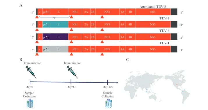
Baseline Characterization of Serum Samples From TDV Recipients Volunteers from dengue-endemic regions who were enrolled in study 203 received 1 dose of TDV on day 0 and a second dose on day 90 (Figure 1). To map the epitopes targeted by vaccine-in-duced nAbs, we selected serum samples from 24 study subjects, collected on day 0 (before vaccination) and day 120 (1 month after the second dose). Of the 24 subjects, 11 had DENV-naive
serum samples and 13 had DENV-preimmune serum samples prior to vaccination.
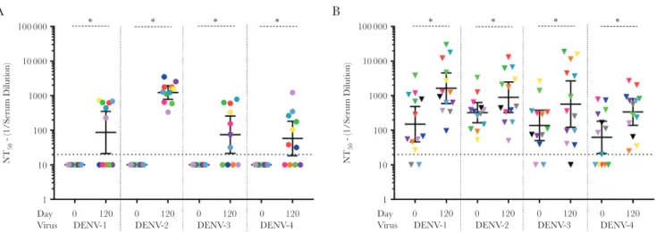
To evaluate epitope-specific responses following vaccination, we first measured levels of serum nAbs against the parental 4 DENV infectious clone–derived recombinant viruses, using flow cytometry and U937 cells expressing DC-SIGN (a DENV attachment receptor; Figures 2 and 3). At day 0, serum from 0 of 11 naive individuals had DENV nAbs (Figure 3A and
Supplementary Table 1). At day 120, serum from 6 naive indi-viduals neutralized DENV-1 West Pac ʹ74 (GMT, 86), serum from 11 neutralized DENV-2 S16803 (GMT, 1233), serum from 7 neutralized DENV-3 Sri Lanka ʹ89 (GMT, 74), and serum from 7 neutralized DENV-4 Sri Lanka ʹ92 (GMT, 58;
Figure 3A). Of the 13 preimmune subjects, before vaccination serum from 11 neutralized DENV-1 (GMT, 150), serum from 13 neutralized DENV-2 (GMT, 322), serum from 11 neutralized DENV-3 (GMT, 138), and serum from 7 neutralized DENV-4 (GMT, 62). By day 120, all vaccinated preimmune subjects had boosted responses, with elevated neutralizing titers against DENV-1 (GMT, 1637), DENV-2 (GMT, 898), DENV-3 (GMT, 571), and DENV-4 (GMT, 338; Figure 3B and Supplementary Table 1).
Mapping the Vaccine-Induced DENV-1 Epitope-Specific Response A chimeric virus was used to determine whether DENV-1 nAbs induced by TDV targeted a type-specific EDI/EDII hinge region epitope defined by human mAb 1F4 (Figure 2). The DENV-1
5′ c prM E NS1 2A 4A 4B –3′
Attenuated TDV-2 A
B C
TDV-1
TDV-3
TDV-4 NS5
2B NS3
–3′
c prM E NS1 2A 2B NS3 4A 4B NS5
–3′
c prM E NS1 2A 2B NS3 4A 4B NS5
–3′
c prM
Immunization Immunization
Day 0 Day 90 Day 120
Sample
Collection CollectionSample
E NS1 2A 2B NS3 4A 4B NS5
5′
5′
5′
Figure 1. Study design. A, The live attenuated tetravalent dengue vaccine (TDV) vaccine is composed of an attenuated dengue virus serotype 2 (DENV-2) backbone with prM and E from DENV-1 for TDV-1, prM and E from DENV-3 for TDV-3, and prM and E from DENV-4 for TDV-4. B, Subjects were immunized with the tetravalent vaccine at days 0 and 90. Serum samples were collected on day 0 before vaccination and on day 120, 30 days after the second dose. C, Study sites were located in the United States (Study 104) and Thailand, Colombia, Puerto Rico, and Singapore (Study 203).
against rDENV-3/1 were significantly higher (GMT, 755) than the nAb titers against the parental DENV-3 (GMT, 178; P< .05;
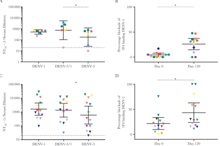
type-specific 1F4 epitope is sensitive to the angle of the EDI/II hinge and is present in DENV-1 virions but not recombinant E protein secreted from cells as a monomer [13]. From the TDV-receiving naive cohort, 6 subjects with DENV-1 nAb titers >1:20 at day 120 were tested for neutralization of DENV-1, DENV-3, and the DENV-3/1 chimera (Figure 4A). Among the 6 naive subjects with a postvaccination DENV-1 response, nAb titers
against rDENV-3/1 were significantly higher (GMT, 755) than the nAb titers against the parental DENV-3 (GMT, 178; P < .05;
Figure 4A). Blockade of 1F4 binding assays also demonstrated a significant increase in mean blockade of 1F4 binding to DENV-1 at a serum dilution of 1:20 between day 0 (2%) and day 120 (33%; P < .05; Figure 4B). In study 104 subjects (all of whom were naive prior to vaccination) [24], neutralization
100 000
A B
* * * *
NT
50
- (1/Serum Dilution)
10 000
1000
100
10
Day
Virus DENV-1
0 120
DENV-2
0 120
DENV-3
0 120
DENV-4
0 120
1
100 000 * * * *
NT
50
- (1/Serum Dilution)
10 000
1000
100
10
Day
Virus DENV-1
0 120
DENV-2
0 120
DENV-3
0 120
DENV-4
0 120
1
Figure 3. Preimmunization and postimmunization neutralizing antibody (nAb) titers against wild-type dengue viruses (DENVs). Neutralization assays were performed and the geometric mean titer (GMT) calculated against DENV-1, DENV-2, DENV-3, and DENV-4 on day 0 and day 120 in subjects who were naive at day 0 (A) or preimmune at day 0 (B). Bars represent the GMT and whiskers the 95% confidence interval. Dashed line indicates the limit of detection for the assay. Nonresponders were assigned a value of half of the limit of detection, for visualization. Within each graph, each individual is assigned the same color, to enable visualization of each person’s nAb titers to the different viruses. Dashed lines indicate the limit of the detection for the assay. *P < .05, by the Wilcoxon test, for comparison of day 0 to day 120. NT50, 50% nAb titer.
Virus DENV-1 DENV-2 DENV-3 DENV-3′
DENV-4′
West Pac ′74 S16803
UNC3001 Sri Lanka ′89 CH-53489
D4ic Sri Lanka ′92 DENV-3/1
DENV-4/2
DENV-4/3
DENV-3 DENV-4
DENV-4
DENV-1
DENV-3/1
DENV-4/2
DENV-4/3 DENV-2
DENV-3 1F4
EDIII
5J7
Strain Backbone Epitope Epitope Donor
findings for DENV-1 and DENV-3 was not statistically different (Supplementary Figure 1A), yet an increase in blockade of 1F4 binding was observed between day 0 (0%) and day 120 (26.9%; P < .05; Supplementary Figure 1C). Preimmune individuals with DENV-1 nAb titers after vaccination (Figure 4C) did not have significantly different levels of nAb to rDENV-3/1 (GMT, 1368) and DENV-3 (GMT, 602). However, in the blockade of binding assay, blockade at day 120 (43%) was significantly greater than blockade at day 0 (22%; P < .05; Figure 4D). These results demonstrate that TDV can elicit type-specific DENV-1 1F4-like nAbs in some individuals.
Mapping the Vaccine-Induced DENV-2 Response
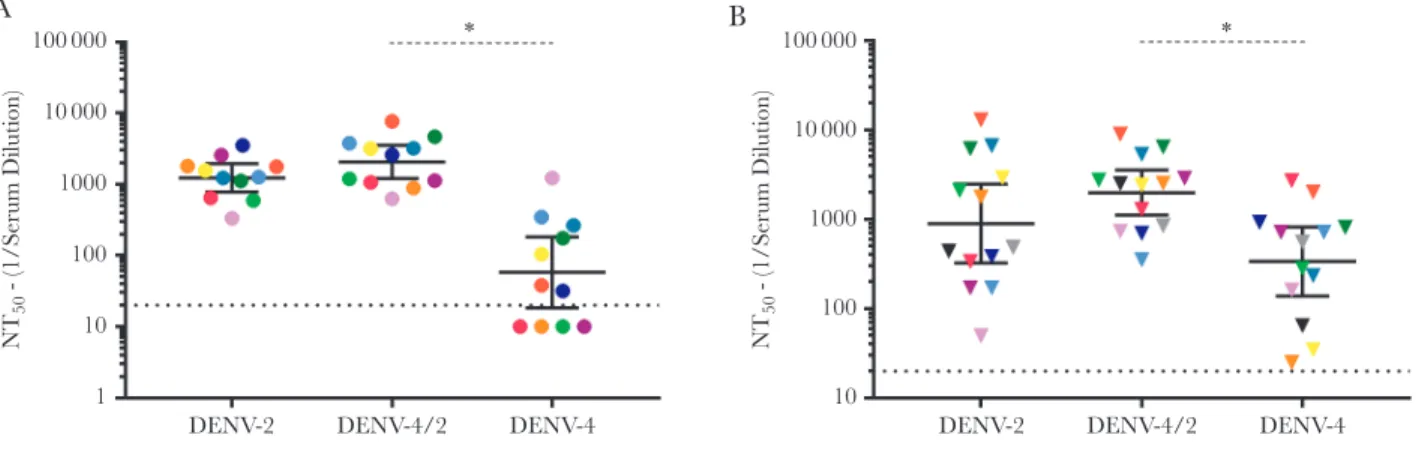
We previously described a recombinant DENV-4/2 chimeric virus containing an E protein with EDI and II from DENV-4 and EDIII from DENV-2 [14, 33]. This chimera displays a DENV-2 quaternary structure epitope centered on EDIII rec-ognized by strongly neutralizing human mAb 2D22 [14]. After
vaccination, all naive subjects had high DENV-2 nAbs titers (GMT, 1233; Figure 5A) and modest DENV-4 nAbs titers (GMT, 58; Figure 5A). DENV-4/2 neutralization (GMT, 2058) was sig-nificantly higher than observed for the parental DENV-4 strain (P < .05; Figure 5A). Study 104 subjects (all of whom were naive prior to vaccination) had higher titers of nAbs to DENV-4/2 as compared to DENV-4, as well (Supplementary Figure 1B; P < .05). DENV-preimmune subjects who received TDV devel-oped higher titers against rDENV-4/2 (GMT, 1993) as com-pared to DENV-4 (GMT, 338; P < .05; Figure 5B). These results demonstrate that, in both naive and DENV-preimmune sub-jects, TDV induces robust DENV-2 nAbs that target type-spe-cific epitopes centered on EDIII of DENV-2.
Mapping the Vaccine-Induced DENV-3 Response
We used a recombinant DENV-4/3 M14 chimeric virus to deter-mine whether DENV-3 nAbs induced by TDV targeted a quater-nary epitope, centered on the EDI/II hinge that spans 2 adjacent
100 000
A B
NT
50
- (1/Serum Dilution)
10 000
1000
100
100
50
Pe
rcentage blockade of
1F4 binding DENV
-1
0 10
DENV-1 DENV-3/1 DENV-3
* *
Day 0 Day 120
1
100 000
C D
NT
50
- (1/Serum Dilution)
10 000
1000
100
100
50
Pe
rcentage blockade of 1F4 binding DENV
-1
0
DENV-1 DENV-3/1 DENV-3
* *
Day 0 Day 120
10
monomers, defined by human mAb 5J7 (Figure 2) [34]. We transplanted the 5J7 epitope core from DENV-3 into DENV-4; this recombinant chimeric virus was efficiently neutralized by mAb 5J7 [29]. The 7 naive subjects with DENV-3 nAb titers of >1:20 after vaccination were tested for neutralization of DENV-3, DENV-4, and DENV-4/3 (Figure 6). In these individuals, the nAb titers were not significantly different between DENV-4/3 (GMT, 243) and the parental DENV-4 (GMT, 81; P < .1841; Figure 6A). From the naive group, immune sera collected after vaccination blocked the binding of mAb 5J7 to DENV-3, demonstrating the presence of serum Abs that bind at or near the epitope (Figure 6B). However, no significant neutralization or blockade was observed with Study 104 subjects (all naive prior to vaccination) after vac-cination (Supplementary Figures 1B and 1D). After vaccination, DENV-3 nAbs in the preimmune group did not significantly track with the 5J7 epitope displayed on the DENV-4/3 chimera (Figure 6C). However, the sera blocked binding of 5J7, suggesting the presence of Abs that bind at or near the epitope (Figure 6D).
Antigenic Cartography
As an alternate strategy to detect the tracking of TDV-induced Ab with transplanted epitopes, we performed antigenic car-tography of the wild-type and recombinant DENVs, using the neutralization data generated from TDV immune sera (Figure 7 and Supplementary Figure 2). In the antigenic map created by this analysis, the antigenic position of the DENV-4/2 recombinant virus moved away from the parental back-bone of DENV-4 toward that of DENV-2, indicating that TDV induces responses directed to the DENV-2 domain displayed on DENV-4 (Figure 7). Cartography findings indicate that TDV induces a robust type-specific response to DENV-2 and variable responses to DENV-1 and 3 epitopes.
DISCUSSION
We have evaluated the epitope specificity of DENV nAbs induced by TDV in naive and dengue-preimmune individuals. Epitope-specific measurements are important because people exposed to primary DENV infection develop a strong nAb response that targets serotype-specific epitopes that protect against homologous-serotype infection. It is unclear whether individuals who receive tetravalent dengue vaccines develop primary serotype-specific responses or broadly cross-reactive Ab responses that target each vaccine component. Also unclear is the role of these responses in durable protection against the 4 serotypes. A recent study demonstrated that dengue-naive indi-viduals who received a tetravalent chimeric yellow fever–den-gue (CYD) vaccine mainly had type-specific nAbs to DENV-4 and cross-reactive nAbs to the other serotypes [9]. The DENV-2 response, in particular, was dominated by nAbs directed to cross-reactive epitopes, and overall CYD vaccine efficacy was lowest against this serotype in clinical trials [9].
Natural DENV-2 infection elicits human mAb 2D22–like type-specific nAbs that bind to a quaternary epitope spanning the EDIII and EDII of a single E protein homodimer [14, 33]. The recombinant chimeric DENV-4/2 displays the 2D22 quater-nary epitope, allowing us to track linear EDIII and quaterquater-nary “2D22-like” responses in human immune sera after infection and vaccination [14, 33]. The Takeda TDV induced robust DENV-2–specific nAbs that tracked with EDIII on DENV-4/2. These studies could not differentiate between nAbs directed to simple epitopes contained with EDIII and more-complex “2D22-like” responses that require both EDIII and residues on adjacent E molecules. Blockade of binding assays with TDV immune sera and human mAb 2D22 could not be performed, as the 2D22 epitope encompasses the highly conserved fusion
100 000
A B
NT
50
- (1/Serum Dilution)
10 000
1000
100
10
DENV-2 DENV-4/2 DENV-4
* *
1
100 000
NT
50
- (1/Serum Dilution)
10 000
1000
100
DENV-2 DENV-4/2 DENV-4
10
100 000
A
NT
50
- (1/Serum Dilution)
100
50
0
Pe
rcentage blockade of 5J7 binding DENV
-3
100
50
0
Pe
rcentage blockade of 5J7 binding DENV
-3
10 000
1000
100
10
DENV-2 DENV-4/2
*
DENV-4 Day 0 Day 120
Day 0 Day 120
* *
* 1
100 000
C
B
D
NT
50
- (1/Serum Dilution)
10 000
1000
100
DENV-2 DENV-4/2 DENV-4
10
Figure 6. Mapping the dengue virus serotype 3 (DENV-3) epitope-specific neutralizing antibody (nAb) response. Immune sera from naive (A and B) and preimmune (C and D) subjects who received the vaccine and developed DENV-3 nAb (50% nAb titer [NT50] >1:20) were selected for mapping DENV-3 responses. Neutralization assays were per-formed using a recombinant chimeric DENV-4/3 and the wild-type (WT) DENV-3 and DENV-4 parental strains of the chimera, and the geometric mean NT50 values (GMTs) were calculated for the naive (A) and preimmune (C) groups after vaccination. Bars represent the GMT and whiskers the 95% confidence interval. Dashed line indicates the limit of detection for the assay. Nonresponders were assigned a value of half the limit of detection, for visualization. Prevaccination and postvaccination immune sera from naive (B) and preimmune (D) subjects were also tested for ability to block the binding of monoclonal Ab 5J7 to DENV-3. Within each graph, each individual is assigned the same color, to enable visualization of each person’s nAb titers to the different viruses. *P < .05, by the Wilcoxon test, for comparison of the parental backbone DENV to the rDENV.
Study 203 Naive Subjects, Day 120
A B
5
0
–5
5
0
–5 DENV-1 DENV-2 DENV-3 DENV-3/1 DENV-4 DENV-4/2 DENV-4/3
–10 –5 0 5 10 –10 –5 0 5 10
Study 203 Preimmune Subjects, Day 120
loop domain of the E glycoprotein, which also elicits binding Abs that interfere with the specificity of the 2D22 blockade assay [33].
We also evaluated subjects with DENV-1 nAbs responses after vaccination. Among naive subjects who developed DENV-1 nAbs after vaccination, Abs that neutralized rDENV-3/1 displaying the 1F4 epitope were significantly greater than the nAb titer against the parental DENV-3 strain. Consistent with these findings, the blockade of binding assay demonstrated the presence of Abs in dengue-naive TDV recipients that pre-vented human mAb 1F4 from binding to DENV-1. Together, these results demonstrate that TDV can elicit nAbs directed to the complex serotype-specific epitope region defined by mAb 1F4 in dengue-naive subjects. The use of the rDENV-3/1 proved more challenging in the preimmune subjects, owing to the high titers of nAbs to both the parental DENV-1 and DENV-3 strains. However, sera from vaccinated dengue-preimmune subjects also blocked 1F4 binding to its epitope on DENV-1, indicating that vaccination elicited Abs that bind to or near the 1F4 epitope. While we recognize that Abs binding close to the 1F4 epitope may sterically hinder 1F4 binding, the combined approaches provide support for the hypothesis that the TDV-1 vaccine component elicited binding and functionally neutraliz-ing Abs directed to the 1F4 epitope in some individuals.
The majority of subjects evaluated in this study from both the dengue-naive and preimmune groups had Abs that neutralized DENV-3. While we were unable to conclusively detect nAbs that tracked with the DENV-3 5J7 epitope, these results should be interpreted with caution. Many individuals selected for this study had high levels of nAbs to both DENV-3 and DENV-4. In rDENV-4/3, a gain of DENV-3 nAbs could be masked by a loss of an epitope targeted by DENV-4 nAbs [35]. Mapping the DENV-3 response is also complicated by the genotype differences between the TDV and UNC DENV strains. As shown by our group pre-viously, DENV-3 genotypes have up to 10-fold significant differ-ences in polyclonal neutralization phenotypes [28]. There are 5 amino acid differences within the mAb 5J7 epitope, 3 of which are contact residues, between the TDV3 and UNC3001strains used for the study. A single contact residue change can dramatically reduce neutralization, complicating interpretation of DENV-3 epitope-specific responses. While 5J7 can bind and neutralize the both the UNC3001 and TDV3 strains, new recombinant viruses may be required to specifically map the 5J7 epitope-specific responses in vaccinees receiving TDV. Recently, several DENV-4 type-specific nAbs have been identified from natural human infections and experimental non-human primate studies [35]. We are currently developing reagents to map DENV-4 type-spe-cific nAb responses and anticipate using these reagents to map TDV responses in the near future.
Conventionally, antigenic cartography has been widely used to compare antigenic relatedness between different strains of viruses [32, 36]. Here we modified the approach to determine
whether the Takeda TDV induced nAbs that tracked with epi-topes displayed on recombinant DENVs. In the cartography map based on nAbs induced by TDV, the DENV-4/2 chimera mapped closer to DENV-2 than to DENV-4, suggesting that most if not all TDV2 Ab responses target the DENV-2 EDIII dis-played on DENV-4. Both rDENV-3/1 and rDENV-4/3 demon-strated shifts toward the donor strain, as well, although the shift was not as pronounced as in the case of rDENV-4/2 strain. In summary our data demonstrate that, in both naive and preim-mune individuals who received TDV, the TDV-2 vaccine com-ponent elicited robust type-specific nAb responses to epitopes on EDIII of DENV-2. TDV also induced nAb responses that tracked with DENV-1 type-specific 1F4 epitope in some sub-jects. While our data demonstrate the presence TDV-induced Abs that bind at or near the DENV-3 5J7 epitope, we were unable to detect functionally nAbs directed to this epitope.
As TDV progresses through its phase 2 and 3 clinical tri-als, the data and methods reported in this study can be used to understand how Abs elicited by vaccination protect people from infection and disease. Our study has some limitations that need to be addressed before large sample sets from large clinical studies can be tested. Many individuals selected for the current study had high levels of nAbs to both the transplant and recipi-ent serotypes. In these individuals, a gain of neutralization due to the transplanted epitope could be masked by a loss of a crit-ical neutralizing site in the recipient strain. This is issue is par-ticularly relevant to rDENV-3/1 and rDENV-4/3 because the known type-specific epitopes on these serotypes are adjacent to each other. In future studies, we plan to deplete all Abs to the recipient strain so that we can unambiguously track the prop-erties of the remaining type-specific Abs. Our studies are also confounded by sequence differences between TDV strains and the recombinant chimeric viruses used for mapping. In partic-ular, the region defined by the DENV-3 5J7 epitope are variable between TDV-3 and the UNC DENV-3 infectious clone. We are currently building recombinant viruses that are matched to epitopes on TDV viruses. Finally, some of the vaccine sera used in this study had detectable DENV-1 or DENV-3 nAbs in the Vero cell assay used at Takeda but not in the U937+DC-SIGN neutralization assay used for epitope mapping. Further studies are needed to understand these cell-type–dependent differences and how they relate to vaccine performance. In con-clusion, this study provides a foundation for the development of high-throughput assays for use in DENV vaccine trials to understand the contribution of Abs to protective immunity.
Supplementary Data
Notes
Financial support. This work was supported by the National Institutes of Health (grant P01AI106695; principal investiga-tor, E. Harris, University of California, Berkeley) and Takeda Vaccines.
Potential conflicts of interest. A. M. d. S. has consulted on dengue vaccines for Takeda Vaccines, Sanofi Pasteur, GSK, and Merck Pharmaceuticals and is an inventor in patents related to dengue vaccines. H. J. D. is an employee of Takeda Vaccines. C. D. P. was an employee of Takeda Vaccines at the time the study was conducted. J. E. O. was an employee of Takeda Vaccines at the time the study was conducted and is an inventor on patents related to dengue vaccines. R. S. B. has consulted on dengue vaccines for Takeda Vaccines and Sanofi Pasteur and is an inventor in patents related to dengue vaccines. All authors have submitted the ICMJE Form for Disclosure of Potential Conflicts of Interest. Conflicts that the editors consider relevant to the content of the manuscript have been disclosed.
References
1. Bhatt S, Gething PW, Brady OJ, et al. The global distribution and burden of dengue. Nature 2013; 496:504–7.
2. Burke DS, Nisalak A, Johnson DE, Scott RM. A prospective study of dengue infections in Bangkok. Am J Trop Med Hyg 1988; 38:172–80.
3. de Alwis R, Williams KL, Schmid MA, et al. Dengue viruses are enhanced by distinct populations of serotype cross-re-active antibodies in human immune sera. PLoS Pathog 2014; 10:e1004386.
4. Clapham HE, Rodriguez-Barraquer I, Azman AS, et al. Dengue virus (DENV) neutralizing antibody kinetics in children after symptomatic primary and postprimary DENV infection. J Infect Dis 2016; 213:1428–35.
5. Wahala WM, Silva AM. The human antibody response to dengue virus infection. Viruses 2011; 3:2374–95.
6. de Alwis R, Smith SA, Olivarez NP, et al. Identification of human neutralizing antibodies that bind to complex epi-topes on dengue virions. Proc Natl Acad Sci U S A 2012; 109:7439–44.
7. Osorio JE, Partidos CD, Wallace D, Stinchcomb DT. Development of a recombinant, chimeric tetravalent den-gue vaccine candidate. Vaccine 2015; 33:7112–20.
8. Durbin AP, Kirkpatrick BD, Pierce KK, et al. A 12-month-in-terval dosing study in adults indicates that a single dose of the national institute of allergy and infectious diseases tetravalent dengue vaccine induces a robust neutralizing antibody response. J Infect Dis 2016; 214:832–5.
9. Henein S, Swanstrom J, Byers AM, et al. Dissecting anti-bodies induced by a chimeric yellow fever-dengue, live-at-tenuated, tetravalent dengue vaccine (CYD-TDV) in naive and dengue-exposed individuals. J Infect Dis 2017; 215:351–8.
10. Smith SA, de Alwis AR, Kose N, Jadi RS, de Silva AM, Crowe JE Jr. Isolation of dengue virus-specific memory B cells with live virus antigen from human subjects following natural infection reveals the presence of diverse novel func-tional groups of antibody clones. J Virol 2014; 88:12233–41. 11. Smith SA, de Alwis R, Kose N, et al. Human monoclonal antibodies derived from memory B cells following live atten-uated dengue virus vaccination or natural infection exhibit similar characteristics. J Infect Dis 2013; 207:1898–908. 12. Beltramello M, Williams KL, Simmons CP, et al. The human
immune response to Dengue virus is dominated by highly cross-reactive antibodies endowed with neutralizing and enhancing activity. Cell Host Microbe 2010; 8:271–83. 13. Fibriansah G, Tan JL, Smith SA, et al. A potent anti-dengue
human antibody preferentially recognizes the conforma-tion of E protein monomers assembled on the virus surface. EMBO Mol Med 2014; 6:358–71.
14. Gallichotte EN, Widman DG, Yount BL, et al. A new qua-ternary structure epitope on dengue virus serotype 2 is the target of durable type-specific neutralizing antibodies. MBio 2015; 6:e01461–15.
15. Fibriansah G, Tan JL, Smith SA, et al. A highly potent human antibody neutralizes dengue virus serotype 3 by binding across three surface proteins. Nat Commun 2015; 6:6341.
16. Teoh EP, Kukkaro P, Teo EW, et al. The structural basis for serotype-specific neutralization of dengue virus by a human antibody. Sci Transl Med 2012; 4:139ra83.
17. Chaudhury S, Gromowski GD, Ripoll DR, Khavrutskii IV, Desai V, Wallqvist A. Dengue virus antibody data-base: systematically linking serotype-specificity with epi-tope mapping in dengue virus. PLoS Negl Trop Dis 2017; 11:e0005395.
18. Hadinegoro SR, Arredondo-García JL, Capeding MR, et al.; CYD-TDV Dengue Vaccine Working Group. Efficacy and long-term safety of a dengue vaccine in regions of endemic disease. N Engl J Med 2015; 373:1195–206.
19. Huang CY, Butrapet S, Tsuchiya KR, Bhamarapravati N, Gubler DJ, Kinney RM. Dengue 2 PDK-53 virus as a chi-meric carrier for tetravalent dengue vaccine development. J Virol 2003; 77:11436–47.
20. Osorio JE, Huang CY, Kinney RM, Stinchcomb DT. Development of DENVax: a chimeric dengue-2 PDK-53-based tetravalent vaccine for protection against dengue fever. Vaccine 2011; 29:7251–60.
21. Osorio JE, Velez ID, Thomson C, et al. Safety and immuno-genicity of a recombinant live attenuated tetravalent den-gue vaccine (DENVax) in flavivirus-naive healthy adults in Colombia: a randomised, placebo-controlled, phase 1 study. Lancet Infect Dis 2014; 14:830–8.
a dengue virus serotype 2 backbone. Expert Rev Vaccines 2016; 15:497–508.
23. George SL, Wong MA, Dube TJ, et al. Safety and immu-nogenicity of a live attenuated tetravalent dengue vac-cine candidate in flavivirus-naive adults: a randomized, double-blinded phase 1 clinical trial. J Infect Dis 2015; 212:1032–41.
24. Rupp R, Luckasen GJ, Kirstein JL, et al. Safety and immuno-genicity of different doses and schedules of a live attenuated tetravalent dengue vaccine (TDV) in healthy adults: a phase 1b randomized study. Vaccine 2015; 33:6351–9.
25. Sirivichayakul C, Barranco-Santana EA, Esquilin-Rivera I, et al. Safety and immunogenicity of a tetravalent dengue vaccine candidate in healthy children and adults in den-gue-endemic regions: a randomized, placebo-controlled phase 2 study. J Infect Dis 2016; 213:1562–72.
26. Sáez-Llorens X, Tricou V, Yu D, et al. Safety and immuno-genicity of one versus two doses of Takeda’s tetravalent den-gue vaccine in children in Asia and Latin America: interim results from a phase 2, randomised, placebo-controlled study. Lancet Infect Dis 2017; 17:615–25.
27. Messer WB, Yount BL, Royal SR, et al. Functional transplant of a dengue virus serotype 3 (DENV3)-specific human monoclonal antibody epitope into DENV1. J Virol 2016; 90:5090–7.
28. Messer WB, Yount B, Hacker KE, et al. Development and characterization of a reverse genetic system for studying dengue virus serotype 3 strain variation and neutralization. PLoS Negl Trop Dis 2012; 6:e1486.
29. Widman DG, Young E, Nivarthi U, et al. Transplantation of a quaternary structure neutralizing antibody epitope from den-gue virus serotype 3 into serotype 4. Sci Rep 2017; 7:17169. 30. de Alwis R, Smith SA, Olivarez NP, et al. Identification of
human neutralizing antibodies that bind to complex epi-topes on dengue virions. Proc Natl Acad Sci U S A 2012; 109:7439–44.
31. Cai Z, Zhang T, Wan XF. A computational framework for influenza antigenic cartography. PLoS Comput Biol 2010; 6:e1000949.
32. Lindesmith LC, Ferris MT, Mullan CW, et al. Broad block-ade antibody responses in human volunteers after immuni-zation with a multivalent norovirus VLP candidate vaccine: immunological analyses from a phase I clinical trial. PLoS Med 2015; 12:e1001807.
33. Fibriansah G, Ibarra KD, Ng TS, et al. DENGUE VIRUS. Cryo-EM structure of an antibody that neutralizes dengue virus type 2 by locking E protein dimers. Science 2015; 349:88–91.
34. Fibriansah G, Tan JL, Smith SA, et al. A highly potent human antibody neutralizes dengue virus serotype 3 by binding across three surface proteins. Nat Commun 2015; 6:6341. 35. Nivarthi UK, Kose N, Sapparapu G, et al. Mapping the
human memory B cell and serum neutralizing antibody responses to DENV4 infection and vaccination. J Virol 2017; 91:doi: 10.1128/JVI.02041-1.