UNIVERSITY OF NORTH CAROLINA AT CHAPEL HILL CHEMISTRY DEPARTMENT
Impact of the Pil-Chp Chemosensory System of Pseudomonas aeruginosa on cyclic AMP Production and Twitching Motility
By Boya Wang
Spring 2016
Table of Contents
Table of Contents 2 Abstract 3-4
I. Introduction 5-8
II. Materials and Methods 9-13 III. Results and Discussion 14-21
A. Results 14-18 B. Discussion 19-21 IV. Contributions 22
V. References 23-24
Abstract
The Chp chemosensory system plays a central role in detecting host signals and activating virulence gene expression, allowing the opportunistic bacterial pathogen Pseudomonsa aeruginosa to cause a wide array of diseases. Specifically, the Chp system
controls production of intracellular cyclic AMP (cAMP) and type IV pilus mediated surface motility (twitching motility). cAMP serves as an allosteric activator of the transcription factor Vfr, a global regulator of virulence gene expression. A central component of the Chp chemosensory system is the hybrid ChpA kinase. In addition to a two stand alone receiver domains PilG and PilH, ChpA is composed of a conserved kinase domain, a serine-containing phosphotransfer domain (Spt 1), a threonine-containing phosphotransfer domain (Tpt 1), six histidine-threonine-containing phosphotransfer domains (Hpt 1-6), and a CheY-like receiver domain (ChpArec). We hypothesized that multiple domains may be necessary to fine-tune signal transduction for twitching motility and/or intracellular cAMP levels.
As a first step in assessing function, we used a biochemical approach to determine which ChpA domains were capable of participating in phosphotransfer events. We found that the Hpt 2 and 3 domains could only reversibly receive phosphoryl groups from ChpArec rather than ATP and Hpt 3 was able to transfer phosphoryl to PilH. Further, we found that ChpArec could both accept phosphoryl groups and transfer phosphoryl groups to Hpt 2-6. Based on these results, we generated point mutations in Hpt 2, 3 and ChpArec domains in all possible combinations and evaluated the impact on cAMP signaling and twitching motility. To assess cAMP signaling, we used a synthetic chromosomal
I. INTRODUCTION
Pseudomonas aeruginosa is an opportunistic, Gram-negative pathogen which
causes infections in animals, plants, and humans (Driscoll, et al., 2007). The hospitalized population, immunocompromised individuals and those with chronic airway diseases such as cystic fibrosis and chronic obstructive pulmonary disease are at the most risk for infection (Giamarellou, 2000). P. aeruginosa is responsible for nearly 10% of all
hospital-acquired infections, which are often life-threatening because of the bacteria’s high levels of antibiotic resistance (NNISS, 2004). P. aeruginosa is found commonly in nature and thrives in moist environments such as respiratory equipment, antiseptic solutions, sink taps, and showers (Giamarellou, 2000).
P. aeruginosa responds to unknown host associated cues by expressing virulence
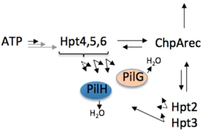
factors. The Pil-Chp chemosensory system, a type of two-component system (TCS), plays a central role in detecting host signals and activating virulence gene expression, allowing P. aeruginosa to cause a wide array of diseases. A central component of the Pil-Chp chemosensory system is the Pil-ChpA kinase, a homologue of the CheA kinase. As illustrated in Figure 1, ChpA is a very large, complex signal transduction protein. In addition to two separate CheY-like receiver domains PilG and PilH, ChpA contains eight Xpt phosphotransfer domains and a C-terminal receiver domain (ChpArec) (Leech, Mattick, 2006). The chpA gene is linked to several other complex virulence pathways in P. aeruginosa, namely cyclic AMP production and type IV pilus (T4P) mediated
Figure 1. The ChpA is a highly complex hybrid histidine kinase.
The second-messenger signaling molecule adenosine 3’, 5’-cyclic monophosphate (cAMP or cyclic AMP) regulates many virulence factors (Wolfgang, et al., 2003). It is synthesized by the adenylate cyclase CyaB, whose activity is controlled by the Chp system (Fulcher, et al., 2010). Once produced, cAMP binds to and activates the transcription factor Vfr (virulence factor regulator) and creates a cAMP-Vfr complex (West et al., 1994). The cAMP-Vfr complex regulates various virulence factors that instigate secretion of toxins and host colonization. Further, the Pil-Chp system, similar to flagellar-based chemotaxis systems, has been shown to regulate twitching motility, resulting from the extension and retraction of T4P (Rauprich, et al., 1996).
The typical TCS features His->Asp phosphotransfer between a signal-sensing histidine kinase and the receiver domain of a partner response regulator, whereby the phosphorylated response regulator executes an output response. In contrast to the E. coli flagellar chemotaxis protein CheA, P. aeruginosa ChpA has nine possible
phosphorylation sites and two potential partner CheY-like receiver proteins that
complicate the signaling strategy of the Chp chemosensory system. Recent research has demonstrated that, of the eight Xpt domains, three (Hpts 4, 5, 6) could be
270kDa 2478aa H1 T H2 H3 S H4 H5 H6 Dimer His Kin CheW D1
ChpA
D2 D3
autophosphorylated with ATP. These same Hpts plus two others- Hpt2 and Hpt3- could become reversibly phosphorylated by the ChpA C-terminal receiver domain (ChpArec). The remaining Xpts (Hpt1, Spt, and Tpt) displayed no detectable phosphotransfer
reactivity. Phosphoryl groups transferred rapidly from all five of the phosphorylated Hpts to ChpArec, but phosphorylation of PilG and PilH was slower. These in vitro studies demonstrated that ChpArec could act as either a phosphate sink or a source of phosphoryl groups for the phosphorylatable Hpts (Silversmith et al., under review).
Figure 2. Scheme summarizing phosphotransfer between Pil-Chp domains. The length of the arrow reflects the relative reactivity. Gray arrows reflect presumed variation of Hpt autophosphorylation by the presence of stimulus. Solid black arrows reflect inherent rates measured here. Dashed arrows indicate the absence of rapid
phosphotransfer (experimental design precluded measurement of slower
II. MATERIALS AND METHODS
Construction of P. aeruginosa ChpA Hpt 2HA and 3HA chromosomal mutants for in vivo studies.
For in vivo studies of chpAHpt2HAand chpAHpt3HAmutants were created via Quickchange methodology (Agilent Technologies) and cloned via Gateway Technology (Invitrogen) into PAKΔcyaAlacP1:ΔlacI-lacZ. First, a large portion of the chpA gene containing the domains in question was amplified using the primers 5’ Hpt2_3_frag (5’ TACAAAAAAGCAGGCTAGCGACGACAACTGGACCCTTG) and 3’ Hpt2_3_frag (3’ TACAAGAAAGCTGGGTCGACACCATTTCCTCGTCCAGC) from a full-length chpA expression plasmid pJNTac4:ChpA. Attachment sites AttB were added to the termini of the amplicon allowing recombination into vector pDONR201 via a Gateway Cloning Technology BP recombination reaction. The Hpt 2 and 3 site were mutated from a histidine to alanine codon via Quikchange methodology reaction and sequence
confirmed.
Then, the mutagenized fragments were moved by LR recombination reaction to the P. aeruginosa destination suicide vector pEXG2GW. Finally, the mutant suicide vector was mated with P. aeruginosa strain PAKΔcyaAlacP1:ΔlacI-lacZ to generate mutants in an otherwise wild type strain with a reporter system to indirectly measure cAMP. Further, Hpt 2 and 3 single and double mutants were cloned into the
PAKΔcyaAchpArecDAlacP1:ΔlacI-lacZ background. Each mutant was confirmed by sequencing and restriction enzyme digest.
A chpArecD2407A (chpArecDA) substitution mutant was cloned via Gateway Technology (Invitrogen) into PAKΔcyaAlacP1:ΔlacI-lacZ. The 3’ region of chpA was amplified as two separate but overlapping sequences, such that the overlapping sequences introduced a D2407A codon substitution. The 5’ fragment was amplified using primers 5’chpArecD2407A (5’
TACAAAAAAGCAGGCTACCTCAGCGATCATGAAGTGCTGC) and
pJNt4chpArec3’ (5’ GCGCGGCATCTCGATGGCCAGCAGGAGGATGTC). The 3’ fragment was amplified using primers 5’ pJNt4chpArec (5’
GACATCCTCCTGCTGGCCATCGAGATGCCGCGC) and 3’ chpArecD2407A (5’ TACAAGAAAGCTGGGTGCGGACGTTCTTCACATGCCACTG). Attachment sites AttB were added to the amplicon and inserted into vector pDONR201 via a BP
recombination reaction.
An LR recombination reaction with the pDONR201 entry clone mutant and destination vector pEXG2GW was performed to generate a suicide vector. Finally, the mutant suicide vector was mated with PAKΔcyaAlacP1:ΔlacI-lacZ to generate mutants in wild type PAK with a reporter system to indirectly measure cAMP. Each mutant was confirmed by sequencing and restriction enzyme digest.
Construction of P. aeruginosa ChpA expression plasmids for in vivo studies.
aeruginosa. To generate pJNtac4, the lacIq-PTOP control region of pMMBV4 (21) was
amplified with primers lacIqtacP 5’
(5’TAATGAATTACAACAGTTTTTATGCATGCTGCTCCCGAACGCCAGCAAGAC G) and lacIqtacP 3’
(5’CTAGTGGATCCCCCGGGCTGCAGGAATTCTGTTTCCTGTGTGAAATTGTTA TC). Underlined nucleotides correspond to pJN105 vector sequence adjacent to and including SphI and EcoRI restriction sites, respectively. The resulting amplicon was cloned by Gibson Assembly (New England Biolabs) into pJN105 linearized by restriction enzyme digest with SphI and EcoRI to remove the araC-PBAD expression control
region. Proper assembly of the resulting pJNtac4 vector was confirmed by sequencing. For complementation, the full-length (ATG-TGA) chpA open reading frame (ORF) was PCR amplified from PAK genomic DNA using primers 5’chpA
(5’ATAACAATTTCACACAGGAAACAGAATTCgaggaggatattcATGGGTGACCGG CACGACTACGTCGCTC) and 3’chpA
(5’CGACTCACTATAGGGCGAATTGGAGCTCTCACTCATGCTGGCCGACCAG GGACTGG). Underlined nucleotides correspond to pJNtac4 vector sequence adjacent to and including EcoRI and SacI restriction sites, respectively. Lowercase italic sequence represents a synthetic ribosome binding site and spacer region. Bold sequences
correspond to the 5’ and 3’ termini of the chpA ORF, respectively. The resulting
amplicon was gel purified and cloned by Gibson Assembly (New England Biolabs) into pJNtac4 linearized by restriction enzyme digest with EcoRI and SacI, such that cloned chpA expression was under the control of the adjacent IPTG controlled PTOP promoter.
stranded sequencing. To generate a ChpArec D2407A substitution mutant, the 3’ region of chpA was amplified as two separate but overlapping fragments, such that the
overlapping sequences introduced a D2407A codon substitution (GAC to GCC). The 5’ fragment was amplified using primers pJNt4chpA5’KpnI
(5’CGCATGGTGCCGTTCGATCGGCTGGTACCG) and pJNt4chpArec 3’ (5’- GCGCGGCA TCTCGA TGGCCAGCAGGAGGA TGTC). The 3’ fragment was
amplified using pJNt4chpArec 5’ (5’- GACA TCCTCCTGCTGGCCA TCGAGA TGCC GCGC) and pJNt4chpA3’Bsu36I (5’ CTCCACGGGGAGAGCCTGAGCAAACTGGC C). The D2407A codon substitution is indicated in bold in the overlapping internal primers. The resulting products were spliced by overlap extension PCR using the flanking primers pJNt4chpA5’KpnI and pJNt4chpA3’Bus36I. The flanking primers correspond to pJNtac4-chpA vector sequence adjacent to and including KpnI and Bsu36I restriction sites, respectively. The spliced product, encompassing the 3’ region of chpA and encoding the D2407A substitution, was cloned by Gibson Assembly into pJNtac4-chpA digested with KpnI and Bsu36I to generate pJNtac4-pJNtac4-chpArec D2407A. The
resulting plasmid was confirmed by sequencing.
In vivo expression of ChpA rec D2407A and P. aeruginosa phenotype assays All chpA in vivo expression and phenotyping experiments utilized
gentamycin. For protein expression of pJNtac4-based expression plasmids, IPTG was included in the growth media at the indicated concentration. Chromosomal mutants were all grown in LB broth or on LB agar without selection. Growth was monitored by OD at 600 nm. For western blots, whole cell lysates were prepared from culture aliquots obtained at OD600 = 0.5-1.0 as previously described (Fulcher, et al., 2010). Proteins were separated on 4-15% gradient SDS- polyacrylamide gels (BioRad), transferred to nitrocellulose and simultaneously probed with rabbit anti-ChpA sera and mouse monoclonal antibody specific for the beta subunit of RNA polymerase (RNAP)
(NeoClone). ChpA anti-serum was generated in rabbits immunized with purified ChpA construct (Cocalico Biologicals). Anti-RNAP antibody was included as a loading and normalization control as previously reported (Topal, H. et al., 2012). Antigen-antibody complexes were detected using goat anti-rabbit IRDye800CW and goat anti- mouse IRDye680LT (LI-COR) and visualized using a LI-COR Odyssey Classic Infrared
III. RESULTS AND DISCUSSION
A. RESULTS
ChpArec acts as a phosphate sink- In P. aeruginosa, twitching motility and intracellular cAMP production are dependent on ChpA and its corresponding, free floating receiver proteins PilG and PilH (Fulcher, et al., 2010). A third receiver, ChpArec, has been shown to both rapidly and reversibly react with ChpA Hpt 2-6 domains and
autodephosphorylates (transfer of the phosphoryl group to water) these domains faster than PilG or PilH (Silversmith et al., under review). The ChpArec domain is also the primary means for phosphorylation of Hpt 2 and Hpt 3. We hypothesized ChpA rec could act as either a phosphate sink, acting to keep phosphoryl groups off PilG and PilH or a source of phosphoryl groups for the phosphorylatable Hpts. In order to test the in vivo effects based upon biochemical analyses, we measured twitching motility and
intracellular cAMP production in a P. aeruginosa strain containing a ChpA substitution mutant that lacked the receiver domain site of phosphorylation (ChpA rec D2407A or ChpArecDA).
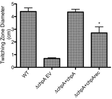
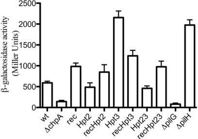
The chpA deletion mutant had drastically reduced levels of intracellular cAMP and twitching motility compared to wildtype, but IPTG induced plasmid-expressed chpA (pJNtac4-chpA) was able to restore wildtype levels. In contrast, plasmid-expressed ChpArecDA showed a significant defect (p<0.001) in twitching motility and about twofold increase in cAMP production (p<0.001) compared to wildtype (Figures 3 and 4). Western blot analysis demonstrates that plasmid expression of ChpA and ChpArecDA resulted in proteins levels that were indistinguishable from that of the wildtype strain (Figure 5).
Subsequently, a chromosomal ChpArecDA mutant was also constructed and resulted in similar defects in cAMP production and twitching (Figures 6 and 7). Finally, strains containing a double mutation with either Hpt2 and ChpA substitutions
(ChpAHArecDA) or Hpt 3 and ChpA substitutions (ChpAHArecDA) were constructed. Both double mutant strains behaved more similarly to the ChpArec single mutant than the wild type strain for both cAMP production and twitching motility (Figures 6 and 7).
production and twitching motility because the ChpArec phenotype dominated in the double mutant strains.
cAMP phenotypes of Hpt2, Hpt3, and ChpArec phosphorylation site mutants reveal both
gain and loss of function- Chemosensory systems specialized to mediate motility by T4P contain multiple phosphotransfer domains within their CheA analogues. ChpA is one of the only studied CheA with eight Xpt domains that can be separated into groups based upon reactivity. Previous in vivo studies indicate that both gain and loss of Pil-Chp function lead to reduced twitching motility. First, chpA and pilG deletion strains both display loss of function (low cAMP and reduced surface pilin relative to wild type),
indicating phosphorylated PilG (PilGp) is necessary for cAMP production via regulation
of the adenylate cyclase CyaB (Fulcher, 2010). In contrast, pilH deletion mutants display
gain of function (elevated cAMP and surface pilin over wild type), indicating that PilH
suppresses cAMP production (Fulcher, 2010).
First, both the Hpt3 and pilH mutant displayed 3-4 fold elevated cAMP production over wild type (Figure 6). This phenotype similarity supports the in vitro findings that Hpt3 rapidly transfers phosphoryl groups to PilH. Additionally, the Hpt 2 mutant displayed reduced cAMP levels between that of the pilG mutant and wild type, indicating partial loss of function (Figure 6). Although in vitro analysis did not indicate phosphoryl group transfer from Hpt 2 to PilG, in vivo cAMP phenotypes support this connection. The observation that the hpt23HA double mutation diminishes cAMP to near the levels seen in the Hpt2HA mutant alone is also consistent with a model in which in
the absence of Hpt3, phosphoryl groups are routed through Hpt2 to PilG and further that
Hpt2 and Hpt3 represent routes of phosphoryl groups to PilG and PilH respectively.
Lastly, the finding that Hpt2HA did not show the same degree of defect as the pilG
deletion mutant is consistent with the fact that Hpt4, 5, 6 are capable of phosphotransfer
to PilG.
The double mutants ChpArecDA, Hpt2HA and ChpArecDA, Hpt3HA and the
triple mutant ChpArecDA, Hpt2HA, Hpt3HA all had levels of cAMP that were
indistinguishable from the ChpArecDA single mutant (Figure 6). This result indicates
that Hpt2 and Hpt3 function downstream from ChpArec, which is consistent with the
scheme assembled in Figure 2 (Introduction) from biochemical results. In this scheme
phosphoryl groups cannot reach Hpt2 or Hpt3 in either the ChpArecDA single mutant or
the Hpt2HA, Hpt3HA double mutant. The increased level of cAMP in the former mutant
compared to the latter is consistent with loss of phosphoryl groups from the system by
rapid autodephosphorylation of ChpArec when present. Lastly, western blot analysis of
performed. In comparison to wild type ChpA expression levels, the chromosomal
mutants displayed similar ChpA levels indicating that the mutant proteins were stable
(Figure 8).
Correlation of twitching and cAMP phenotypes- The same set of chromosomal mutants
was evaluated for Type-IV pili mediated twitching motility. Twitching motility is a
relatively insensitive measure of Pil-Chp output as it involves a complex interplay
between pilus extension and retraction dynamics, such that gain-of-function and
loss-of-function mutations both result in suboptimal or reduced motility (Bertrand, 2010).
Grouping mutants based on twitching motilitygave the same groupings described above
for cAMP phenotypes.
The Hpt3HA and pilHmutants, which shared strong gain of function cAMP
phenotypes (Figure 6), exhibited similar twitching defects with zone expansion rates
30-40% of the wild type strain (Figure 7). Likewise, all of the mutants containing the
ChpArecDA substitution (ChpArecDA, ChpArecDA,Hpt2HA, ChpArecDA,Hpt3HA,
and ChpArecDA,Hpt2HA,Hpt3HA), which shared similar modest gain of function cAMP
phenotypes,gave the similar twitching rates of 50-60% of wildtype (Figure 7). Thus the
twitching data is also consistent with the ChpArec mutant being epistatic (upstream) to
both Hpt2 and Hpt3. Finally, the Hpt2HA and Hpt2HAHpt3A mutants, which had similar
cAMP phenotypes (diminished relative to wild type), also had similar twitching of
B. DISCUSSION
Bacterial chemosensory signal transduction systems that regulate motility by type IV pili often contain a CheA kinase with numerous potential sites of phosphorylation (Whitchurch, et al., 2004). Recently, novel signal mechanisms regarding a complex CheA homologue, the ChpA histidine kinase in P. aeruginosa are shown in Figure 2. The ChpA kinase is a central component of the Pil-Chp chemosensory system in P.
aeruginosa which regulates cAMP production and T4P-mediated twitching motility, both of which play roles in pathogenesis. The ChpA kinase contains eight Xpt domains, a C-terminal receiver domain (ChpArec) and interacts with two stand-alone receiver domains (PilG and PilH), each involved in a complex series of phosphotransfer steps. Notably, ChpArec reacted rapidly and reversibly with Hpt 2-6 and could potentially act as a phosphate sink due to its efficient autodephsophorylation or a source of phosphoryl groups for Hpt 2-6 or PilH. Moreover, the Hpt 2 and 3 domains could only receive phosphoryl groups from ChpArec rather than ATP, and appeared to transfer them to PilG and PilH, with Hpt3 to PilH being the fastest observed route (Silversmith et al., under review).
This is in contrast to previous data which showed there was no twitching in a ChpArec mutant (it behaved the same as a ΔchpA mutant) in a different strain PAO1 containing a chromosomal deletion mutant of chpA complemented with point mutated ChpArec in an expression plasmid (Leech, Mattick, 2006). However, partial twitching ability is expected because a mutant in another phosphate sink receiver domain PilH resulted in highly elevated cAMP levels but still displayed partial twitching and because there are still available phosphotransfer routes in ChpA (Fulcher, et al., 2010).
Subsequently, single point mutants of the Hpt 2 and 3 domains were created on the chromosomal chpA gene. The Hpt2HA mutant displayed slightly lower cAMP levels with almost wild type levels of twitching while the Hpt3HA mutant displayed highly elevated cAMP levels with partial twitching ability. These phenotypes suggest the Hpt2 domain aids in cAMP levels by providing phosphoryl groups for PilG. In contrast, the Hpt3 domain most likely supplies phosphoryl groups to PilH (and is consistent with biochemical analysis) or remove or prevent phosphoryl groups from reaching PilG. Lastly, the both double mutants of Hpt2HArecDA and Hpt3HArecDA mimic the
ChpArecDA single mutant phenotype with about a twofold increase in cAMP production. This indicates the dominant role ChpArec plays over either Hpt 2 or 3 domain as it is upstream in the phosphotransfer mechanism of ChpA and further demonstrates either domain is not necessary for cAMP production.
Taken together, previously reported genetic data (Fulcher, et al., 2010; Bertrand,
et al., 2010) as well as the phenotypes of seven new mutants reported here are consistent
with multiple fundamental features of the Chp/Pil signaling network deduced from our
phosphoryl group flow within ChpA proposed here. However, some aspects of the
genetic data suggest the scheme based on biochemical results alone is incomplete. In
particular, the preferred in vivo routes of phosphotransfer between Hpts 2-6 and
PilG/PilH may not have been identified by our in vitro experiments. We are currently
IV. Contributions
Thanks to the Wolfgang lab: Dr. Matthew C Wolfgang, Dr. Cindy Gode, Dr. Justin Graham, Bryan Zorn, Mark Baumgarten, Stephen Lanhan, and Joseph Hatch.
V. REFERENCES
Bertrand, J. J., West, J. T., and Engel, J. N. (2010) Genetic analysis of the regulation of type IV pilus function by the Chp chemosensory system of Pseudomonas aeruginosa. J. Bacteriol. 192, 994-1010
Driscoll JA, Brody SL, Kollef MH. The epidemiology, pathogenesis and treatment of Pseudomonas aeruginosa infections. Drugs. 2007; 67:351–368. [PubMed:
17335295]
Fulcher NB, Holliday PM, Klem E, Cann MJ, Wolfgang MC. 2010. The Pseudomonas aeruginosa Chp chemosensory system regulates intracellular cAMP levels by modulating adenylate cyclase activity. Mol. Microbiol. 76:889 –904
Giamarellou, H. (2000) Therapeutic guidelines for Pseudomonas aeruginosa infections. Int J Antimicrob Agents 16: 103–106.
Leech AJ, Mattick JS (2006) Effect of site-specific mutations in different phosphotransfer
domains of the chemosensory protein ChpA on Pseudomonas
aeruginosa motility. J Bacteriol 188:8479–8486.
National Nosocomial Infection Surveillance System. National Nosocomial Infection
Surveillance (NNIS) System report, data summary from January 1992 through
June 2004, issued October 2004. Am. J. Infect. Control 32:470-485.
Schneider, C. A., Rasband, W. S., and Eliceiri, K. W. (2012) NIH Image to ImageJ: 25 years of image analysis. Nat. Methods 9, 671-675.
Silversmith R. E., Wang, B., Fulcher, N. B., Wolfgang, M. C., and Bourret R. B. (under review). Phosphoryl group flow within the Pseudomonas aeruginosa Pil-Chp chemosensory system: differential function of the eight phosphtransferase and three receiver domains.
Topal, H., Fulcher, N.B., Bitterman, J., Salazar, E., Buck, J., Levin, L.R., Cann, M. J., Solfgang, M.C., and Steegborn, C. (2012). Crystal Structure and Regulation Mechanisms of the CyaA Adenylyl Cyclase from the Human Pathogen Pseudomonas aeruginosa. J. Mol. Biol. 416: 271-286.
West SE, Sample AK, Runyen-Janecky LJ. The vfr gene product, required for
Pseudomonas aeruginosa exotoxin A and protease production, belongs to the
cyclic AMP receptor protein family. J Bacteriol. 1994; 176:7532–7542. [PubMed: 8002577]
Whitchurch CB, Leech AJ, Young MD, Kennedy D, Sargent JL, Bertrand JJ, et al. Characterization of a complex chemosensory signal transduction system which controls twitching motility in Pseudomonas aeruginosa. Mol Microbiol. 2004; 52:873–893. [PubMed: 15101991]
VI. SUPPLEMENTAL DATA
Figure 3. The ChpA receiver domain suppresses cAMP synthesis in P. aeruginosa. Mutation of the ChpA receiver domain site of phosphorylation from an aspartic acid to an alanine increase cAMP synthesis. Phenotypes were assayed for wildtype P. aeruginosa strain PAK (WT) carrying empty vector (pJNtac4) and an isogenic chpA deletion strain
(ΔchpA) carrying empty vector or complemented with either plasmid-encoded wildtype
ChpA (pJNtac4-chpA) or ChpA with an alanine substitution at the receiver domain site phosphorylation, ChpArec D2407A (pJNtac4-chpArec D2407A). Relative intracellular cAMP levels were determined by β-galactosidase assay of strains harboring a quantitative cAMP-dependent transcriptional reporter (lacP1ΔlacI-lacZ) (Fulcher, et al., 2010). Assay was performed in the presence of 75 µg/ml gentamycin for plasmid maintenance and 25 µM IPTG for induction of plasmid expression of chpA alleles with three replicates. Asterisk represents significant difference form wild type (p<0.001).
WT
ΔchpA EV
Figure 4. The ChpA receiver domain modulates twitching motility. Mutation of the ChpA receiver domain decreased twitching motility. The same strains were assayed as in Figure 3 with five replicates. Twitching motility measurements represent the diameter of subsurface zones of expansion 40 hours post inoculation of bacteria to the agar-plastic interface (Fulcher, et al., 2010). Asterisk represents significant difference form wild type (p<0.001).
WT
ΔchpA EV
Figure 6. The ChpA phosphotransfer Hpt 2 and 3 domains and receiver domains work in conjunction to modulate cAMP phenotypes. Double and triple mutations of the Hpt2 and 3 domains in tandem with the receiver domain resemble the phenotype of the single receiver domain mutant. Single mutation of the Hpt 2 domain decreased cAMP, similar to a PilG mutant, and mutation of the Hpt 3 domain increased cAMP production, similar to a PilH mutant. A Hpt 23 double mutant phenocopies a Hpt 2 single mutant. Phenotypes were assayed for wild type PAK (WT), an isogenic chpA deletion strain (ΔchpA), a chromosomal ChpArec mutation (rec), a ChpA Hpt2 mutation (Hpt2),
and ChpA Hpt 3 mutation (Hpt3) in all seven possible combinations. The PilG and PilH mutants were reported before but assayed here as a comparison. Relative intracellular cAMP levels were determined by β-galactosidase assay of strains harboring a quantitative cAMP-dependent transcriptional reporter (lacP1ΔlacI-lacZ) (Fulcher, et al., 2010).
wt
ΔchpA rec Hpt2recHpt2Hpt3recHpt3Hpt23 recHpt23
Figure 7. The Hpt 2, 3, and rec domains modulate twitching motility in correlation with cAMP phenotypes. All seven combinations of Hpt 2, 3, and rec domain mutants were assayed as in Figure 5. The same phenotype conclusions can be drawn and demonstrates there may need to be an optimal level of intracellular cAMP in order to display wild type twitching ability. Assay represents seven replicates.
wt
ΔchpArecDAHpt2HA
recDAHpt2HA Hpt3HA
recDAHpt3HA Hpt23HA
recDAHpt23HA
ΔpilGΔpilH
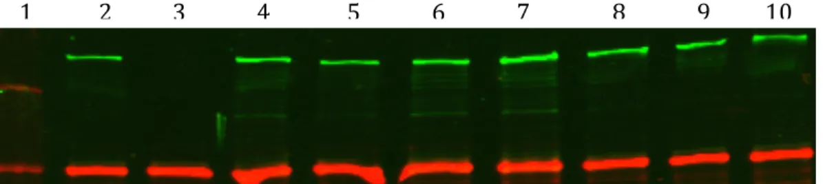
Figure 5. The ChpA receiver domain mutant does not alter ChpA protein
expression. Lane one is Precision Plus Protein Kaleidoscope ladder with 250kDA top band. Lane 2 is wild type PAK carrying empty vector (pJNtac4), lane two is an isogenic
chpA deletion strain (ΔchpA) carrying empty vector, lane 3 is a chpA deletion strain
complemented with plasmid-encoded wildtype ChpA (pJNtac4-chpA), lane 4 is a chpA deletion strain complemented with an alanine substitution at the receiver domain site phosphorylation, ChpArec D2407A (pJNtac4-chpArec D2407A). Green bands represent ChpA (260kDA) and red bands are an RNA polymerase standard. ChpA expression levels in lanes 3 and 4 are complemented to wild type ChpA in the presence of 75 µg/ml gentamycin for plasmid maintenance and 25 µM IPTG for induction of plasmid
expression.
Figure 8. Mutation of multiple ChpA domains may modulate ChpA protein expression. Lane one is Precision Plus Protein Kaleidoscope ladder with 250kDA top band. Lane 2 is wild type PAK, lane 3 is an isogenic chpA deletion strain (ΔchpA), lane 4 chromosomal ChpArecDA mutant, lane 5 is a chromosomal ChpA Hpt 2 mutant, lane 6 is a chromosomal ChpA recDA and Hpt 2 double mutant, lane 7 is a chromosomal ChpA Hpt 3 mutant, lane 8 is a chromosomal ChpA recDA and Hpt 3 double mutant, lane 9 is a Hpt23 double mutant, lane 10 is a Hpt23 rec triple mutant. Green bands were probed with a ChpA specific monoclonal antibody (260kDA) green bands were probed with a anti E. coli RNA polymerase anitbody. Strains were grown in plain LB and are whole cell
lysates of those assayed in Figure 5.