ScholarWorks @ Georgia State University
ScholarWorks @ Georgia State University
Chemistry Theses Department of Chemistry
Spring 4-27-2011
Conformational Bias in 2'-Selenium-Modified Nucleosides and the
Conformational Bias in 2'-Selenium-Modified Nucleosides and the
Effect on Helical Structure and Extracellular Recombinant Protein
Effect on Helical Structure and Extracellular Recombinant Protein
Production: Current Systems and Applications
Production: Current Systems and Applications
Richard A. Thompson
Follow this and additional works at: https://scholarworks.gsu.edu/chemistry_theses
Recommended Citation Recommended Citation
Thompson, Richard A., "Conformational Bias in 2'-Selenium-Modified Nucleosides and the Effect on Helical Structure and Extracellular Recombinant Protein Production: Current Systems and Applications." Thesis, Georgia State University, 2011.
https://scholarworks.gsu.edu/chemistry_theses/37
This Thesis is brought to you for free and open access by the Department of Chemistry at ScholarWorks @ Georgia State University. It has been accepted for inclusion in Chemistry Theses by an authorized administrator of
EFFECT ON HELICAL STRUCTURE AND EXTRACELLULAR RECOMBINANT
PROTEIN PRODUCTION: CURRENT SYSTEMS AND APPLICATIONS
by
RICHARD ADAM THOMPSON
Under the Direction of Dr. Markus W. Germann
ABSTRACT
Part One. X-ray crystallography has benefited from the synthetic introduction of selenium to different positions within nucleic acids by easing the solving of the phase problem. Interestingly,
its addition to the 2' position of the ribose ring also significantly enhances crystal formation.
This phenomenon was investigated to describe the effect of selenium-based and other 2'
modifications to the ribose ring of nucleosides in solution, as well as the incorporation of the
selenium-modified nucleotides into a helical structure. This work correlates the difference in
conformation propensity between the selenium containing nucleosides and oligomers towards a
rationale behind the enhanced crystal forming behavior. Part Two. Recombinant protein production is a critical tool in laboratories and industries, and inducing extracellular transport of
these products to the culture medium shows potential for improving cases where the yields are
not sufficient in quality or quantity. This review incorporates current practices and systems with
future perspectives.
EFFECT ON HELICAL STRUCTURE AND EXTRACELLULAR RECOMBINANT
PROTEIN PRODUCTION: CURRENT SYSTEMS AND APPLICATIONS
by
RICHARD ADAM THOMPSON
A Thesis Submitted in Partial Fulfillment of the Requirements for the Degree of
Master of Science
in the College of Arts and Sciences
Georgia State University
Copyright by Richard Adam Thompson
ON HELICAL STRUCTURE AND EXTRACELLULAR RECOMBINANT PROTEIN
PRODUCTION: CURRENT SYSTEMS AND APPLICATIONS
by
RICHARD ADAM THOMPSON
Committee Chair: Dr. Markus W. Germann
Committee: Dr. Zhen Huang
Dr. Jenny Yang
Electronic Version Approved:
Office of Graduate Studies
College of Arts and Sciences
Georgia State University
DEDICATION
ACKNOWLEDGEMENTS
Countless thanks to Dr. Germann for being accessible and accommodating to me throughout
the pursuit of this degree, my fellow group members for their various assistance in my practical
education, and to my family, friends, and colleagues who make up the support system I've relied
TABLE OF CONTENTS
ACKNOWLEDGEMENTS ... v
LIST OF TABLES ... x
LIST OF FIGURES ...xi
1 INTRODUCTION ... 1
1.1 Introduction to Nucleic Acid Structure ... 1
1.2 The Concept of Pseudorotation ... 1
1.3 Nucleic Acids in X-ray Crystallography ... 2
1.4 Selenium Modifications in Biopolymers ... 3
1.5 Goals of This Study ... 4
2 MATERIALS AND METHODS ... 5
2.1 Nucleoside Studies ... 5
2.2 Computational Parameters ... 5
2.3 Melting Temperature Assays ... 6
2.4 Ethidium Bromide Fluorescence ... 7
2.5 Imino Proton Observation ... 7
2.6 Model Development ... 7
3 RESULTS ... 8
3.1 Data Fitting and NMR Assignments ... 8
3.2 Nucleoside Characterization via Pseudorotation Calculations ...11
3.3 Duplex Stability ...14
4 CONCLUSIONS ...20
5 REFERENCES ...22
6 Appendix ...25
6.1 Appendix A - Reconstructed PSEUROT Batch File ...25
6.2 Appendix B – Inputs and Results for PSEUROT 6.0 Calculations ...26
6.2.1 2'-Deoxyuridine ...26
6.2.2 Uridine ...29
6.2.3 2'-Methoxy-Uridine ...30
6.2.4 2'-Fluoro-Deoxyuridine ...31
6.2.5 2'-Methylthio-Deoxyuridine ...33
6.2.6 2'-Selenomethyl-Deoxyuridine ...35
6.3 Appendix C - Compiled Matlab Results ...37
6.3.1 2'-Deoxyuridine ...37
6.3.2 Uridine ...39
6.3.3 2-Methoxy-Uridine ...41
6.3.4 2'-Fluoro-Deoxyuridine ...43
6.3.5 2’-Methylthio-Deoxyuridine ...45
6.3.6 2'-Selenomethyl-Deoxyuridine ...47
7 INTRODUCTION ...50
7.1 The Central Dogma of Molecular Biology ...50
7.3 Diversity in Environment and Function ...52
7.4 Commercial Protein Production and the Perspective of this Manuscript ..54
8 PROKARYOTIC PROTEIN EXPORT ...56
8.1 Prokaryotic Cell Biology ...56
8.2 Prokaryotic Secretion Mechanisms ...57
8.2.1 Type I Secretion ...57
8.2.2 Type II Secretion ...59
8.2.3 Type III Secretion ...62
8.2.4 Types IV - VII ...65
9 EUKARYOTIC PROTEIN EXPORT ...68
9.1 Basic Eukaryotic Cell Biology ...68
9.2 Protein Sorting/ Targeting ...70
9.2.1 Signal Recognition Protein ...72
9.2.2 Tail-Anchored Proteins ...73
9.2.3 Post-translational Modification and Regulation ...74
10 CURRENT SYSTEMS FOR RECOMBINANT PROTEIN SECRETION ...75
10.1 Escherichia coli ...76
10.2 Streptomyces lividans ...78
10.3 Saccharomyces cerevisiae ...79
10.4 Pichia pastoris ...81
10.6 Insect and Mammalian Platforms ...84
11 CHALLENGES OF RECOMBINANT PROTEIN PRODUCTION ...85
11.1 Protein Misfolding ...85
11.2 Disulfide Bond Formation ...86
11.3 Codon Usage and Discrepancies ...87
11.4 Other Machinery Bottlenecks ...88
11.5 Scale-Up ...90
12 PRACTICAL APPLICATIONS ...93
12.1 Pharmaceutical Production ...94
12.2 Live-Vaccine Therapeutics ...97
12.3 Energy Production ...98
12.4 Spider Silk Monomers ... 102
13 POTENTIAL AVENUES FOR FUTURE RESEARCH ... 103
LIST OF TABLES
Table 1.1 Duplex sequences for NMR and TM studies, USe is compound 6... 5
LIST OF FIGURES
Figure 1.1 Pseudorotation Wheel and Sugar Puckering Conventions. ... 2
Figure 1.2 Modified Nucleosides used in this study. ... 4
Figure 3.1 COSY spectrum of 2'-methylseleno-deoxyuridine ... 9
Figure 3.2 Simulation vs. NMR Data. ...10
Figure 3.3 Relationship between 3J3’-4’ coupling constant and %S conformation ...14
Figure 3.4 Duplex stability from ethidium bromide fluorescence.. ...15
Figure 3.5 Duplex stability from UV Melting curves. ...16
Figure 3.6 Imino proton spectra of sequences II and III at 288 K. ...17
Figure 3.7 2’ Se-CH3 groups incorporated in A and B helical structures...19
Figure 7.1 Central Dogma of Molecular Biology...51
Figure 7.2 Co and Post-Translational Secretion Mechanisms. ...54
Figure 8.1 Mechanism of protein export by T1SS. ...58
Figure 8.2 Comparison of T2SS mechanisms ...62
Figure 8.3 Illustration of the T3SS/ Host Cell Conjugate Apparatus ...64
Figure 8.4 Overview of Known Bacterial Secretion Systems ...68
Figure 9.1 Overview of Eukaryotic Cell Biology. ...69
0
PART ONE
1 INTRODUCTION
1.1 Introduction to Nucleic Acid Structure
Nucleic acids are a vital component of any biological system and as such they are
very widely studied. The cellular processes that nucleic acids and their constituents are
a part of span from information or energy storage to catalysis or intercellular signaling.
The driving force behind many nucleic acid functions, from drug or protein binding4-6 to
regulation of transcription or replication7, is its structure. A monomer nucleotide is
characterized by a nucleosidic base, a ribose sugar and a phosphate linker moiety, all
of which can have an effect on the macroscopic structure of the molecule, but as the
ribose units connect the base to the phosphate backbone, and being a five-membered
ring prone to dynamic switching, or puckering, between conformations, the character of
the sugar has a heavy influence on the overall configuration, especially in a
double-stranded helix.
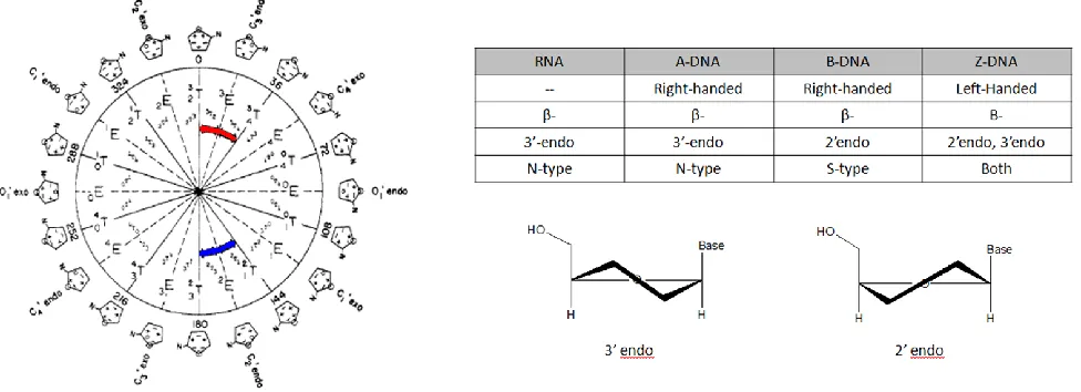
1.2 The Concept of Pseudorotation
The characterization of this non-planarity of a ring system using the concept of
pseudorotation was done on cyclopentane first by Kilpatrick et al.8 and followed by
others9 who deduced the dynamic nature of these rings through various thermodynamic
data. The concept was expanded by Altona and Sundaralingam who combined this
concept with X-ray crystallography data to relate the five intracyclic torsion angles of a
nucleosidic sugar in two pseudorotation parameters: phase angle (P) and puckering
amplitude (Φm). They showed the rings essentially exist as two main types, North
(3'-endo) and South (2'-(3'-endo), as designated by their phase angle1 (Fig. 1.1). They further
values10-11 to describe the two state equilibrium that furanose rings exhibit and through
various refinements have allowed for quantification of the percentage of either form that
exists at equilibrium.12 No experimental data has suggested the use of a third
pseudorotation parameter, and it has since also been shown that different forms of
nucleic polymers prefer different sugar conformations.13 A brief overview of trends in
different nucleic acid structures is given in Figure 1.1. The Altona-Sundaralingam (AS)
formalism is a powerful tool for extracting structural information based on coupling
constants, which will be employed later.
1.3 Nucleic Acids in X-ray Crystallography
Since many techniques are used to investigate structural properties of nucleic
[image:16.612.64.552.78.255.2]develop a clear understanding of molecular processes. X-ray crystallography has been
used to much success to study structural characteristics of many biological
macromolecules, but to overcome the difficulties of crystallization and phase
determination force crystallographers to alter the natural structure of these
macromolecules with heavy-atom soakings or modifications. An additional problem in
this case is that methods that are very effective with proteins have been proven to be
much more difficult in DNA and RNA.14 For instance, bromine derivatization can be
problematic because it acts as a good leaving group and can attract nucleophilic attack
if it is positioned anywhere on the furanose ring. Bromine addition to the base can lead
to decomposition when exposed to UV light, as exhibited in photo-crosslinking of nucleic
acids to proteins in order to determine contact points.15
1.4 Selenium Modifications in Biopolymers
Interestingly, the incorporation of selenium atoms into a macromolecule, which has
been shown to work well in protein crystal samples,16 has recently been explored by
Huang et al. as a method for DNA or RNA structural investigation.17-19 They have
reported minimal disruption of structure between crystal structures of unmodified DNA
oligomers and derivatives with 2'-selenomethyl and 5-bromine modifications, and the
sugar pucker of all of these molecules are found to be A-form DNA, having a 3'-endo
conformation.14 More remarkably, they also report a much quicker rate of crystal
formation with the selenomethyl modification than the bromine derivative or the
unmodified control,20 which raises questions about the effect the methyl-selenium
modification has on crystal stability or desolvation rates considering the sugar pucker is
that in solution-based biophysical studies of nucleic acids, a 2' substituent is a
determinant of the sugar conformation and its dynamics,21-22 so its effect on crystal
formation could be a result of this.
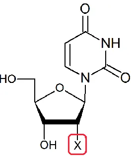
1.5 Goals of This Study
This work looks at the 2'-methylseleno derivative of uridine free in solution to
determine its propensity towards one conformation or the other and to compare this
behavior to that of other 2'-uridine substitutions. It also addresses the structural origin
for the facilitated crystal formation by investigating the duplex structures containing
selenium modifications using NMR, melting temperatures, ethidium bromide
fluorescence and molecular modeling. The nucleoside behavior is then compared to the
crystal structures and other data of the selenium modified oligomers. The library of 2'
substituted uridines is described in Figure 1.2, while the sequences used in the oligomer
[image:18.612.242.374.77.235.2]studies are presented in Table 1.1.
Table 1.1 Duplex sequences for NMR and TM studies, USe is compound 6
I 5‟-d(CATGCATG)
II 5‟-d(GCGAATTCGC)
III 5‟-d(GCGAAUSeTCGC)
IV 5‟-d(CGCGAATTCGCG)
2 MATERIALS AND METHODS
2.1 Nucleoside Studies
The sulfur and selenium based modifications were prepared as reported,14,23 and all
other compounds were purchased from Tech Chem. Nucleoside experiments were
performed with a Bruker Avance 500 MHz spectrometer equipped with a TBI
triple-resonance broadband capable probe head at 298K. Samples were prepared to be 1.0
mM nucleoside in D2O with 10 mM sodium phosphate adjusted to pH* 6.0. DSS was
used as an internal standard. Routine 1D 1H NMR experiments with water presaturation
pulses were performed on each nucleoside in order to confirm purity of the samples and
to measure coupling constants. Double quantum filter COSY experiments (32 scans)
were recorded to confirm assignments. A low-flip angle COSY was recorded for
deoxyuridine (1) to clarify couplings caused by the 2' and 2" protons.
2.2 Computational Parameters
DAISYSIM, a component of Topspin 2.1 (Bruker), was used to simulate spectra
from the acquired NMR data in order to precisely determine the individual couplings and
chemical shifts. DAISYSIM refines coupling and chemical shift estimates by a
user-directed iteration algorithm. The refined coupling constants were used as the input into
practices. Also, in an attempt to move from a command line style of input to a more
modern, user-friendly, GUI-based computational method, a Matlab-based (Mathworks)
pseudorotation program was used for further substantiation.25 PSEUROT 6.0 has been
used to much success to calculate pseudorotation parameters of pentose rings from
NMR data in several instances26-28 and was provided by Altona and de Leeuw. The
Matlab program was provided through a GNU General Public License by Hendrickx and
Martins. The computation for each compound was initially set up with the conditions
described in the user's manual of PSEUROT 6.0. The initial %S conformer was varied in
subsequent trials in order to alleviate any bias built into the program with respect to
conformational preference. Each of these initial states was refined during the
computation by each program to give a theoretical pure N- and S-conformer population
which was used to fit the data. The change in electronegativity of the 2' substitutions
was accounted for in the input file; the values are derived from a Huggin's based
electronegativity scale referenced to hydrogen specifically for use with generalized
Haasnoot-Karplus equation as suggested by the authors of PSEUROT 6.0.29-32 The
Matlab program, since it was designed with the same computational premises,
suggested the same values in the User's Manual.25 The input and output data from each
program are compiled in the Appendices.
2.3 Melting Temperature Assays
The melting assay was performed on the control and modified duplexes (II and III,
respectively), through absorbance monitoring at 274 nm. The buffer was prepared to
400 mM sodium chloride, 10 mM sodium phosphate, and 0.1 mM EDTA at pH 6.5. The
prepared in the same buffer with 32 μM DNA in order to quantify the effect of
concentration on the formation of a duplex. During the TM assay, the temperature was
reversibly ramped from 20oC to 90oC at 0.3oC/ min, controlled by a Cary spectrometer
and heating block.
2.4 Ethidium Bromide Fluorescence
Oligomer samples of increasing length (octamer: I, decamer: II, Se-decamer: III,
dodecamer: IV) were 15 µM in nucleotides or ~0.8 µM in duplex concentration and
contained 1 µg/mL ethidium bromide, 100 mM NaCl, 10 mM sodium phosphate and 0.1
mM EDTA at pH 6.5. Samples were individually placed into PCR tubes and imaged on a
Typhoon 9400 Variable Mode Imager from Amersham Biosciences. Excitation for
imaging occurred at 532 nm and emission was measured at 610 nm.
2.5 Imino Proton Observation
NMR samples of sequences II and III were prepared at 50mM sodium chloride, 10
mM sodium phosphate, 0.1 mM EDTA and pH 6.4 in 9:1 H2O:D2O. Imino proton spectra
were recorded on an Avance 600 MHz spectrometer using jump and return water
suppression according to established practice.33 Selenium samples (sequence III) were
prepared at strand concentrations of 100 and 20 µM (designated high and low,
respectively). The control sequence was prepared at 250 µM, in order to minimize
acquisition time.
2.6 Model Development
Standard A- and B-form DNA helical models of sequence III were built within
secondary structure. The A-form set the sugar puckering as N–type and with a rise and
twist of 2.548 Å and 32.7o per base, respectively, as described in the Spartan manual.
The second model was made to be B-form (S-type, 3.375 Å, 36o). After the models
were built, the modification was inserted into the 2‟ position.
3 RESULTS
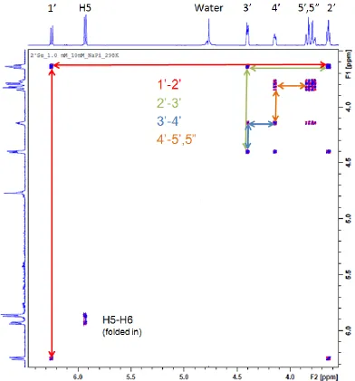
3.1 Data Fitting and NMR Assignments
In order to obtain an accurate description of the relevant coupling constants from
the NMR data, the spectra were simulated from within TOPSPIN 2.1. In order for the
simulation to be properly designed, the proper assignment of residues is critical. 2-D
COSY spectra for each compound in Figure 1.2 were obtained to fully assign the peaks
with high confidence. The correlation of resonances seen in the spectra was used to
fully assign the sugar ring protons. The diagonal peaks arise from the peaks in each
dimension seeing themselves (i.e. y = x), and from the diagonal one can determine
which other peaks are within 3 bonds of the peak of interest. Knowing that the 1‟ proton
should only see one resonance, one can follow the rest of the correlation pathway
around the ring. Figure 3.1 shows the COSY spectrum of compound 6, with the pathway
highlighted. This strategy was repeated for each compound in this study and the full
assignment of the ring protons was determined. Using the assignments, the inputs for
the simulations were created.
Using DAISYSIM, a spin system simulation was fit to the NMR data according to
a qualitative assessment by the user, i.e. if the simulation has not been accurately laid
fashion until the simulation fits the data appropriately. This trial and error type method
worked well in this situation but might not be the most effective way to determine
obscure coupling constants from complicated systems. Nevertheless, this method was
able to simulate the data to a high level of accuracy, although there was not an RMSD
value returned by the fitting program, the experimental data and the simulation
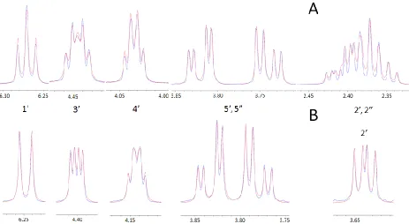
[image:23.612.110.502.71.492.2]1-D 1H spectra of compounds 1 and 6. Upon first looking at the spectra, without
considering the COSY spectra, the splitting patterns make sense when comparing the
two. The 1‟ proton is split by two signals in the spectrum of compound 1, corresponding
to the 2‟ and 2” protons. In compound 6‟s spectrum, the 1‟ peak is only split by one
proton, at the 2‟ position, because the 2” proton has been replaced by the methylseleno
group in compound 6. The 3‟ signal in 1‟s spectrum is split by an extra signal as well, as
evident when comparing to 6, following the same logic. This observation combined with
the COSY spectra gives a high level of confidence in the data obtained from the
simulations.
[image:24.612.75.538.72.326.2]3.2 Nucleoside Characterization via Pseudorotation Calculations
The data from the NMR experiments is the backbone of the subsequent study,
and all of the coupling values obtained by fitting the raw data in DAISYSIM were used in
the pseudorotational calculations. The experimental data is compiled in Table 3.1 and
shows reasonable correlation with literature reports. The trends in 3J values and
chemical shift with respect to substituent identity begin to reveal themselves even
before pseudorotation parameters are calculated, and hint at the behavior of the sugar
ring and the effects of the different modifications. Specifically, the 3J1‟-2‟ and 3J3‟-4‟ values,
which arguably are the most affected by a change from 2‟-endo to 3‟-endo
conformations, are the most dynamic of the data collected and sets the stage for
explanation through a pseudorotation perspective. The fitted coupling data was used as
the input parameters for the calculation of pseudorotation values as described in
Section 2.2. Since all endocyclic coupling constants were known, the discrepancies
arising from the mathematical determination of pseudorotation parameters, i.e. five
torsion angle expressions with five variables, were minimized by eliminating solutions
which did not fit within the whole set of equations. The optimized conformations and the
percent of each were similar between PSEUROT and Matla and correlated with
published results.23,34-37 In Table 3.1, the top portion tabulates the NMR data, while the
bottom portion shows the output from PSEUROT 6.0 (PS) and the Matlab program
(ML). The pseudorotation data is also compiled in Table 3.1. It is interesting to see how
the two starting conformations do not differ much between compounds, (the range is
roughly 50o) but the percent S conformation varies significantly. The data is believable
electro-12
Table 3.1 Compiled NMR and Pseudorotation Data
X H OH OCH3 F* SCH3 SeCH3
Exp Lit35 Exp Lit35 Exp Lit34 Exp Lit35 Exp Lit23 Exp
J1'-2' 7.2 6.3 4.5 4.2 3.9 3.6 1.4 1.5 8.3 8.5 8.7
J 1'-2'' 6.1 6.4 19.7 19.7
J 2'-3' 6.9 6.3 5.3 5.3 5.2 5.0 5.1 5.8 5.5 5.7
J2''-3' 3.9 4.3 21.5 21.6
J 3'-4' 3.9 4.0 5.5 5.7 5.7 8.6 8.7 2.8 2.0 2.9
J H5-H6 8.1 8.1 8.1 8.0 8.1 8.3 8.1 8.1 8.1 8.1
δ2' 2.4 2.4 4.3 4.3 4.1 5.2 5.2 3.6 3.4 3.6
δ4' 4.0 4.0 4.1 4.1 4.1 4.2 4.1 4.2 3.9 4.1
PS ML Lit35 PS ML Lit 35 PS ML Lit 34 PS ML Lit 35,36 PS ML Lit 23,37 PS ML
I.
N Type
P 18.0 2.3 18 32.4 13.5 18 12.5 34.4 11 28.6 36.1 21 -22.5 50.6 -- -13.3 7.2
ΦM 38.0 33.5 -- 32.0 30.5 -- 32.0 35.5 35 32.0 34.2 -- 32.0 16.7 -- 32 20.3
II.
S type
P 141.5 149.7 162 156.6 129.40 162 144.7 162.7 171 38.6 -4.0 159 138.7 127.1 -- 137.6 134.7
ΦM 32.3 22.9 -- 35.0 41.70 -- 35.0 30.9 37 35.0 34.2 -- 35.0 45.1 -- 35.0 40.4
negativity will drive the system into a state with a higher %N conformation,38 as 1 favors
a predominately S mixture while 2, 3, and 4 prefer an increasingly N mixture.
Compound 4, despite varying the starting conditions with different starting mixtures,
converged to „equilibrium‟ where both conformers were of N-type, essentially implying
that the S-type conformer does not exist free in solution under these conditions. Both
PSEUROT and Matlab returned this output. The literature shows that chlorine and
bromine substitutions fit the overall relationship between electronegativity and percent
S; the report of a 50-50 mixture of conformers makes sense36 as these atoms have an
electronegativity value less than oxygen but more than hydrogen. This trend is no
longer observed, however, when considering the sulfur and selenium based
compounds. Compounds 5 and 6 are found to more strongly prefer the S conformation
than compound 1 in solution, which is the opposite of what the electronegativity or
crystal structures suggest. Since the programs correlate well with literature results, a
computational error is unlikely and an inference can be made that steric effects between
the 2' substituents and the base drive the preference of the S conformation. There is
strong correlation between various NMR data points and the %S value, which is an
intrinsic principle of the programs themselves, but suggests that reasonable prediction
of sugar puckering dynamics can be made from raw NMR data. As stated above, the
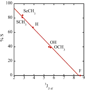
3
J1‟-2‟ and 3J3‟-4‟ values are the most dynamic because they are the most affected by a
change from 2‟-endo to 3‟-endo conformations. Especially relevant is the 3
J3‟-4‟ couplings
because they are affected by the ring dynamics, and would be only minimally impacted
by the 2'-substituent identity and when plotted against %S, as in Figure 3.3, show linear
and works when individual couplings are known. These results clearly establish that
2'-SeCH3-modified nucleosides strongly prefer a 2'-endo conformation in solution.
However, this is exactly the opposite of what is observed in the crystal structures. This
discrepancy was an interesting revelation and prompted further investigation of
selenium-containing nucleosides within a duplex in solution.
3.3 Duplex Stability
In order to gain perspective on the physical effects of the 2‟-SeCH3-modification
[image:28.612.144.472.73.418.2]acquired and examined. The results of the ethidium bromide intercalation study (Figure
3.4) showed a clear discrepancy in the fluorescence of the different samples. The
fluorescence intensity typically increases as a function of oligonucleotide length as more
intercalation sites are possible in longer sequences. The unmodified octamer, decamer,
and dodecamer exhibited this behavior. However, the Se-decamer displayed a lower
fluorescence than the octamer, alluding to the destabilization effects of the modification.
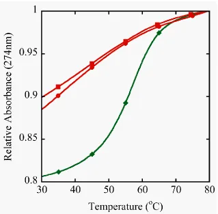
The difference in melting curves between the unmodified decamer and the
selenium decamer is also immediately noticed. The stability of the self complementary
sequence III containing one 2‟-SeCH3-modification and its control II was determined by
UV melting (Figure 3.5). The control duplex forms a standard B-type helical structure
and exhibits a regular melting profile with an expected stability.40-41 On the other hand,
the shifted and shallow melting curve for the DNA strand III containing a single
2‟-SeCH3-modification demonstrates through a change in hyperchromicity that duplex
formation was seriously destabilized. The observation that the denaturation was not
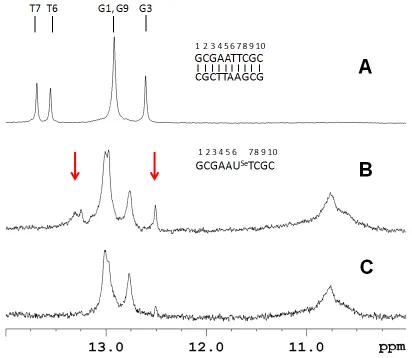
[image:29.612.83.537.74.216.2]hairpin structures. This data makes sense when compared to the imino proton spectra.
The unmodified decamer II is shown to have five imino proton resonances, consistent
with the C2 rotational symmetry of a duplex structure (Figure 3.6A). In contrast
sequence III, at 100 µM strand concentration, showed more imino proton resonances
than would be expected for a duplex (Figure 3.6B). This strongly indicates the presence
of multiple structures. Of note, there are resonances near 10.8 ppm that are generally
associated with unpaired hairpin loop resonances.42-43 If duplex III is examined at 20 μM
strand concentration the spectrum simplifies and essentially only 3 GC base pairs are
observed in addition to the hairpin loop resonances (Figure 3.6C). Under these Figure 3.5 Duplex stability from UV Melting curves. Samples were prepared in 400 mM NaCl, 10 mM NaPi, 0.1 mM EDTA at pH 6.5. The unmodified control decamer (II,♦) at 8.5 µM
showed a TM of 59oC and two different concentrations of the selenium decamer (III) were
[image:30.612.148.467.70.383.2]conditions the predominant species is a hairpin structure with a stem consisting of 3 GC
base pairs. Salon et al. have previously demonstrated complete base pairing for a
non-self-complementary duplex containing a single 2‟-Se-modification.44
However the
stability was compromised in this construct as well and homoduplex formation of the
individual strands was observed at elevated temperatures. Taken in context, all of this
[image:31.612.104.516.76.434.2]solution, even though this modification has an intrinsic preference for a southern sugar
conformation, as demonstrated by the nucleoside study.
3.4 Crystal Structures and Models
Zhen Huang's group has made several selenium modified nucleosides and
solved crystal structures with these analogs incorporated into a DNA helix (pdb: 1MA8,
3IFF, 2NSK, 2HC7, 2DLJ).14,20 In every case, whether the 2'-selenomethyl-modification
is present or not, the helix exhibits A-type, predominately N-sugar behavior. Even in
1MA8, where the selenomethyl groups are opposite and adjacent to each other, which
was shown above to not exist in solution at room temperature, they situate themselves
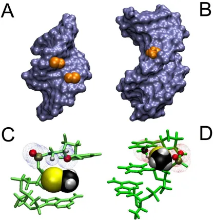
into the minor groove in a complete duplex (Figure 3.7a). Considering how deoxyuridine
and 2'-methylseleno-uridine both prefer the S-conformation in solution, there has to be a
driving force behind this change in overall sugar conformation. To obtain further insight
why 2‟-methylseleno-uridine (6) adopts a northern conformation when part of a DNA
duplex, as seen in crystal structures, standard A- and B- type DNA helical models45 with
the appropriate modifications (i.e. sequence III) were investigated. The A-type helix
containing the Se-modification was homologous to the crystal structures published in
the literature. There are no steric clashes with the backbone, neighboring bases or
deoxyribose ring. In contrast, in the B-helical model the modification is situated in the
major groove, but the 3‟ phosphate as well as the base on the 3‟ side of the modification
clash with selenomethyl group. This is especially apparent when the 3‟ base is thymine
whose methyl group is also in the major groove. Therefore, a base with a smaller
footprint in the major groove would be expected to be less perturbed which agrees with
Figure 3.7 2’ Se-CH3 groups incorporated in A and B helical structures. Panels A and B
represent Connelly surfaces with the Se-CH3 group depicted as VdW representation in orange.
In panel C and D the bases and sugars are shown in green and Se-CH3 is depicted as VdW
spheres. A) The group is nestled comfortably in the minor groove of an A-type helix (pdb: 1MA8). C) No clashes are apparent between the 3‟ neighboring residue and Se-CH3 . The blue
dotted spheres depict VdW spheres of close atoms. B) In a B-type helical model (dGCGAAUSeTCGC) the Se-CH3 group points away from the major groove but experiences
[image:33.612.99.516.93.522.2]reiterate, the B-type model is intended to be a qualitative picture of how the selenium
modification affects the stability of a B-type helix. There were no molecular dynamics
simulations because there is no experimental data from which restraints could be
obtained.
4 CONCLUSIONS
The notion that the southern sugar conformation is not tolerated well in a B-helix
because of steric interactions explains the solution-based structural data but could also
rationalize the enhanced crystal growth. DNA∙RNA hybrids have been shown to form
A-type structures, and alkylation of the 2‟-hydroxyl group has been shown to increase the
stability of these structures by lowering the intrinsic nucleophilicity of the hydroxyl group
and altering the hydrogen bonding pattern. Addition of a methyl group no longer allows
the hydroxyl group to act as a donor in a hydrogen bonding pair, and can now only
accept hydrogen bonds. This effect has been said to drive local structure towards an
A-type helix.21 Divalent selenium atoms, as in compound 6, can form hydrogen bonds with
donor atoms, but because selenium has limited ability for induced dipoles due to its
size, these bonds are very weak46 and most likely has an influence on the hydrogen
bonding network around the modification. Also, it has been proposed that the hydration
of the minor groove will be affected by the pattern of purines and pyrimidines when a
2‟-modification is made,21 but recent data showed that the nature of the base of the
modified nucleotide is not a determining factor as enhanced crystal growth was also
observed for other 2‟-selenomethyl-modified nucleotides.47-49
Thus, the following can be
A single 2‟-selenomethyl group narrows the conformational space by destabilizing
the B-helical form while promoting A-helix formation. Moreover, the 2‟-methylseleno
group fits snuggly into the minor groove of an A-helix and can serve as the origin for a
B- to A- conversion, which is also aided by dehydration during crystallization. In
addition, the 2‟-selenomethyl group locally dehydrates the minor groove which further
facilitates the crystallization process. The impact of this behavior suggests that this
modification could be used in samples that have been difficult to crystallize for their
structural determination, yet consideration must be given to the fact that the
conformational bias imparted by the modification could disrupt the normal behavior of
the sample in solution. Positioning the modification in a place within a hairpin loop or on
the 5‟ side of a less-bulky purine base are the least likely to distort the structure by steric
5 REFERENCES
1 Altona, C. & Sundaralingam, M. Conformational analysis of the sugar ring in nucleosides and nucleotides. New description using the concept of pseudorotation. JACS94, 8205-8212, (1972).
2 Rich, A., Nordheim, A. & Wang, A. H. The chemistry and biology of left-handed Z-DNA.
Annu Rev Biochem53, 791-846, (1984).
3 Sinden, R. R. DNA Structure and Function. (Academic Press, Inc., 1994).
4 Du, Y. H., Huang, J., Weng, X. C. & Zhou, X. Specific Recognition of DNA by Small Molecules. Curr. Med. Chem.17, 173-189 (2010).
5 Segal, D. J., Dreier, B., Beerli, R. R. & Barbas, C. F. Toward controlling gene expression at will: Selection and design of zinc finger domains recognizing each of the 5 '-GNN-3 ' DNA target sequences. PNAS USA96, 2758-2763 (1999).
6 Tolstorukov, M. Y., Jernigan, R. L. & Zhurkin, V. B. Protein-DNA hydrophobic recognition in the minor groove is facilitated by sugar switching. J. Mol. Biol.337, 65-76, (2004). 7 Hiratani, I. & Gilbert, D. M. Replication timing as an epigenetic mark. Epigenetics4,
93-97 (2009).
8 Kilpatrick, J. E., Pitzer, K. S. & Spitzer, R. The Thermodynamics and Molecular Structure of Cyclopentane. JACS69, 2483-2488, (1947).
9 Adams, W. J., Geise, H. J. & Bartell, L. S. Structure, equilibrium conformation, and pseudorotation in cyclopentane. An electron diffraction study. JACS92, 5013-5019, (1970).
10 Altona, C. & Sundaralingam, M. Conformational analysis of the sugar ring in nucleosides and nucleotides. Improved method for the interpretation of proton magnetic resonance coupling constants. JACS95, 2333-2344 (1973).
11 Karplus, M. Vicinal Proton Coupling in Nuclear Magnetic Resonance. JACS85, 2870-2871 (1963).
12 Vanwijk, J., Huckriede, B. D., Ippel, J. H. & Altona, C. Furanose Sugar Conformations in DNA from NMR Coupling Constants. Methods Enzymol.211, 286-306 (1992).
13 Leslie, A. G., Arnott, S., Chandrasekaran, R. & Ratliff, R. L. Polymorphism of DNA double helices. J Mol Biol143, 49-72, (1980).
14 Jiang, J., Sheng, J., Carrasco, N. & Huang, Z. Selenium derivatization of nucleic acids for crystallography. Nucleic Acids Res35, 477-485 (2007).
15 Meisenheimer, K. M. & Koch, T. H. Photocross-linking of nucleic acids to associated proteins. Crit. Rev. Biochem. Mol. Biol.32, 101-140 (1997).
16 Hendrickson, W. A., Horton, J. R. & LeMaster, D. M. Selenomethionyl proteins produced for analysis by multiwavelength anomalous diffraction (MAD): a vehicle for direct
determination of three-dimensional structure. EMBO J9, 1665-1672 (1990). 17 Du, Q. et al. Internal Derivatization of Oligonucleotides with Selenium for X-ray
Crystallography Using MAD. JACS124, 24-25, (2001).
18 Sheng, J., Jiang, J., Salon, J. & Huang, Z. Synthesis of a 2'-Se-thymidine
phosphoramidite and its incorporation into oligonucleotides for crystal structure study.
Org Lett9, 749-752, (2007).
19 Sheng, J. & Huang, Z. Selenium derivatization of nucleic acids for X-ray crystal-structure and function studies. Chem Biodivers7, 753-785, (2010).
20 Sheng, J. & Huang, Z. Selenium Derivatization of Nucleic Acids for Phase and Structure Determination in Nucleic Acid X-ray Crystallography. Int J Mol Sci9, 258-271 (2008). 21 Lubini, P., Zürcher, W. & Egli, M. Stabilizing effects of the RNA 2'-substituent: crystal structure of an oligodeoxynucleotide duplex containing 2'-O-methylated adenosines.
22 Rozners, E. Carbohydrate Chemistry for RNA Interference: Synthesis and Properties of RNA Analogues Modified in Sugar-Phosphate Backbone. Curr. Org. Chem.10, 675-692 (2006).
23 Fraser, A., Wheeler, P., Cook, P. D. & Sanghvi, Y. S. Synthesis and conformational properties of 2'-deoxy-2'-methylthiopyrimidine and 2'-deoxy-2'methylthio-purine nucleosides: Potential antisense applications. J. Heterocycl. Chem.30, 1277-1287 (1993).
24 de Leeuw, F. A. A. M., Altona, C. Computer-Assisted Pseudorotation Analysis of Five-membered Rings by Means of Proton Spin-Spin Coupling Constants: Program
PSEUROT. J. Comput. Chem.4, 428-437 (1983).
25 Hendrickx, P. & Martins, J. A user-friendly Matlab program and GUI for the
pseudorotation analysis of saturated five-membered ring systems based on scalar coupling constants. Chemistry Central Journal2, 20 (2008).
26 Houseknecht, J. B., Altona, et al. Conformational Analysis of Furanose Rings with PSEUROT: Parametrization for Rings Possessing the Arabino, Lyxo, Ribo, and Xylo Stereochemistry and Application to Arabinofuranosides. J. Org. Chem. 67, 4647-4651, (2002).
27 Rosemeyer, H. et al. Stereoelectronic effects of modified purines on the sugar
conformation of nucleosides and fluorescence properties. Nucleosides Nucleotides Nucl. Acids16, 821-828 (1997).
28 Watts, J. K., Sadalapure, K., et al. J. Synthesis and Conformational Analysis of
2„-Fluoro-5-methyl-4„-thioarabinouridine (4„S-FMAU). J. Org. Chem.71, 921-925 (2006). 29 Altona, C. et al. Empirical group electronegativities for vicinal NMR proton-proton
couplings along a C-C bond: solvent effects and reparameterization of the Haasnoot equation. Magn. Reson. Chem.32, 670-678 (1994).
30 Altona, C. et al. Relationship between proton-proton NMR coupling constants and substituent electronegativities. V. Empirical substituent constants deduced from ethanes and propanes. Magn. Reson. Chem.27, 564-576 (1989).
31 Haasnoot, C. A. G., De, L. F. A. A. M., De, L. H. P. M. & Altona, C. The relationship between proton-proton NMR coupling constants and substituent electronegativities. Part II. Conformational analysis of the sugar ring in nucleosides and nucleotides in solution using a generalized Karplus equation. Org. Magn. Reson.15, 43-52 (1981).
32 Donders, L. A., Leeuw, F. A. A. M. d. & Altona, C. Relationship Between Proton-Proton NMR Coupling Constants and Subsituent Electronegativites IV: An Extended Karplus Equation Accounting for Interactions Between Substituents and its Application to Coupling Constant Data Calculated by the Extended Hueckel Method. Magn. Reson. Chem.27, 556-563 (1989).
33 Plateau, P. & Gueron, M. Exchangeable proton NMR without base-line distorsion, using new strong-pulse sequences. JACS104, 7310-7311 (1982).
34 Davies, D. B. & Danyluk, S. S. Nuclear magnetic resonance studies of 5'-ribo- and deoxyribonucleotide structures in solution. Biochemistry13, 4417-4434 (1974). 35 Guschlbauer, W. & Jankowski, K. Nucleoside Conformation is determined by the
Electronegativity of the Sugar Substituent. Nucleic Acids Res8, 1421-1433 (1980). 36 Joecks, A., Koeppel, H., Schleinitz, K. D. & Cech, D. NMR-spektroskopische
Untersuchengen zum Konformationsverhalten von 2'- und 3'-halogensubstituierten Pyrimidinnucleosiden. Journal fuer praktische Chemie325, 881 (1983).
38 Uesugi, S., Miki, H., Ikehara, M., Iwahashi, H. & Kyogoku, Y. A linear relationship between electronegativity of 2'-substituents and conformation of adenine nucleosides.
Tetrahedron Lett.20, 4073-4076 (1979).
39 Rinkel, L. T. & Altona, C. Conformational Analysis of the Deoxyribofuranose Ring in DNA by means of sums of proton-proton coupling constants: A Graphical Method. J. Biomol. Struct. Dyn.4, 621-649 (1987).
40 Aramini, J. M., Kalisch, B. W., Pon, R. T., vandeSande, J. H. & Germann, M. W. Structure of a DNA duplex that contains alpha-anomeric nucleotides and 3'-3' and 5'-5' phosphodiester linkages: Coexistence of parallel and antiparallel DNA. Biochemistry35, 9355-9365 (1996).
41 Aramini, J. M., Mujeeb, A. & Germann, M. W. NMR solution structures of d(GCGAAT-3 '-3 '-alpha T-5 '-5 '-CGC)(2) and its unmodified control. Nucleic Acids Res.26, 5644-5654 (1998).
42 Germann, M. W., Kalisch, B. W., Lundberg, P., Vogel, H. J. & van de Sande, J. H. Perturbation of DNA hairpins containing the EcoRI recognition site by hairpin loops of varying size and composition: physical (NMR and UV) and enzymatic (EcoRI) studies.
Nucleic Acids Res.18, 1489-1498 (1990).
43 Haasnoot, C. A. G., der Hartog, J. H. J., de Rooij, J. F. M., van Boom, J. H. & Altona, C. Loopstructures in synthetic oligonucleotides. Nucleic Acids Res.8, 169-181 (1980). 44 Salon, J., Chen, G., Portilla, Y., Germann, M. W. & Huang, Z. Synthesis of a
2„-Se-uridine Phosphoramidite and Its Incorporation into Oligonucleotides for Structural Study.
Org. Lett.7, 5645-5648 (2005).
45 Egli, M. Structural Aspects of Nucleic Acid Analogs and Antisense Oligonucleotides.
Angewandte Chemie International Edition in English35, 1894-1909 (1996). 46 Madzhidov, T. I. & Chmutova, G. A. The nature of hydrogen bonds with divalent
selenium compounds. Journal of Molecular Structure: THEOCHEM959, 1-7 (2010). 47 Salon, J., Sheng, J., Gan, J. & Huang, Z. Synthesis and crystal structure of
2'-Se-modified guanosine containing DNA. J Org Chem75, 637-641 (2010).
48 Sheng, J., Salon, J., Gan, J. H. & Huang, Z. Synthesis and crystal structure study of 2 '-Se-adenosine-derivatized DNA. Science China-Chemistry53, 78-85 (2010).
6 Appendix
6.1 Appendix A - Reconstructed PSEUROT Batch File
::This batch file was reconstructed from the version contained in a degraded copy of PSEUROT ::that had been copied multiple times over a few years. As I am not a computer programmer, ::I'm not entirely sure why it works, but it does. When using the command line context
::described in the manual, this .bat file correctly renames inputs and outputs for use in the ::PSEUROT program, and the output files are competent. PSEUROT is able to run to completion, ::which is stated at the end of the output files. However, the‘MANY’functionality does not ::work to completion.
@ECHO OFF
if '%1'=='' goto Usage copy %1 %1.inp
copy %1.inp pseurot6.inp
psrot62 <pseurot6.inp >pseurot6.out
if exist pseurot6.out copy pseurot6.out %1.out >NUL if exist pseurot6.mn1 copy pseurot6.mn1 %1.mn1 >NUL if exist pseurot6.mn2 copy pseurot6.mn2 %1.mn2 >NUL if exist pseurot6.mn3 copy pseurot6.mn3 %1.mn3 >NUL if exist pseurot6.mn4 copy pseurot6.mn4 %1.mn4 >NUL if exist pseurot6.mn5 copy pseurot6.mn5 %1.mn5 >NUL
:pkzip %1.zip %1.inp %1.mn1 %1.mn2 %1.mn3 %1.mn4 %1.mn5 :goto Einde :Usage
echo Usage: PS62 filename
echo where filename does not have an extension echo.
6.2 Appendix B – Inputs and Results for PSEUROT 6.0 Calculations
6.2.1 2'-Deoxyuridine
Trial 1 Input
dU
CTRL MAXIT 25 TRIM 0.1 RCNV 0.5 PRINT 1 DATA 5
1'-2' -144.0 1.030 121.4 0.72 1.27 0.00 0.62 1'-2" -144.0 1.020 0.9 0.72 1.27 0.62 0.00 2'-3' 0.0 1.060 2.4 0.62 0.00 1.26 0.62 2"-3' 0.0 1.060 122.9 0.00 0.62 1.26 0.62 3'-4' 144.0 1.090 -124.0 0.72 1.26 1.27 0.68 TSET 1
298 7.19 6.14 3.89 6.9 3.89
Trial 1 Output
+++++++++++++++++++++++++++++++++++++++++++++++++++++++ + PSEUROT v 6.0 March 1993 + + John van Wijk FAAM de Leeuw + + Gorlaeus Laboratories, State University of Leiden + +++++++++++++++++++++++++++++++++++++++++++++++++++++++
++++ CASE : 1 +++++++++++++++++++++++++++++++++++++++++++++ PSEUROT 6.0 +++++
TITLE: dU
The minimization has converged.
============================================================================== F I N A L O U T P U T ============================================================================== Total number of iterations: 9
CONFORMER I: CONFORMER II:
P == 18.0 ( .314 RAD) P == 184.6 ( 3.222 RAD) PHIM == 38.0 ( .663 RAD) PHIM == 59.7 ( 1.041 RAD)
PHIHH = 98.4 ==> JHH = 1.22 PHIHH = 168.0 ==> JHH = 11.24 PHIHH = -21.9 ==> JHH = 7.70 PHIHH = 47.1 ==> JHH = 4.11 PHIHH = 40.7 ==> JHH = 6.56 PHIHH = -60.6 ==> JHH = 2.04 PHIHH = 161.2 ==> JHH = 10.01 PHIHH = 59.9 ==> JHH = 3.50 PHIHH = -163.4 ==> JHH = 8.81 PHIHH = -68.5 ==> JHH = 1.58
TEMP SET 298 JEXP JCAL JDIF 1'-2' 7.19 7.07 .12 1'-2" 6.14 5.60 .54 2'-3' 3.89 3.92 -.03 2"-3' 6.90 6.21 .69 3'-4' 3.89 4.59 -.70 X(1)X(2) .42 .58 .505
ERROR ANALYSIS:
ROOT MEAN SQUARE DEVIATION OF THE FIT: .505 STANDARD DEVIATION IN PARAMETERS:
0 .208 .112 .061
CORRELATION MATRIX OF PARAMETERS
PAR. 1 2 3 1 1.000
2 .305 1.000
3 .509 .212 1.000
Trial 2 Input
dU_databse parameters
CTRL MAXIT 25 TRIM 0.1 RCNV 0.5 PRINT 1 DATA 5
1'-2' -144.0 1.030 121.4 0.56 1.26 0.00 0.62 1'-2" -144.0 1.020 0.9 0.56 1.26 0.62 0.00 2'-3' 0.0 1.060 2.4 0.62 0.00 1.26 0.62 2"-3' 0.0 1.060 122.9 0.00 0.62 1.26 0.62 3'-4' 144.0 1.090 -124.0 0.67 1.26 1.26 0.68 TSET 1
298 7.19 6.14 3.89 6.9 3.89
START 18.0 38.0 162.0 33.0 .78 FITF 00111
Trial 2 Output
The minimization has converged.
=============================================================================== F I N A L O U T P U T =============================================================================== Total number of iterations: 8
CONFORMER I: CONFORMER II:
P == 18.0 ( .314 RAD) P == 181.0 ( 3.159 RAD) PHIM == 38.0 ( .663 RAD) PHIM == 59.1 ( 1.032 RAD)
PHIHH = 98.4 ==> JHH = 1.29 PHIHH = 170.0 ==> JHH = 11.63 PHIHH = -21.9 ==> JHH = 8.03 PHIHH = 49.0 ==> JHH = 3.87 PHIHH = 40.7 ==> JHH = 6.56 PHIHH = -60.2 ==> JHH = 2.08 PHIHH = 161.2 ==> JHH = 10.01 PHIHH = 60.3 ==> JHH = 3.45 PHIHH = -163.4 ==> JHH = 8.86 PHIHH = -71.2 ==> JHH = 1.43
TEMP SET 298 JEXP JCAL JDIF 1'-2' 7.19 7.18 .01 1'-2" 6.14 5.66 .48 2'-3' 3.89 4.01 -.12 2"-3' 6.90 6.27 .63 3'-4' 3.89 4.63 -.74 X(1)X(2) .43 .57 .487
ERROR ANALYSIS:
ROOT MEAN SQUARE DEVIATION OF THE FIT: .487 STANDARD DEVIATION IN PARAMETERS:
0 .224 .107 .057
CORRELATION MATRIX OF PARAMETERS
PAR. 1 2 3 1 1.000
2 .187 1.000
3 .503 .148 1.000
6.2.2 Uridine
Trial 1 Input
rU_database_parameters
ctrl maxit 25 trim 0.1 rcnv 0.5 print 1 data 3
1'-2' -144.0 1.102 123.3 0.56 1.26 1.26 0.62 2'-3' 0.0 1.090 0.2 0.62 1.26 1.26 0.62 3'-4' 144.0 1.095 -124.9 0.62 1.26 1.26 0.68 tset 1
298 4.5 5.3 5.5
start 18.0 32.0 153.6 35.0 .20 fitf 10101
Trial 1 Output
rU_database_parameters MAXIMUM NUMBER OF ITERATIONS 25 REACHED.
=============================================================================== F I N A L O U T P U T =============================================================================== Total number of iterations: 25
CONFORMER I: CONFORMER II:
P == 32.4 ( .566 RAD) P == 156.6 ( 2.733 RAD) PHIM == 32.0 ( .559 RAD) PHIM == 35.0 ( .611 RAD)
PHIHH = 110.3 ==> JHH = 1.53 PHIHH = 160.9 ==> JHH = 8.89 PHIHH = 29.6 ==> JHH = 5.47 PHIHH = -34.8 ==> JHH = 5.03 PHIHH = -159.9 ==> JHH = 8.53 PHIHH = -105.4 ==> JHH = 1.04
TEMP SET 298 JEXP JCAL JDIF 1'-2' 4.50 4.50 .00 2'-3' 5.30 5.29 .01 3'-4' 5.50 5.50 .00 X(1)X(2) .60 .40 .004
ROOT MEAN SQUARE DEVIATION OF THE FIT: .004
FIT 3 OBS TO 3 PARS -> ERROR ANALYSIS OMITTED
6.2.3 2'-Methoxy-Uridine
Trial 1 Input
OMe_database_parameters
ctrl maxit 25 trim 0.1 rcnv 0.5 print 1 data 3
1'-2' -144.0 1.102 123.3 0.56 1.26 1.26 0.62 2'-3' 0.0 1.090 0.2 0.62 1.26 1.26 0.62 3'-4' 144.0 1.095 -124.9 0.62 1.26 1.26 0.68 tset 1
298 3.9 5.2 5.7
start 18.0 32.0 153.6 35.0 .20 fitf 10101
Trial 1 Output
OMe_database_parameters MAXIMUM NUMBER OF ITERATIONS 25 REACHED.
============================================================================= F I N A L O U T P U T ============================================================================= Total number of iterations: 25
CONFORMER I: CONFORMER II:
P == 12.5 ( .218 RAD) P == 144.7 ( 2.526 RAD) PHIM == 32.0 ( .559 RAD) PHIM == 35.0 ( .611 RAD)
PHIHH = 99.9 ==> JHH = .80 PHIHH = 161.9 ==> JHH = 8.99 PHIHH = 34.3 ==> JHH = 5.08 PHIHH = -30.9 ==> JHH = 5.37 PHIHH = -157.0 ==> JHH = 8.18 PHIHH = -112.6 ==> JHH = 1.64
TEMP SET 298 JEXP JCAL JDIF 1'-2' 3.90 3.90 .00 2'-3' 5.20 5.19 .01 3'-4' 5.70 5.70 .00 X(1)X(2) .62 .38 .007
ROOT MEAN SQUARE DEVIATION OF THE FIT: .007
FIT 3 OBS TO 3 PARS -> ERROR ANALYSIS OMITTED
6.2.4 2'-Fluoro-Deoxyuridine
Trial 1 Input
::F, trial 1, electronegativity changes based on table V B in full description
2F_database_parameters
ctrl maxit 25 trim 0.1 rcnv 0.5 print 1 data 3
1'-2' -144.0 1.102 123.3 0.56 1.26 1.37 0.62 2'-3' 0.0 1.090 0.2 0.62 1.37 1.26 0.62 3'-4' 144.0 1.095 -124.9 0.62 1.26 1.26 0.68 tset 1
298 1.38 5.0 8.6
start 18.0 32.0 153.6 35.0 .20 fitf 10101
Trial 1 Output
2F_database_parameters
The minimization has converged.
=============================================================================== F I N A L O U T P U T =============================================================================== Total number of iterations: 9
CONFORMER I: CONFORMER II:
P == 30.5 ( .533 RAD) P == 150.6 ( 2.629 RAD) PHIM == 32.0 ( .559 RAD) PHIM == 35.0 ( .611 RAD)
PHIHH = 109.3 ==> JHH = 1.28 PHIHH = 161.6 ==> JHH = 8.71 PHIHH = 30.2 ==> JHH = 5.14 PHIHH = -33.0 ==> JHH = 5.26 PHIHH = -159.8 ==> JHH = 8.52 PHIHH = -108.9 ==> JHH = 1.30
TEMP SET 298 JEXP JCAL JDIF 1'-2' 1.38 1.28 .10 2'-3' 5.00 5.14 -.14 3'-4' 8.60 8.52 .08 X(1)X(2) 1.00 .00 .111
ROOT MEAN SQUARE DEVIATION OF THE FIT: .111
FIT 3 OBS TO 3 PARS -> ERROR ANALYSIS OMITTED
Trial 2 Input
::"F, trial 2, predominately S"
F_database_parameters_predom_S
ctrl maxit 25 trim 0.1 rcnv 0.5 print 1 data 3
1'-2' -144.0 1.102 123.3 0.56 1.26 1.37 0.62 2'-3' 0.0 1.090 0.2 0.62 1.37 1.26 0.62 3'-4' 144.0 1.095 -124.9 0.62 1.26 1.26 0.68 tset 1
298 1.4 5.0 8.6
start 18.0 32.0 153.6 35.0 .80 fitf 10101
Trial 2 Output
F_database_parameters_predom_S
MAXIMUM NUMBER OF ITERATIONS 25 REACHED.
=============================================================================== F I N A L O U T P U T =============================================================================== Total number of iterations: 25
CONFORMER I: CONFORMER II:
P == 28.6 ( .499 RAD) P == 38.6 ( .673 RAD) PHIM == 32.0 ( .559 RAD) PHIM == 35.0 ( .611 RAD)
PHIHH = 108.2 ==> JHH = 1.19 PHIHH = 113.0 ==> JHH = 1.63 PHIHH = 30.8 ==> JHH = 5.09 PHIHH = 30.0 ==> JHH = 5.16 PHIHH = -159.6 ==> JHH = 8.50 PHIHH = -163.2 ==> JHH = 8.89
TEMP SET 298 JEXP JCAL JDIF 1'-2' 1.40 1.33 .07 2'-3' 5.00 5.11 -.11 3'-4' 8.60 8.63 -.03 X(1)X(2) .68 .32 .079
ROOT MEAN SQUARE DEVIATION OF THE FIT: .079
FIT 3 OBS TO 3 PARS -> ERROR ANALYSIS OMITTED
6.2.5 2'-Methylthio-Deoxyuridine
Trial 1 Input
::"SMe, trial 1, electronegativity from table V B"
SMe_database_parameters
ctrl maxit 25 trim 0.1 rcnv 0.5 print 1 data 3
1'-2' -144.0 1.102 123.3 0.56 1.26 0.7 0.62 2'-3' 0.0 1.090 0.2 0.62 0.7 1.26 0.62 3'-4' 144.0 1.095 -124.9 0.62 1.26 1.26 0.68 tset 1
298 8.3 5.7 2.83
start 18.0 32.0 153.6 35.0 .20 fitf 10101
Trial 1 Output
SMe_database_parameters MAXIMUM NUMBER OF ITERATIONS 25 REACHED.
=============================================================================== F I N A L O U T P U T =============================================================================== Total number of iterations: 25
CONFORMER I: CONFORMER II:
P == -22.5 ( -.393 RAD) P == 138.7 ( 2.421 RAD) PHIM == 32.0 ( .559 RAD) PHIM == 35.0 ( .611 RAD)
PHIHH = 89.0 ==> JHH = .69 PHIHH = 161.7 ==> JHH = 10.08 PHIHH = 32.4 ==> JHH = 6.46 PHIHH = -28.5 ==> JHH = 5.52 PHIHH = -143.2 ==> JHH = 6.12 PHIHH = -116.5 ==> JHH = 2.06
TEMP SET 298 JEXP JCAL JDIF 1'-2' 8.30 8.30 .00 2'-3' 5.70 5.70 .00 3'-4' 2.83 2.83 .00 X(1)X(2) .19 .81 .000
ROOT MEAN SQUARE DEVIATION OF THE FIT: .000
FIT 3 OBS TO 3 PARS -> ERROR ANALYSIS OMITTED
Trial 2 Input
::"SMe, trial 2, predominately S starting cond"
SMe_database_parameters_predom_S
ctrl maxit 25 trim 0.1 rcnv 0.5 print 1 data 3
1'-2' -144.0 1.102 123.3 0.56 1.26 0.7 0.62 2'-3' 0.0 1.090 0.2 0.62 0.7 1.26 0.62 3'-4' 144.0 1.095 -124.9 0.62 1.26 1.26 0.68 tset 1
298 8.3 5.8 2.8
start 18.0 32.0 153.6 35.0 .80 fitf 10101
Trial 2 Output
SMe_database_parameters_predom_S MAXIMUM NUMBER OF ITERATIONS 25 REACHED.
=============================================================================== F I N A L O U T P U T =============================================================================== Total number of iterations: 25
CONFORMER I: CONFORMER II:
P == -30.8 ( -.538 RAD) P == 136.9 ( 2.390 RAD) PHIM == 32.0 ( .559 RAD) PHIM == 35.0 ( .611 RAD)
PHIHH = 88.2 ==> JHH = .67 PHIHH = 161.6 ==> JHH = 10.07 PHIHH = 30.2 ==> JHH = 6.65 PHIHH = -27.7 ==> JHH = 5.60 PHIHH = -138.7 ==> JHH = 5.38 PHIHH = -117.6 ==> JHH = 2.20
TEMP SET 298 JEXP JCAL JDIF 1'-2' 8.30 8.30 .00 2'-3' 5.80 5.80 .00 3'-4' 2.80 2.80 .00 X(1)X(2) .19 .81 .000
ROOT MEAN SQUARE DEVIATION OF THE FIT: .000
FIT 3 OBS TO 3 PARS -> ERROR ANALYSIS OMITTED
6.2.6 2'-Selenomethyl-Deoxyuridine
Trial 1 Input
::"SeMe, trial 1, the electronegativity for S from table V B used, since it was an extra- ::polated value and the Pauling negativity of S and Se only differ by 0.03. The Pauling ::scale is broader than the Altona scale, which is used here and is correlated to coup- ::ling constants
SeMe_database_parameters
ctrl maxit 25 trim 0.1 rcnv 0.5 print 1 data 3
1'-2' -144.0 1.102 123.3 0.56 1.26 0.68 0.62 2'-3' 0.0 1.090 0.2 0.62 0.68 1.26 0.62 3'-4' 144.0 1.095 -124.9 0.62 1.26 1.26 0.68 tset 1
298 8.65 5.7 2.86
start 18.0 32.0 153.6 35.0 .20 fitf 10101
Trial 1 Output
SeMe_database_parameters MAXIMUM NUMBER OF ITERATIONS 25 REACHED.
=============================================================================== F I N A L O U T P U T =============================================================================== Total number of iterations: 25
CONFORMER I: CONFORMER II:
P == -14.8 ( -.258 RAD) P == 137.6 ( 2.402 RAD) PHIM == 32.0 ( .559 RAD) PHIM == 35.0 ( .611 RAD)
PHIHH = 90.4 ==> JHH = .72 PHIHH = 161.6 ==> JHH = 10.11 PHIHH = 33.9 ==> JHH = 6.37 PHIHH = -28.0 ==> JHH = 5.58 PHIHH = -147.1 ==> JHH = 6.74 PHIHH = -117.2 ==> JHH = 2.15
TEMP SET 298 JEXP JCAL JDIF 1'-2' 8.65 8.65 .00 2'-3' 5.70 5.70 .00 3'-4' 2.86 2.86 .00 X(1)X(2) .16 .84 .000
ROOT MEAN SQUARE DEVIATION OF THE FIT: .000
FIT 3 OBS TO 3 PARS -> ERROR ANALYSIS OMITTED
Trial 2 Input
::"SeMe, trial 2, starting predom S"
SeMe_database_parameters_predom_S
ctrl maxit 25 trim 0.1 rcnv 0.5 print 1 data 3
1'-2' -144.0 1.102 123.3 0.56 1.26 0.68 0.62 2'-3' 0.0 1.090 0.2 0.62 0.68 1.26 0.62 3'-4' 144.0 1.095 -124.9 0.62 1.26 1.26 0.68 tset 1
298 8.6 5.7 2.9
start 18.0 32.0 153.6 35.0 .80 fitf 10101
Trial 2 Output
SeMe_database_parameters_predom_S MAXIMUM NUMBER OF ITERATIONS 25 REACHED.
=============================================================================== F I N A L O U T P U T =============================================================================== Total number of iterations: 25
CONFORMER I: CONFORMER II:
P == -13.3 ( -.233 RAD) P == 137.6 ( 2.402 RAD) PHIM == 32.0 ( .559 RAD) PHIM == 35.0 ( .611 RAD)
PHIHH = 90.8 ==> JHH = .73 PHIHH = 161.6 ==> JHH = 10.11 PHIHH = 34.1 ==> JHH = 6.35 PHIHH = -28.0 ==> JHH = 5.58 PHIHH = -147.7 ==> JHH = 6.85 PHIHH = -117.2 ==> JHH = 2.15
TEMP SET 298 JEXP JCAL JDIF 1'-2' 8.60 8.60 .00 2'-3' 5.70 5.70 .00 3'-4' 2.90 2.90 .00 X(1)X(2) .16 .84 .000
ROOT MEAN SQUARE DEVIATION OF THE FIT: .000
FIT 3 OBS TO 3 PARS -> ERROR ANALYSIS OMITTED
6.3 Appendix C - Compiled Matlab Results
6.3.1 2'-Deoxyuridine
//////////////////////////////////////////////////////////////
START Pseudorotational calculation
////////////////////////////////////////////////////////////// Local minimum possible. Constraints satisfied.
No active inequalities.
--- Optimized parameters
---
Conformation 1 P : 2.314 Phi_m : 33.513
Conformation 2 P : 149.771 Phi_m : 22.999
Temperature Coefficients %Conformation1 : 34.682
--- Endocyclic torsion angles
--- Conf.1 Conf.2
Phi0: 33.500 -19.602
Phi1: -27.455 8.793
Phi2: 10.924 5.374
Phi3: 9.781 -17.489
--- Final couplings
---
Temperature 1:
--- Conf1 Conf2 Avg. Exp. Diff. --- 7.70 6.92 7.19 7.19 -0.00 0.99 8.88 6.14 6.14 0.00 6.84 6.93 6.90 6.90 0.00 9.64 0.84 3.89 3.89 -0.00 7.71 1.87 3.89 3.89 -0.00 --- RMSD : 0.00 Hz
--- ERROR ANALYSIS
---
i dP1/dPi dP2/dPi dP3/dPi dP4/dPi dP5/dPi dRMSD/dPi 1 1.000 -0.704 0.566 0.380 0.405 0.015 2 -0.625 1.000 -0.415 -0.470 -0.485 0.015 3 1.267 -0.955 1.000 0.531 0.574 0.021 4 1.294 -1.724 0.745 1.000 1.038 0.025 5 1.272 -1.677 0.745 0.949 1.000 0.023
i dP1/dCi dP2/dCi dP3/dCi dP4/dCi dP5/dCi dRMSD/dCi 1 80.440 -34.693 73.888 35.526 56.410 0.017 2 95.746 -37.006 34.880 46.578 64.161 0.024 3 -9.106 -27.014 -23.388 14.642 27.562 0.058 4 62.868 -38.223 32.906 51.052 66.891 0.067 5 119.476 -38.679 25.426 74.002 87.071 0.153
--- RMSD : 0.00 Hz
Total time : 0.92 s
6.3.2 Uridine
//////////////////////////////////////////////////////////////
START Pseudorotational calculation
////////////////////////////////////////////////////////////// //////////////////////////////////////////////////////////////
Local minimum possible. Constraints satisfied.
No active inequalities.
--- Optimized parameters
---
Conformation 1 P : 169.600 Phi_m : 30.892
Conformation 2 P : 46.669 Phi_m : 39.158
Temperature Coefficients %Conformation1 : 43.120
--- Endocyclic torsion angles
---
Conf.1 Conf.2
Phi0: -30.377 27.327
Phi1: 21.043 -38.670
Phi2: -3.671 35.242
Phi3: -15.103 -18.353