http://dx.doi.org/10.4236/crcm.2016.55034
Endomyocardial Fibrosis:
Echocardiographic Profile
Ramachandran MuthiahThoothukudi Medical College Hospital, Thoothukudi, India
Received 13 April 2016; accepted 24 May 2016; published 27 May 2016
Copyright © 2016 by author and Scientific Research Publishing Inc.
This work is licensed under the Creative Commons Attribution International License (CC BY).
http://creativecommons.org/licenses/by/4.0/
Abstract
Evolution of two-dimensional and color Doppler echocardiography identified the typical features of Endomyocardial Fibrosis (EMF) such as dense ventricular apical fibrosis, dysfunction of atrio-ventricular valve and sub-valvular apparatus and cavity dimensions. This is the tool used most for the diagnosis of EMF in areas where the disease is endemic in Africa. Today echocardiography is used as a screening tool to identify cases of EMF at the community level and it could be confirmed at the bed side. Background of these cases highlighted the echocardiographic features of EMF in different age groups and the oldest one reported at the age of 85 years in a female in advanced stage at this coastal district of Thoothukudi in India.
Keywords
Apical Fibrosis, African Heart, Pericardial Effusion, EMF Syndrome, Mushroom Sign
1. Introduction
Endomyocardial Fibrosis (EMF) is an idiopathic disorder of the tropical and sub-tropical regions of the world that is characterized by the development of restrictive cardiomyopathy [1]. In EMF, the underlying process pro-duces patchy fibrosis of the endocardial surface of the heart. Fibrosis increases the stiffness of the heart, result-ing in the restrictive physiology. Ventricular stiffness along with atrioventricular (AV) valvular regurgitation results in atrial enlargement, which has been linked to atrial arrhythmias such as atrial fibrillation. Fibrosis also reduces conduction velocity, impairs activation pattern and may provide the substrate for wave breaks and reen-try [2]. Atrial fibrillation has been reported in more than 30% of patients with EMF followed by other rhythm or conduction abnormalities like junctional rhythm, heart blocks, and intraventricular conduction delay [3].
A 54 years old woman presented with puffiness of face and bilateral pedal edema of one month duration. Blood chemistry revealed normal. ECG showed Twave inversions in right precordial leads. X-ray chest revealed normal. Transthoracic 2D echocardiographic images revealed RV apical fibrosis with an appearance of mu-shroom in apical four chamber view as shown in Figure 1 and parasternal long axis view as shown in Figure 2.
Case 2 (Right ventricular EMF in a male aged 47 years)
The clinical features are similar to case-1. Transthoracic 2D echocardiographic imaging showing RV apical fibrosis with mild tricuspid regurgitation and a dilated atria as shown in Figure 3.
Case 3 (85 years old female with advanced RV EMF)
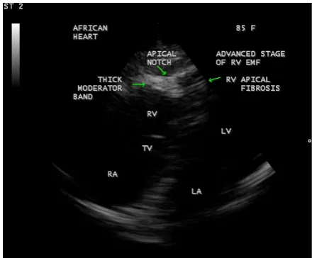
Clinical features are similar to case 1 and in addition, the patient had conduction disturbances such as heart block (IIIrd degree). 2D echocardiographic imaging revealed RV apical fibrosis with a thick, calcified moderator band engulfed in fibrosis, causing ventricular wall retraction and resulting an apical notch in the RV wall, giving an appearance of the map of Africa suggesting the advanced stage of the RV EMF as shown in Figure 4.
Case 4 (Left ventricular (LV) EMF in a 10 year old boy)
Clinical features are similar to case 1 and masquerading as acute nephritis and nephritic syndrome, idiopathic dilated cardiomyopathy, rheumatic heart disease, but auscultation of the heart and renal parameters were normal. 2D echocardiographic imaging revealed LV apical obliteration with fibrosis and a dilated left ventricle suggest-ing a subacute stage of LV EMF as shown in Figure 5.
Case 5 (Left ventricular EMF in an adult aged 54 years)
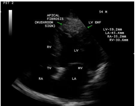
[image:2.595.197.432.510.693.2]The clinical features are similar to case 1 and 2D echocardiographic imaging showing the dense fibrosis in LV apex and a dilated left ventricle with an ejection fraction of 50% as shown in Figure 6.
Figure 2. Parasternal long axis view showing a dense RV apical fibrosis.
Figure 3. Apical 4 chamber view showing RV apical fibrosis,
mild tricuspid regurgitation and a dilated atria.
Figure 4. Tilted apical four chamber view showing RV apical fibrosis, thick and calcified moderator band, apical notch on the
[image:3.595.202.425.292.463.2] [image:3.595.202.426.501.683.2]Figure 5. This figure shows LV apical fibrosis, dilated left
[image:4.595.202.426.83.272.2]ventricle and normal left atrium.
Figure 6. This figure shows the LV apical fibrosis with dilated
cardiac chambers in LV EMF.
Case 6 (Left ventricular EMF with pericardial effusion in a female aged 23 years)
23 years old female presented with puffiness of face and chest discomfort during breathing with a history of 2 to 3 episodes of febrile illness during childhood. Echocardiographic screening revealed Left ventricular EMF with mild pericardial effusion as shown in Figure 7and typical mushroom sign of apical fibrosis in RV EMF in a 30-year old female as shown in Figure 8,in Biventricular EMF in a 8-year old boy as shown in Figure 9. Screening of family members revealed normal.
These cases are responded to diuretics and improvement in their life style occurred and they are advised periodic follow up. There is no in-hospital mortality so far among EMF cases detected and now flourishing as two to three cases monthly in this endemic area of Thoothukidi similar to the endemic rheumatic heart diseases in this region. It is a coastal district with hot and humid climatic conditions and majority are having a history of febrile illness years back before the onset this illness. Screening of family members revealed no abnormalities and so it is not familial.
3. Discussion
3.1. Echocardiographic Features
[image:4.595.201.428.310.488.2]Figure 7. This figure shows endocardial fibrosis on left
[image:5.595.204.425.83.259.2]ventricular wall with mild pericardial effusion.
Figure 8. Apical four chamber view showing RV EMF in a 30-year-old female.
Figure 9. This figure shows the mushroom sign in a 8-year
[image:5.595.203.426.297.472.2] [image:5.595.206.423.511.694.2]EMF, the trabecular portion of the ventricle is separated from the remaining cavity by a large fibrotic endocardi-al plaque, underneath which there is myocardium of apparently normendocardi-al texture [11]. This large fibrotic plaque obliterating the apex of the ventricle giving a typical appearance of mushroom and designated as “mushroom sign” which is a classical feature of EMF in echocardiography as shown in Figures 1-3, Figure 5, Figure 6 and Figure 8. The mechanism of right ventricular cavity obliteration corresponds to compaction of trabeculae be-tween the thickened fibrous endocardium and the epicardium, excluding the trabecular portion of the ventricle from the circulation [12]. The fibrotic process produces an “artificial floor” to the right ventricle, separating the inflow cavity from the obliterated muscular trabecular part, which does not contain thrombus or blood. The moderator band is lost, engulfed in the endocardial fibrosis as shown in Figure 4. The progressive fusion of tra-beculae between the thickened endocardium and epicardium results in retraction of the ventricular cavity. There is a reduction in the distance between the epicardium and the false floor and this process is associated with pull-ing of the wall by the retracted tricuspid valve apparatus, resultpull-ing in the distinctive findpull-ing of advanced right- sided EMF called “apical notch” as shown in Figure 6. The “apical notch” gives the heart a shape that resem-bles the map of Africa, hence the designation “Heart of Africa” [13].
Left ventricular obliteration affects both the apex and recesses of the posterior mitral leaflet excluding these parts from the ventricular cavity. It is thought that obliteration by thrombi and subsequent scarring and fibrosis are the mechanisms involved, both leading to reduction of the diastolic properties of the ventricles. Also, throm-bi may involve the sub-valvular apparatus, leading to scarring and fibrosis of the leaflets to the ventricular wall, therefore resulting in leaflet movement restriction and AV valve dysfunction. Left ventricular apex is never re-tracted, it becomes thicker leading to considerable reduction of the longitudinal diameter of the ventricle, result-ing in a spherical ventricular cavity as shown in Figure 5. The fibrotic process of EMF involves the atrioventri-cular (AV) valve and sub-valvular apparatus and masquerading as rheumatic heart disease in endemic areas for both diseases. In chronic rheumatic heart disease, leaflet thickening is usually restricted to the tip of the valve, extends to the chordae, and is not associated with obliteration of the ventricle [14][15]. Both the restriction to the ventricular filling and the AV valve regurgitation results in increase in atrial pressure, leading to progressive atrial dilatation. Outflow tracts of the ventricles and semilunar valves are spared from the disease process of EMF.
3.3. Echocardiographic Criteria
Mocumbi et al. have described a set of criteria for the diagnosis and classification of endomyocardial fibrosis as shown in Table 1 and this can apply only to the disease in the advanced stage.
A definite diagnosis of EMF was made in the presence of two major criteria or one major criterion associated with two minor criteria.
3.4. Clinical Features
Table 1. Criteria for diagnosis and assessment of the severity of Endomyocardial fibrosis.
Criterion Score
Major criteria
Endomyocardial plaques > 2 mm in thickness 2
Thin (≤1 mm) endomyocardial patches affecting more than one ventricular wall 3 Obliteration of right ventricular or left ventricular apex 4 Thrombi or spontaneous echo contrast without severe ventricular dysfunction 4 Retraction of the right ventricular apex (right ventricular apical notch) 4 Atrioventricular-valve dysfunction due to adhesion of the valvular apparatus to the
ventricular wall (The score is assigned according to the severity atrioventricular regurgitation)
1 - 4
Minor criteria
Thin endomyocardial patches localized to one ventricular wall 1 Restrictive flow pattern across mitral or tricuspid valves 2
Pulmonary-valve diastolic opening 2
Diffuse thickening of the anterior mitral leaflet 1
Enlarged atrium with normal-size ventricle 2
M-movement of the interventricular septum and flat posterior wall 1 Enhanced density of the moderator or other intraventricular bands 1 A total score of <8 = mild EMF; 8 to 15 = moderate EMF; >15 = severe EMF.
urticaria [16]. This illness may disappear, or it may leads to rapidly developing cardiac failure or it may evolve into established and apparently inactive EMF with predominant right ventricular or left ventricular disease. The exudative pericardial effusion of variable degree is a common presentation as illustrated in Figure 7. These ob-servations suggest that EMF is a systemic inflammatory disease, which has been called the “EMF syndrome” [17].
4. Conclusion
Right and Left ventricular Endomyocardial Fibrosis in different age groups have been described by Transtho-racic 2D Echocardiographic imaging. A right ventricular EMF at the age of 85 years in an elderly woman and a Left ventricular EMF with mild pericardial effusion at the age of 23 years in a young woman were detected in this coastal district of Thoothukudi in India.
References
[1] Dato, I. (2014) How to Recognize Endomyocardial Fibrosis? Journal of Cardiovascular Medicine (Hagerstown). [2] Rossi, S., Baruffi, S., Bertuzzi, A., Miragoli, M., Corradi, D., Maestri, R., et al. (2008) Ventricular Activation Is
Im-paired in Aged Rat Hearts. American Journal of Physiology, Heart and Circulatory Physiology, 295, H2336-H2347. http://dx.doi.org/10.1152/ajpheart.00517.2008
[3] Gupta, P.N., Valiathan, M.S., Balakrishnan, K.G., et al. (1989) Clinical Course of Endomyocardial Fibrosis. British Heart Journal, 62, 450-454.
[4] Freers, L., Mavania-Kizza, M., Ziegler, H. and Rutaceinnio, M. (1996) Echocardiographic Dignosis of Heart Disease in Uganda. Tropical Doctor Journal, 26, 125-128.
[5] Mocumbi, A.O., Carrilho, C., Sarathchandra, P., Ferreira, M.B., Yacoub, M.H. and Burke, M. (2010) Echocardiogra-phy Accurately Assesses the Pathological abnormalities of Chronic Endomyocardial Fibrosis. International Journal of Cardiovscular Imaging, 6,445-452.
[6] Mocumbi, A.O., Ferreiro, M.B., Sidi, D. and Yacoub, M.H. (2008) A Population Study of Endomyocardial Fibrosis in a Rural Area of Mozambique. New England Journal of Medicine, 369, 43-49.
http://dx.doi.org/10.1056/NEJMoa0708629
Heart Journal, 19, 600-630. http://dx.doi.org/10.1016/0002-8703(60)90464-6
[14] Saraiva, L.R., Carneiro, R.W., Arruda, M.B., Brindeiro Filho, D. and Lira, V. (1999) Mitral Valve Disease with Rheumatic Appearance in the Presence of Left Ventricular Endomyocardial Fibrosis. Arquivos Brasileiros de Cardi-ologia, 72, 327-332. http://dx.doi.org/10.1590/S0066-782X1999000300006
[15] Metras, D., Quezzin, Coulibaly, A., Quattara, K., Bertrand, E. and Chauvet, J. (1983) Endomyocardial Fibrosis Mas-querading as Rheumatic Mitral Incompetence, A Report of Six Surgical Cases. Journal of Thoracic Cardiovascular Surgery, 86, 753-756.
[16] Freers, I., Hakim, J., MyanjaKizza, H. and Parry, F. (2004) The Heart, Endomyocardial Fibrosis, Still a Mystery after 60 Years. Principles of Medicine in Africa, 837-886.
[17] Freers, J., Masembe, V., Schmauz, R. and Mayanja, H., (2000) Endomyocardial Fibrosis (EMF) Syndrome in Uganda.