Molecular single-cell analysis reveals that
CD5-positive peripheral blood B cells in healthy
humans are characterized by rearranged
Vkappa genes lacking somatic mutation.
M Fischer, … , U Klein, R Küppers
J Clin Invest.
1997;
100(7)
:1667-1676.
https://doi.org/10.1172/JCI119691
.
B cells expressing the CD5 cell surface antigen are involved in certain B cell malignancies
and autoimmune diseases. From studies in the mouse, it emerged that CD5+ B cells
represent a separate lineage of B lymphocytes that, in contrast to conventional (CD5-) B
cells, are not driven into T cell-dependent immune responses in which rearranged variable
(V) region genes are diversified by somatic hypermutation. Against this background it came
as a surprise that human disease-involved CD5-positive autoreactive B cells as well as B
cell chronic lymphocytic leukemias can harbor somatically mutated V region genes. Recent
V gene analyses on CD5+ B cells in healthy adults did not give rise to a clear picture about
the fraction of somatically mutated among all CD5+ B cells. In this work we used a
molecular single-cell analysis to determine reliably the frequency of mutated CD5+ B cells
in healthy humans: single, kappa light chain-expressing CD5+ peripheral blood B cells
were isolated by flow cytometry, and rearranged Vkappa genes were amplified by PCR.
From one donor, CD5+CD19+ B cells were analyzed. Since CD5+ B cells were found
among IgM+IgD+ and IgM+IgD- cells (but almost not among class-switched cells) from two
other donors, individual cells corresponding to these IgM-expressing subsets were
investigated separately. The sequence analysis of rearranged Vkappa genes revealed that
most if not all […]
Research Article
Find the latest version:
J. Clin. Invest.
© The American Society for Clinical Investigation, Inc. 0021-9738/97/10/1667/10 $2.00
Volume 100, Number 7, October 1997, 1667–1676 http://www.jci.org
Molecular Single-cell Analysis Reveals that CD5-positive Peripheral Blood B Cells
in Healthy Humans are Characterized by Rearranged V
kGenes Lacking
Somatic Mutation
Matthias Fischer, Ulf Klein, and Ralf Küppers
Institute for Genetics, University of Cologne, 50931 Cologne, Germany
Abstract
B cells expressing the CD5 cell surface antigen are involved in certain B cell malignancies and autoimmune diseases. From studies in the mouse, it emerged that CD51
B cells represent a separate lineage of B lymphocytes that, in con-trast to conventional (CD52
) B cells, are not driven into T cell–dependent immune responses in which rearranged variable (V) region genes are diversified by somatic hyper-mutation. Against this background it came as a surprise that human disease–involved CD5-positive autoreactive B cells as well as B cell chronic lymphocytic leukemias can harbor somatically mutated V region genes. Recent V gene analyses on CD51 B cells in healthy adults did not give rise
to a clear picture about the fraction of somatically mutated among all CD51 B cells. In this work we used a molecular
single-cell analysis to determine reliably the frequency of mutated CD51 B cells in healthy humans: single, k light
chain–expressing CD51
peripheral blood B cells were iso-lated by flow cytometry, and rearranged Vk genes were
am-plified by PCR. From one donor, CD51
CD191
B cells were analyzed. Since CD51 B cells were found among IgM1IgD1
and IgM1
IgD2
cells (but almost not among class-switched cells) from two other donors, individual cells corresponding to these IgM-expressing subsets were investigated sepa-rately. The sequence analysis of rearranged Vk genes
re-vealed that most if not all CD51
B cells in healthy humans carry unmutated V region genes. From one of the donors, a novel polymorphic Jk2 gene segment was identified. To
ex-plain the discrepancy between the frequent occurrence of disease-associated somatically mutated CD51
B cells and the low incidence or absence of somatic mutation in normal CD51
B cells, we speculate that CD51
B cells usually do not participate in germinal center reactions, but if they occa-sionally do so, they may be at an increased risk to become involved in autoimmune diseases or B cell malignancies. (J. Clin. Invest. 1997. 100:1667–1676.) Key words: B1 cells•
CD5 • single cell PCR • somatic hypermutation • variable
re-gion genes
Introduction
B lymphocytes expressing the CD5 antigen are the predomi-nant B cell population early in life (1, 2). Later, their number is reduced, and CD51
B cells account for z 10–25% of
periph-eral blood (PB)1 B cells in man (1–3). These cells are also
found in spleen and tonsil at a comparable frequency (1–3). One characteristic feature of CD51
B cells (also called B1a cells) is that many of them produce autoreactive antibodies, which are usually polyreactive (4). Numbers of CD51
B cells are elevated in certain autoimmune diseases, and in some of these cases CD51
B cells producing high-affinity autoantibod-ies have been detected (4–7). A relationship between CD51
B cells and autoimmunity is further suggested by the finding that mouse strains harboring elevated levels of CD51
B cells are prone to autoimmune diseases (1). CD51
B cells are also in-volved in certain B cell malignancies. More than 95% of cases of B cell chronic lymphocytic leukemia (B-CLL), the most common leukemia of adults in the Western world, express CD5 (1). In addition, CD5 is regularly found on tumor B cells in small lymphocytic lymphoma (1).
The origin of CD51
B cells in humans and mice is much de-bated (1, 8). Since CD5 expression can be induced in vitro on CD5-negative B cells, it has been suggested that CD5 repre-sents an activation marker (9). In the induction/selection model, B cells recognizing autoantigens with low affinity are selected to become CD51
B cells (10). On the other hand, transfer experiments in mice support a dual-lineage model that states that CD5-positive and CD5-negative (conventional) B cells represent distinct lineages of B cells (11). Those transfer experiments also revealed that CD51
B cells repesent a self-replenishing population of long-lived lymphocytes (12, 13).
Immunoglobulin V genes expressed by CD51
B cells have been the subject of a large number of studies. A sequence analysis of immunoglobulin V region genes allows one to gain insight into the stage of differentiation of the respective B cell. Immature and naive B cells carry unmutated variable (V) gion genes (14–17). Somatic mutations are introduced into re-arranged V genes in the course of T cell–dependent immune reactions (14, 18). This process takes place in the microenvi-ronment of the germinal center (GC) (19–21). Consequently, somatically mutated V region genes are found in GC B cells and their descendants, i.e., memory B cells and GC-derived plasma cells (16, 17, 21). Whereas murine CD51
B cells ex-press unmutated V region genes (22–24), the situation appears to be different in the human: somatically mutated V genes have been described in cases of CD5-expressing B-CLL (25– 28) and in CD51
B cells producing high-affinity autoantibodies
M. Fischer and U. Klein contributed equally to this work.
Address correspondence to Dr. Ralf Küppers, University of Co-logne, University Clinics, LFI E4 R706, Joseph-Stelzmannstr. 9, 50931 Cologne, Germany. Phone: 4490; FAX: 49-221-478-6383; E-mail: [email protected]
Received for publication 9 April 1997 and accepted in revised form 7 August 1997.
(5, 6). Moreover, somatic mutations have been reported to be present in V genes of CD51 B cells isolated from healthy do-nors (16, 29, 30). These findings suggest that in terms of so-matic mutation, there is no difference between CD51 B cells and conventional B cells in humans.
Two findings prompted us to reinvestigate the issue of the occurrence and frequency of CD51 B cells carrying somatically mutated V genes in healthy individuals: first, most CD51 B cells are found among IgM1IgD1 B lymphocytes (2, 3). The vast majority of these naive B cells, however, carry unmutated V region genes (15–17, 31), which also implies that most CD51 B cells express unmutated V genes. Obviously, this result con-trasts with the V gene studies on CD51 B cells in healthy indi-viduals (16, 29, 30). A possible explanation for this discrepancy might be that a small subset of cells with mutated V region genes among IgM1IgD1 B cells could have been missed in the analyses of IgM1IgD1 B cells, or that the mutated V genes in those studies (16, 29, 30) were derived from IgD2 CD51 B cells. Second, we recently showed that human peripheral blood B lymphocytes carrying somatically mutated V region genes, i.e., IgM1IgD2 (IgM-only) and class-switched B cells, harbor elevated levels of mRNA coding for immunoglobulin when compared with naive IgM1IgD1 B cells (31). Since the studies on CD51 B cells of healthy donors were carried out by amplifying V region gene transcripts from cell populations (16, 29, 30), those investigations might be biased towards somati-cally mutated V genes.
To overcome these problems, we sorted single CD51 B cells by flow cytometry, and amplified rearranged Vk genes from genomic DNA of individual cells. PCR products were se-quenced directly. This approach should allow one to deter-mine reliably the frequency of CD51 B cells carrying mutated V region genes.
Methods
Isolation of single cells and flow cytometry. 60–100 ml PB was ob-tained from three male, healthy donors ages 27–35. PB mononuclear cells (MC) were purified by Ficoll-Isopaque density centrifugation, and were incubated with unconjugated antihuman k light chain (IgG1-isotype, Becton Dickinson, Mountain View, CA) for 15 min on ice. After washing, the cells were incubated with goat anti–mouse-IgG1 microbeads (Miltenyi Biotec Inc., Bergisch Gladbach, Ger-many) for 20 min at 48C. Subsequently, k-expressing B cells were en-riched to . 90% by magnetic cell separation using the MiniMACS™ system (Miltenyi Biotec Inc.) (15). For isolation of single CD51CD191 B cells from donor HK, the cell suspension was incubated with anti-CD5-PE and anti-CD19-FITC (Becton Dickinson). The cell suspen-sions from the other two donors were stained with anti-CD5-FITC (Becton Dickinson), goat anti–human (GaH)-IgM-PE (Sigma Chem-ical Co., München, Germany), and biotinylated GaH-IgD (Southern Biotechnology Associates, Birmingham, AL). After washing, GaH-IgD was developed with Streptavidin-CyChrome (PharMingen, San Diego, CA). All staining procedures were performed for 15 min on ice. Using a FACS 440 (Becton Dickinson), single k-expressing B cells of the following B cell fractions were sorted directly into PCR reaction tubes containing 20 ml PCR buffer and 20 ng 5S rRNA: CD51CD191 B cells (donor HK), CD52IgM1IgD2 B cells, CD51IgM1IgD2 B cells, and CD51IgM1IgD1 B cells (donors RK and AB). In addition, a frac-tion of peripheral blood mononuclear cells (PBMC) was stained with anti-CD3-FITC (Becton Dickinson), and single T cells (CD31) were sorted into reaction tubes as described above.
The size of CD51IgM1IgD1 as well as CD51IgM-only PB B cells and their CD5-negative counterparts was determined by forward
scatter analysis. The populations did not differ from each other in this respect.
For analysis of isotype-switched B cells with regard to CD5-expres-sion, PBMC of seven healthy adult donors were incubated with anti-CD19 microbeads (Miltenyi Biotec Inc.) for 20 min at 48C, and CD191 B cells were enriched to 99% by MiniMACS™. The B cell– enriched cell suspensions were stained with anti-CD5-PE, GaH-IgD, and with either GaH-IgG-FITC (Southern Biotechnology Associ-ates) or rabbit anti–human-IgA F(ab9)2-FITC (Dako Corp.,
Ham-burg, Germany) as described above. Samples were analyzed on a FACScan (Becton Dickinson).
To analyze tonsillar non-GC B cells consisting of naive as well as memory B cells (17) for expression of CD5, this fraction was enriched by depleting GC B cells (CD381) and T cells using the MACS system (Miltenyi Biotec Inc.). Tonsillar MC were incubated with anti-CD3 microbeads (Miltenyi Biotec Inc.) and unconjugated anti-CD38 (IgG1-isotype; Becton Dickinson) for 20 min at 48C, and after washing with anti-IgG1 microbeads. After magnetic cell separation, tonsillar non-GC B cells were enriched to z 85%. This fraction was analyzed for
surface expression of CD5 on IgM1IgD1 and IgM-only cells as well as on isotype-switched memory B cells using the staining procedures de-scribed above.
Single cell PCR and sequencing of PCR products.Single cells were incubated in 20 ml PCR buffer containing 0.5 mg/ml proteinase K for 1 h at 508C. The enzyme was inactivated by denaturation at 958C (10 min). For the first round of amplification, a primer mix consisting of four Vk family–specific primers, which recognize sequences in the framework region I of the members of the Vk1–4 gene families, and a downstream primer specific for a sequence 39 of Jk5 was used (see Ta-ble I). This approach prevents amplification of Vk-Jk1,2,3 & 4 rear-rangements that have been located away from the Ck locus by inver-sion (32), and therefore presumably do not undergo somatic hypermutation due to their large distance from both the k intron and the k 39-enhancer (33). Owing to the small distance between the five Jk gene segments, however, VkJk rearrangements lying in the proper location in the Igk locus can be efficiently amplified with a primer 39 of the last Jk gene. The first round of amplification was carried out in the same reaction tube in a 50-ml volume containing Expand™ High Fidelity buffer (Boehringer Mannheim, Mannheim, Germany), 1.75 mM MgCl2, 100 mM dATP, dGTP, dTTP, and dCTP, respectively, 50
nM of each primer, and 2.5 U Expand™ High Fidelity polymerase (Boehringer Mannheim). The amplification program consisted of 35 cycles of 60 s at 958C, 30 s at 618C, and 80 s at 728C, followed by a final incubation step at 728C for 5 min. Enzyme was added after the first denaturation step. For the second round of amplification, the same Vk family–specific primers were used together with three internal Jk -specific primers hybridizing to the five human Jk gene segments (see Table I). The second round of amplification was performed in sepa-rate reactions for each of the four Vk primers using 1 ml of the first-round reaction mixture in a 50-ml volume containing 10 mM Tris-HCl, pH 8.3 (208C), 50 mM KCl, 2.0 mM MgCl2, 100 mM dATP,
dGTP, dTTP, and dCTP, respectively, 125 nM of each primer, and 0.7 U Taq DNA polymerase (Boehringer Mannheim). The amplification conditions were as described above, except that the elongation step at 728C was carried out for 60 s, and 40 cycles were applied. PCR prod-ucts were purified by gel electrophoresis. An aliquot of the isolated DNA was sequenced directly with 32P-labeled oligonucleotides of the
second round using the dsDNA Cycle Sequencing System (GIBCO BRL, Eggenstein, Germany) as recommended by the supplier. Nucle-otide sequences were analyzed using DNASIS™ software (Pharma-cia, Freiburg, Germany). In some cases, it was not possible to deter-mine by sequence analysis which of the five Jk gene segments had been rearranged. In these cases, the second round of PCR was re-peated using the respective Vk family-specific primer together with each of the Jk-specific primers in separate reactions (Jk typing).
(CDR) III lengths . 9 amino acids between IgM-only CD52, IgM-only CD51, and IgM1IgD1CD51 B cells were assessed statistically using Fisher’s exact test.
Results
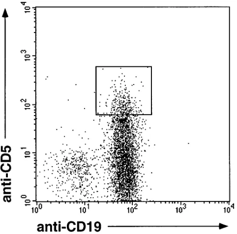
Vk gene sequences of CD191CD51 B cells. After enrichment of k light chain–expressing PB B cells from donor HK by mag-netic cell separation, single cells of the CD191CD51 fraction, which amounted to z 20% of the B cell compartment, were
isolated by fluorescence-activated cell sorting (Fig. 1). Rear-ranged Vk genes of individual cells were amplified from ge-nomic DNA using a seminested PCR strategy (31). The exper-iment was restricted to analysis of Vk region genes since the human immunoglobulin k locus is well characterized, and hu-man Vk germline genes seem to exhibit only little polymor-phism, which allows unambiguous identification of somatic
mutations in rearranged Vk genes (34, 35). Negative controls consisted of one single T cell, plus one reaction mixture with-out cells per seven B lymphocytes analyzed. None of these negative controls gave rise to a PCR product.
[image:4.612.57.560.74.192.2]Of 14 CD51 B cells analyzed by PCR, 11 cells gave rise to a total of 14 amplificates, 12 of which were sequenced (Fig. 2). All sequences represented unique VkJk joints. Cell X8 carried one in-frame and one out-of-frame rearrangement, whereas cell X9 showed only a single out-of-frame rearrangement. Cell X12 gave rise to two potentially functional Vk region genes. This result may be explained by the fact that two cells had been sorted into the reaction tube at the isolation step. An-other possibility is that one of the k light chains, though the gene segments had been functionally assembled, was unable to pair properly with the H chain expressed by the cell, and there-fore an additional rearrangement occurred in the second Igk locus (36, 37). From each of the remaining seven cells, a single in-frame rearrangement was obtained. Whereas Vk gene rear-rangements X5K4 and X8K1 showed two basepairs difference each compared to the most homologous Vk germline genes, the other ten Vk sequences turned out to be unmutated (Fig. 2 and Table II). This result indicates that the majority of human CD51 PB B cells carry unmutated Vk region genes. It does not, however, exclude the possibility that a small subset of mutated CD51 B cells might exist in human PB.
Table I. Sequences of Oligonucleotides Used as Primers for the Amplification of Rearranged Vk Genes
Oligonucleotide Sequence
Vk1 59-GACATCC,AG.G,AT.TGACCCAGTCTCC,AT.TC-39
Vk2 59-CAG,AT.CTCCACTCTCCCTG,CT.CCGTCA-39
Vk3 59-TTGTG,AT.TGAC,AG.CAGTCTCCAG,GC.CACC-39
Vk4 59-AGACTCCCTGGCTGTGTCTCTGGGC-39
39Jk5 59-CTCTAAA,AG.GTCAATACTGGCCATC-39
59Jk1,2,4 59-ACTCACGTTTGAT,CT.TCCA,GC.CTTGGTCC-39
59Jk3 59-GTACTTACGTTTGATATCCACTTTGGTCC-39
59Jk5 59-GCTTACGTTTAATCTCCAGTCGTGTCC-39
The Vk primers hybridize to the framework region I of the respective family. ,. denotes a nucleotide mix at this position.
[image:4.612.58.299.452.690.2]Figure 1. Flow cytometric analysis of k-expressing B cells (after MACS enrichment) derived from PB of a 27-yr-old person (donor HK). Cells were stained with anti-CD19-FITC and anti-CD5-PE. The gate set for sorting of single CD51CD191 B cells is indicated.
Table II. Summary of Mutations in Vk Region Genes Amplified from CD51CD191, CD52IgM-only, CD51IgM-only, and CD51IgM1IgD1 PB B Cells Derived from Three Donors
Cell population Donor n* Unmutated‡ Range§ % Mutationi
CD51 CD191 HK 12 10 0–2 0.1
CD51 IgM1 IgD2 RK 11 10 0–4 0.1
AB 10 8 0–4 0.2
CD51 IgM1 IgD1 RK 11 11 0 0
AB 13 13 0 0
CD52 IgM1 IgD2 RK 11 2 0–12 2.5
AB 7 1 0–16 2.0
*Number of VkJk rearrangements analyzed; ‡number of unmutated VkJk
rearrangements; §range of mutations per gene. iMutation frequencies
were calculated by considering the nucleotide differences in the Vk
re-gion genes relative to the corresponding Vk and Jk germline genes,
[image:4.612.315.557.572.682.2]Vk gene sequences of IgM1IgD1 and IgM1IgD2 CD5-expressing B cells.The expression of mutated V genes in hu-man B lymphocytes is mainly restricted to isotype-switched and IgM-only (IgM1IgD2) B cells (15–17, 31). These two B
cell subsets represent z 15% and 10% of human PB B cells,
[image:5.612.101.530.55.234.2] [image:5.612.59.298.372.663.2]respectively, whereas naive IgM1IgD1 B cells usually com-prise more than 70% of all PB B lymphocytes (15, 31, 38, 39). Consequently, the question arose as to whether CD51 B cells Figure 2. Nucleotide sequences of VkJk junctions amplified from CD51CD191 PB B cells by single-cell PCR. Sequences are designated by an X and a sample number of the cell, followed by the corresponding Vk family to which the sequence was assigned. Shown are the 39 end of the re-spective Vk gene, N-region nucleotides (N), and the 59 end of Jk. In addition, the corresponding Vk and Jk germline genes are indicated. Jk genes identified by Jk typing (see Methods) are denoted by *. The sum of nucleotide differences relative to the corresponding germline genes is shown in the right column. Codons are numbered according to Kabat et al. (74). Full-length sequences are available from EMBL/GenBank under ac-cession nos. Z98605–Z98679.
Figure 3. Peripheral blood. Flow cytometric analysis of MACS-enriched CD191 PB B cells derived from a healthy adult person. Cells were stained with anti-CD5-PE, anti-IgD-CyChrome, and either (left top) anti-IgA-FITC or (left bottom) anti-IgG-FITC. (Left) Windows were set around (a) the IgA1, (b) the IgG1, and (c) the IgD1 popula-tions. (Right) CD5-expression of (top) IgA1, (middle) IgG1, and (bottom) IgD1 cells.
[image:5.612.313.555.374.642.2]occur among class-switched and/or IgM-only B cells, and, if this was the case, whether those CD5-positive B lymphocytes harbor mutated V genes. Although class-switched cells are thought to be negative for CD5 expression, in the literature one finds surprisingly little data on this subject (40). To clarify this matter, MACS-enriched CD191 PB B cells derived from seven healthy adult donors were stained with anti-IgD and anti-CD5, and with either anti-IgA or anti-IgG. FACScan
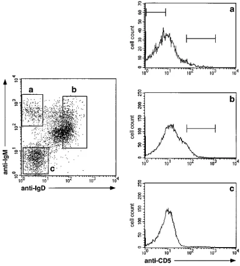
[image:6.612.57.551.67.620.2]analysis revealed in all cases that CD5-expressing cells were barely detectable (i.e., , 1%) among isotype-switched B cells (Fig. 3). On the other hand, we reported in a previous study on a small fraction of CD51 B lymphocytes within the PB IgM-only compartment (31). This result was confirmed by flow-cyto-metric analysis of PB B cells (from donors RK and AB) that were enriched by magnetic cell separation and stained with anti-IgM, anti-IgD, and anti-CD5 (Fig. 4). The fraction of
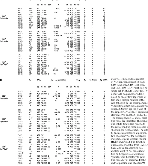
Figure 5. Nucleotide sequences of VkJk junctions amplified from CD52IgM-only, CD51IgM-only, and CD51IgM1IgD1 PB B cells by single-cell PCR. (A) Donor RK; (B) donor AB. Sequences are desig-nated by one or two uppercase let-ters and a sample number of the cell, followed by the corresponding Vk family to which the sequence was assigned. Shown are the 39 end of the respective Vk gene, N-region nu-cleotides (N), and the 59 end of Jk. The corresponding Vk and Jk germ-line genes are indicated. The sum of nucleotide differences relative to the corresponding germline genes is shown in the right column. The C to G nucleotide exchange at position two of codon 97 of the novel poly-morphic Jk2 gene segment (donor RK) is underlined. Full-length se-quences are available from EMBL/ GenBank under accession nos. Z98605–Z98679. *Jk genes identi-fied by Jk typing (see Methods);
§pseudogene; ‡homology to
CD51 B cells among the IgM-only subset accounted for
z 10% each, whereas the fraction of CD51 B cells among the
IgM1IgD1 compartment amounted to
z 20% in both cases.
To determine the level of somatic mutation within rear-ranged Vk genes, single k-positive B cells of both donors were isolated from the CD51IgM1IgD1, the CD51IgM1IgD2, and, as a control, from the CD52IgM1IgD2 subset. From donor RK, single cells of all three fractions were isolated twice in two separate experiments. Single-cell PCR and sequencing were performed as described above. Taking both donors together, a total of 106 cells were analyzed by PCR. 52 cells gave rise to a single, and 10 cells to two distinct PCR products. 63 amplifi-cates from 55 cells were sequenced, all of which represented unique VkJk rearrangements (Fig. 5). Eleven cells showed a single out-of-frame rearrangement, whereas six cells carried both a productive and a nonproductive VkJk joint. Again, two cells (C6 and F23) gave rise to two distinct in-frame rearrange-ments each. The remaining 36 cells revealed a single poten-tially functional Vk region gene each.
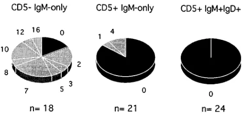
15 out of 18 rearrangements of the CD52IgM-only com-partment turned out to be mutated (range: 2–16 mutations, Figs. 5 and 6 and Table II). Interestingly, the three unmutated sequences represented out-of-frame VkJk joints (Fig. 5). These three genes may be derived from cells that carry somatic muta-tions in the heavy chain and functional light chain genes: in some k light chain–expressing B cells, the second (presumably nonfunctional) rearranged k allele is inactivated by deletion of the Ck gene and the k enhancers, leaving the VkJk joint un-touched (41). This deletion most likely abolishes somatic mu-tation in the respective VkJk joint (33, 42). The frequency of so-matic mutation of CD52IgM-only PB B lymphocytes was calculated to be 2.5% for donor RK, and 2.0% for donor AB (Table II). Both values are in a similar range to mutation fre-quencies observed for IgM-only PB B cells in previous studies (15, 31). In contrast, only 3 out of 21 rearrangements of the CD51IgM-only subset harbored somatic mutations (1, 4, and 4 nucleotide exchanges), whereas the other 18 sequences were identical to the corresponding Vk and Jk germline genes (Figs. 5 and 6 and Table II). In addition, among 24 rearrangements obtained from CD51IgM1IgD1 PB B cells, not a single point mutation was found (Figs. 5 and 6 and Table II).
Evidence for a novel polymorphic Jk2 gene segment. The Jk2 gene segment was found to be rearranged in eleven clonally in-dependent Vk region genes obtained from cells of donor RK
(Fig. 5 A). Position two of codon 97 of the respective rear-ranged Jk2 genes differed from the published Jk2 germline gene (43) by a C to G nucleotide exchange in nine of these re-arrangements. Since six of those V region genes carried no mu-tation in the rest of their sequence, it is unlikely that the C to G exchanges represent somatic mutations. The result rather sug-gests that donor RK is heterozygous for a previously unidenti-fied polymorphic Jk2 gene which replaces a threonine (en-coded by ACT) by a serine (AGT) at the last position of the CDRIII. A search in the European Molecular Biology Labora-tories database (release 95) revealed that a Jk2 gene with AGT at codon 97 was previously sequenced from three other indi-viduals, although this was not identified as a polymorphism in those studies (44–46). Thus, we identified a novel, albeit rare Jk2 gene polymorphism.
Vk gene usage of CD51 B cells. We obtained 43 potentially functional VkJk joints from CD51 B cells. The sequences could be assigned to a total of 16 different Vk germline genes of the Vk gene families 1, 2, 3, and 4. Nine sequences showed highest homology to the Vk3 gene A27, five sequences each to the sin-gle Vk4 (B3) gene and the A3/A19 (Vk2) gene, and four se-quences to O2/O12 (Vk1). Three sese-quences each could be as-signed to L12A (Vk1), L2 (Vk3), and L6 (Vk3). Nine Vk germline genes were found to be rearranged either once or twice (Figs. 2 and 5). The Vk genes that have been used fre-quently by CD51 B cells in this study, in particular A27, were also found to be overrepresented in compilations of rear-ranged Vk sequences, the majority of which were derived from conventional B cells (35, 47). Thus, it appears that human CD51 PB B cells do not differ considerably from conventional B cells with regard to Vk gene usage.
N-region additions at VkJk junctions of CD51 B cells. A hall-mark of murine B cells developing early in ontogeny, a sub-stantial fraction of which belongs to the CD5-expressing subset (1, 48), is the rare occurrence of non-germline-encoded nucle-otides at the VHDHJH junctions (49, 50). On the other hand,
N-region additions have frequently been detected in CD51 B cells obtained from adult mice, even though less frequently than in conventional B cells (62% versus 93%) (49–51).
In contrast, N sequences are a common feature of human B lymphocytes already in the fetus, and they appear to be consis-tently present at VHDHJH junctions of B cells in the adult (52,
53). Moreover, IgH CDRIII sequences derived from CD51 and CD52 human cord blood B cells revealed no significant difference with respect to the frequency of N-region additions (54). In our collection of 57 Vk gene rearrangements amplified from CD51 B cells of three adult donors, 34 contained non-germline-encoded nucleotides at their VkJk junctions (60%), whereas N sequences were found at 10 out of 18 VkJk junctions derived from CD52 B cells (56%) (Figs. 2 and 5). These data, therefore, support the notion that N-region additions occur frequently in human CD51 B cells, and that the frequency, un-like in the mouse, does not differ from that of conventional B cells.
[image:7.612.57.299.569.682.2]CDRIII lengths of potentially functional Vk region genes. Comparison of the CDRIII lengths of potentially functional Vk region genes demonstrated distinct patterns between CD52 and CD51 B cells: whereas none of the 13 rearrangements obtained from CD52IgM-only B cells revealed CDRIIIs ex-ceeding 9 amino acids, those CDRIIIs were found in 7 of 15 re-arrangements (10 codons each) of the CD51IgM-only and in 7 of 18 rearrangements (6 with 10 and one with 11 codons) of the Figure 6. Distribution of Vk region genes with respect to the number
CD51IgM1IgD1 subpopulation (Fig. 5). The difference be-tween the CD52 subset on the one hand, and the CD51 IgM-only as well as the CD51IgM1IgD1 subset on the other, was statistically significant (P5 0.005 and P5 0.012, respectively). In contrast, the two CD51 B cell fractions did not differ signifi-cantly from each other (P5 0.461).
Since it has been suggested that CD5-expressing B cells are involved in autoimmune diseases, particularly in rheumatoid arthritis (1, 5–7), it is interesting to note that Bridges et al. re-cently reported on increased proportions of Vk transcripts with unusually long CDRIIIs amplified from synovia and PB of pa-tients with rheumatoid arthritis as compared to healthy indi-viduals (55). This overrepresentation, however, was rather due to an increase in VkJk joints containing CDRIIIs of 11 amino acids, than to an increase in those with CDRIIIs of 10 amino acids, the latter being overrepresented in the CD51 subsets of this study. Thus, the relationship between CD5-positive B cells carrying Vk region genes with extended CDRIII lengths and B cells expressing Vk region genes with long CDRIIIs in patients suffering from rheumatoid arthritis remains to be clarified.
Analysis of tonsillar B cell subsets with regard to CD5 ex-pression. To determine whether the distribution of CD5 on B cell subsets derived from secondary lymphoid organs resem-bles that seen in the PB, we analyzed tonsillar B cells for ex-pression of CD5 on IgM-expressing, as well as on isotype-switched B lymphocytes. Besides naive and memory B cells, the tonsil contains a large fraction of GC B cells (17). These cells are known to be CD5-negative (56). To avoid masking in the flow cytometric analysis of tonsillar memory and naive B cells for CD5 expression by the numerous GC B cells, we de-pleted the latter, and, in addition, T cells, by magnetic cell sep-aration. Concerning the pattern of IgM and IgD expression on the enriched cells, we were surprised to find only two of seven tonsils showing a clearly discernible IgM-only subpopulation. Whereas most tonsils were derived from children or young adults (ages 5–19), these two tonsils were from donors over 30 years old, suggesting that IgM-expressing memory cells in this tissue accumulate with age. In one of these cases, expression of CD5 on IgM1IgD1 on the one hand, and IgM-only cells on the other, resembled that seen on their counterparts in the PB (data not shown). In the other case, the sample size was too small to determine the fraction of CD5-positive IgM-only B cells. The IgM1IgD1 cells of the remaining tonsils, like IgM1IgD1 PB B cells, comprised
z 20% CD51 cells (data not
shown). Additionally, fractions enriched for non-GC B cells from three tonsils were analyzed for the CD5 expression on class-switched B cells. Fig. 7 shows that, whereas a sizeable proportion of IgD-positive B cells expressed CD5, few if any CD5-positive cells could be detected among IgG1 and IgA1 B cells. Thus, at least in the case of the tonsil, the expression pat-tern of CD5 on B cell subsets of secondary lymphoid organs mirrors that seen on the corresponding PB B lymphocyte sub-sets (Figs. 3, 4, and 7).
Discussion
Previous investigations suggested that CD51 B cells in healthy individuals regularly acquire somatic mutations. In three RNA-based studies, somatic mutations were found in 12/ 16 (29), 16/23 (30), and 5/8 (16) V gene transcripts amplified from populations of CD51 B cells isolated by flow cytometry. In yet another RNA-based study, the ratio of mutated to
un-mutated V genes (3/9) was lower (46). Moreover, 16 cell lines generated by EBV transformation of CD51 B cells from healthy adult donors have been described in the literature (25, 57–59). Among those lines, 12 have been reported to be mu-tated. Collectively, these studies suggested that the majority of CD51 B cells in healthy humans express somatically mutated V genes.
Despite the conformity of these data, the following obser-vations prompted us to reinvestigate V genes of human CD51 B cells for somatic mutation: first, the finding that most CD51 B cells are found among IgM1IgD1 B lymphocytes, the vast majority of which carry unmutated V region genes (15–17, 31), implies that most CD51 B cells express unmutated V region genes. Second, we recently observed that IgM-only and class-switched B cells harbor elevated levels of mRNA for k light chains when compared with IgM1IgD1 naive B cells (31). Since IgM-only as well as class-switched B cells carry somati-cally mutated V genes (15, 16, 31), the presence of suchcells in a given sample will bias population-based studies of V gene transcripts towards mutated sequences. This consideration is particularly important in analyses of CD51 B cells: since CD5 is expressed on B cells only weakly, the populations of conven-tional and CD51 B cells overlap in flow cytometric analyses (see Figs. 1, 3, 4, and 7) (2, 3, 16, 29, 30). Thus, sorted CD51 populations are likely to be contaminated by conventional B cells, z 20–30% of which are IgM-only or class-switched cells.
[image:8.612.317.554.56.344.2]not represent a suitable approach to determine the frequency of CD51 B cells harboring mutated V genes.
Furthermore, the collection of EBV lines derived from CD51 B cells is clearly not representative of the CD51 B cell population; two of the lines harboring mutated V genes ex-press isotypes other than IgM (57), which is atypical for CD51 B cells, as we and others have shown (Figs. 3 and 7) (40, 60). In addition, in the work of Schutte et al. (59), the two lines ana-lyzed were the only ones that expressed CD5 among 500 lines established from adult PB. This underrepresentation of CD5-expressing EBV lines is not surprising, since sorted CD51 B cells usually lose CD5 expression upon EBV immortalization (59, 61). Under these circumstances, the two CD5-positive lines can hardly be regarded as being representative of CD51 B cells. Nevertheless, analyses of the EBV lines indicate that some CD51 B cells carrying somatic mutations can be found in healthy adults, although their frequency cannot be reliably de-termined from those investigations (25, 57–59).
The vast majority of CD51 PB B cells in healthy humans carry unmutated V region genes. In this study, CD5-expressing PB B cells derived from healthy adults were analyzed for the level of somatic mutation in rearranged Vk genes by single-cell PCR. This approach has several advantages when compared with the analysis of cell lines or cDNA-libraries generated from cell populations (62); a single-cell experiment is obvi-ously not biased by varying transcript levels or by preferential EBV transformation of particular subsets of cells. By analyz-ing sanalyz-ingle cells, the frequency of B cells harboranalyz-ing somatically mutated V region genes within a B cell subset can be reliably determined. Furthermore, single-cell PCR allows unambigu-ous identification of somatic mutations in contrast to cell pop-ulation analyses that involve cloning of PCR products. Amplif-icates obtained from single cells can be sequenced directly without being cloned, and therefore sequence errors due to misincorporation by Taq DNA polymerase are negligible.
In a first experiment, Vk region genes of single CD51 CD191 PB B cells were investigated. Among 10 cells analyzed, 2 VkJk joints showed 2 mutations each, whereas the remaining rearranged Vk genes were identical to their corresponding germ-line genes (Fig. 2 and Table II). This result indicates that the majority of CD51 B cells in healthy humans carry unmutated V genes.
Since the finding of two slightly mutated V genes in the analysis of CD51CD191 PB B cells might imply that a subset of CD51 B cells carries mutated V genes, we sought a way to identify such a putative subpopulation. In man, CD51 B cells are found among IgM-expressing, but not among isotype-switched PB B lymphocytes (Figs. 3 and 4) (40). The IgM1 PB B cell compartment consists of z 90–95% IgM1IgD1 B cells
that harbor predominantly unmutated V region genes (15, 17), and of about 5–10% IgM-only B cells, the majority of which were shown to carry mutated V gene rearrangements (31). Both of these subsets contain a fraction of CD5-expressing B lymphocytes (z 20 and 10%, respectively) (Fig. 4). Thus, if
CD51 B cells harboring mutated V genes exist, one might ex-pect to find them within the IgM-only population.
Single IgM-only and single IgM1IgD1 B cells of the CD5-positive fraction were isolated from PB of two donors, and re-arranged Vk genes were amplified and sequenced. Whereas all 19 cells of the IgM1IgD1CD51 B cell subset showed unmu-tated Vk rearrangements, 3 of the 19 IgM-only CD51 B cells analyzed carried slightly mutated VkJk joints (Figs. 5 and 6 and
Table II). In contrast, mutated Vk region genes were obtained from 14 out of 17 cells of the control population, i.e., IgM-only CD52 B cells. The fraction of B cells harboring mutated Vk genes of the latter subset was significantly larger than were the respective fractions of the IgM-only CD51 and IgM1IgD1 CD51 subsets (P, 0.001 and P, 0.001, respectively). On the other hand, the difference in the proportion of mutated among all cells between the IgM-only CD51 and the IgM1IgD1CD51 subset (3/19 versus 0/19) did not reach statistical significance (P5 0.115). Owing to the overlap of CD51 and CD52 B cell subsets, we consider it likely that the mutated cells of the IgM-only CD51 fraction are due to cellular contamination of IgM-only CD52 memory B cells.
Nevertheless, we cannot exclude the possibility that mu-tated CD51 B cells exist infrequently in humans. On the as-sumption that the mutated CD51 B cells in this analysis do not represent contaminating conventional IgM-only memory B cells, the frequency of mutated CD51 B cells in human PB, based on the results of this study, can be estimated as follows: 90–95% of CD51 B cells express both IgM and IgD; all of those cells analyzed carried unmutated Vk region genes (Figs. 5 and 6 and Table II); and 5–10% of CD51 B lymphocytes represent IgM-only B cells, 16% of which were mutated (Figs. 5 and 6 and Ta-ble II). As a result, the frequency of mutated cells would be , 2% of all CD51 PB B cells. We conclude, therefore, that the vast majority of PB CD51 B cells express unmutated V region genes. The finding that tonsillar CD51 B cells show the same isotype distribution as do PB CD51 B cells (see Figs. 3, 4, and 7) implies that this conclusion also holds true for CD51 B cells of secondary lymphoid organs.
This sequence analysis shows that IgM-only PB B cells in humans are composed of two separate subsets; CD52 (conven-tional) memory B cells harboring mutated V genes, and a small fraction of CD51 B cells carrying unmutated V genes. Whether the two subsets of CD51 B cells analyzed in this study, i.e., IgM-only and IgM1IgD1CD51 B cells, differ in other aspects than the level of IgD expression, remains un-clear.
CD51 B cells involved in diseases can be somatically mu-tated. B-CLL, small lymphocytic lymphoma, and some cases of prolymphocytic leukemia are considered to represent ma-lignancies of CD51 B cells. Although early studies of antibody genes expressed in B-CLL and small lymphocytic leukemia suggested that the tumor B cells carry unmutated V genes (63– 67), it has become clear that a considerable fraction of cases of CD5-positive B-CLL and prolymphocytic leukemia harbor so-matically mutated V region genes (25–28, 68–70). In addition, mutated V genes were also found in each of six EBV-immor-talized, flow-cytometrically isolated CD51 B cells producing high-affinity, monoreactive autoantibodies derived from pa-tients suffering from autoimmune diseases (5, 6). Thus, CD51 B cells involved in autoimmunity and/or B cell malignancy fre-quently harbor somatically mutated V genes.
CD51 B cells drawn into a GC reaction in rare instances may be at an increased risk to become involved in autoimmu-nity and/or B cell malignancy.The frequent occurrence of mu-tated CD51 B cells in disease contrasts with the finding of this analysis, that the CD51 B cell population of healthy individu-als is characterized by unmutated V region genes. How can this apparent discrepancy be explained?
rep-resent conventional B cells that began to express CD5 on the cell surface after they left the GC. This hypothesis finds sup-port in the observation that CD5 expression can be induced on conventional B cells in vitro (9).
One can also, however, envision an alternative scenario (see reference 8). CD51 B cells represent a population of cells, many of which produce low-affinity, poly- and autoreactive an-tibodies (4). These cells usually do not participate in T cell– dependent immune reactions, and hence do not establish GCs. In rare cases, however, CD51 B cells may be drawn into a GC reaction, and within this microenvironment, undergo somatic hypermutation. Through this process, low-affinity autoanti-bodies may give rise to high-affinity, pathogenic autoantibod-ies, as they have been detected in patients suffering from au-toimmune diseases (5, 6). The vigorous clonal expansion of GC B cells and the processes of somatic hypermutation and class switch recombination may put the long-lived CD51 B cells (71) at an increased risk for malignant transformation, be-cause these activities may raise the chance to acquire trans-forming events. As a consequence, cases with mutated V genes would be overrepresented among B-CLLs.
The assumption that CD51 B cells normally do not partici-pate in GC reactions is supported by transfer experiments in the mouse that showed that CD51 B cells only poorly reconsti-tute GCs (8, 72). In addition, these results argue against the possibility that CD51 B cells may establish GCs, and upon do-ing so, lose CD5 expression, thereby generatdo-ing CD52 GC de-scendants. The scenario discussed in the previous paragraph finds further support by many similarities between the tumor cells in CD51 B-CLL and CD51 B cells, indicating their deri-vation from CD51 and not from conventional B cells (73). Col-lectively, the aspects mentioned here support the view that CD51 B cells, only in rare instances proliferate and mutate within a GC, but if they do so, they are at an increased risk to become involved in immune dysfunctions.
Acknowledgments
We thank Christoph Göttlinger for help with the FACS, Andreas Thiel for help with the FACS-figures, Christoph Fischer for assis-tance with statistical analysis, and Klaus Rajewsky for stimulating dis-cussion and critical reading of the manuscript.
This work was supported by the Deutsche Forschungsgemein-schaft through grants Di184/9, SFB243, and SFB502. Matthias Fischer was recipient of a scholarship of the Studienstiftung des deutschen Volkes.
References
1. Kipps, T.J. 1989. The CD5 B cell. Adv. Immunol. 47:117–185.
2. Bhat, N.M., A.B. Kantor, M.M. Bieber, A.M. Stall, L.A. Herzenberg, and N.N.H. Teng. 1992. The ontogeny and functional characteristics of human B-1
(CD51 B) cells. Int. Immunol. 4:243–252.
3. Gadol, N., and K.A. Ault. 1986. Phenotypic and functional
characteriza-tion of human LEU1 (CD5) B cells. Immunol. Rev. 93:23–34.
4. Casali, P., and A.L. Notkins. 1989. Probing the human B-cell repertoire
with EBV: polyreactive antibodies and CD51 B lymphocytes. Ann. Rev.
Im-munol. 7:513–535.
5. Harindranath, N., I.S. Goldfarb, H. Ikematsu, S.E. Burastero, R.L. Wilder, A.L. Notkins, and P. Casali. 1991. Complete sequence of the genes en-coding the VH and VL regions of low- and high-affinity monoclonal IgM and
IgA1 rheumatoid factors produced by CD51 B cells from a rheumatoid
arthri-tis patient. Int. Immunol. 3:865–875.
6. Mantovani, L., R.L. Wilder, and P. Casali. 1993. Human rheumatoid B-1a
(CD51 B) cells make somatically hypermutated high affinity IgM rheumatoid
factors. J. Immunol. 151:473–488.
7. Kasaian, M.T., and P. Casali. 1995. B-1 cellular origin and VH segment structure of IgG, IgA, and IgM anti-DNA autoantibodies in patients with
sys-temic lupus erythematosus. Ann. NY Acad. Sci. 764:410–423.
8. Stall, A.M., S.M. Wells, and K.P. Lam. 1996. B-1 cells: unique origins and
functions. Semin. Immunol. 8:45–59.
9. Wortis, H.H., M. Teutsch, M. Higer, J. Zheng, and D.C. Parker. 1995. B-cell activation by crosslinking of surface IgM or ligation of CD40 involves
al-ternative signal pathways and results in different B-cell phenotypes. Proc. Natl.
Acad. Sci. USA. 92:3348–3352.
10. Arnold, L.W., C.A. Pennell, S.K. McCray, and S.H. Clarke. 1994. De-velopment of B-1 cells: segregation of phosphatidyl choline-specific B cells to
the B-1 population occurs after immunoglobulin gene expression. J. Exp. Med.
179:1585–1595.
11. Hayakawa, K., R.R. Hardy, L.A. Herzenberg, and L.A. Herzenberg. 1985. Progenitors for Ly-1 B cells are distinct from progenitors for other B cells.
J. Exp. Med. 161:1554–1568.
12. Hayakawa, K., R.R. Hardy, A.M. Stall, L.A. Herzenberg, and L.A. Herzenberg. 1986. Immunoglobulin-bearing B cells reconstitute and maintain
the murine Ly-1 B cell lineage. Eur. J. Immunol. 16:1313–1316.
13. Kantor, A.B., A.M. Stall, S. Adams, K. Watanabe, and L.A.
Herzen-berg. 1995. De novo development and self-replenishment of B cells. Int.
Immu-nol. 7:55–68.
14. Kocks, C., and K. Rajewsky. 1989. Stable expression and somatic
hyper-mutation of antibody V regions in B-cell developmental pathways. Ann. Rev.
Immunol. 7:737–759.
15. Klein, U., R. Küppers, and K. Rajewsky. 1993. Human IgM1IgD1 B
cells, the major B cell subset in the peripheral blood, express Vk genes with no
or little somatic mutation throughout life. Eur. J. Immunol. 23:3272–3277.
16. Klein, U., R. Küppers, and K. Rajewsky. 1994. Variable region gene analysis of B cell subsets derived from a 4-year-old child: somatically mutated
memory B cells accumulate in the peripheral blood already at young age. J.
Exp. Med. 180:1383–1393.
17. Pascual, V., Y.J. Liu, A. Magalski, O. de Bouteiller, J. Banchereau, and J.D. Capra. 1994. Analysis of somatic mutation in five B cell subsets of human
tonsil. J. Exp. Med. 180:329–339.
18. Kelsoe, G. 1995. In situ studies of the germinal center reaction. Adv.
Im-munol. 60:267–288.
19. Jacob, J., G. Kelsoe, K. Rajewsky, and U. Weiss. 1991. Intraclonal
gen-eration of antibody mutants in germinal centres. Nature (Lond.). 354:389–392.
20. Berek, C., A. Berger, and M. Apel. 1991. Maturation of the immune
re-sponse in germinal centers. Cell. 67:1121–1129.
21. Küppers, R., M. Zhao, M.L. Hansmann, and K. Rajewsky. 1993. Tracing B cell development in human germinal centres by molecular analysis of single
cells picked from histological sections. EMBO J. 12:4955–4967.
22. Förster, I., H. Gu, and K. Rajewsky. 1988. Germline antibody V regions as determinants of clonal persistence and malignant growth in the B cell
com-partment. EMBO J. 7:3693–3703.
23. Tarlinton, D., A.M. Stall, and L.A. Herzenberg. 1988. Repetitive usage
of immunoglobulin VH and D gene segments in CD51 Ly-1 B clones of (NZB 3
NZW)F1 mice. EMBO J. 7:3705–3710.
24. Pennell, C.A., T.J. Mercolino, T.A. Grdina, L.W. Arnold, G. Haughton, and S.H. Clarke. 1989. Biased immunoglobulin variable region gene expression
by Ly-1 B cells due to clonal selection. Eur. J. Immunol. 19:1289–1295.
25. Cai, J., C. Humphries, A. Richardson, and P.W. Tucker. 1992. Extensive and selective mutation of a rearranged VH5 gene in human B cell chronic
lym-phocytic leukemia. J. Exp. Med. 176:1073–1081.
26. Hashimoto, S., M. Dono, M. Wakai, S.L. Allen, S.M. Lichtman, P. Schulman, V.P. Vinciguerra, M. Ferrarini, J. Silver, and N. Chiorazzi. 1995. So-matic diversification and selection of immunoglobulin heavy and light chain
variable region genes in IgG1CD51 chronic lymphocytic leukemia B cells. J.
Exp. Med. 181:1507–1517.
27. Efremov, D.G., M. Ivanovski, N. Siljanovski, G. Pozzato, L. Cevreska, F. Fais, N. Chiorazzi, F.D. Bastista, and O.R. Burrone. 1996. Restricted immu-noglobulin VH region repertoire in chronic lymphocytic leukemia patients with
autoimmune hemolytic anemia. Blood. 87:3869–3876.
28. Johnson, T.A., L.Z. Rassenti, and T.J. Kipps. 1997. Ig VH1 genes ex-pressed in B cell chronic lymphocytic leukemia exhibit distinctive molecular
features. J. Immunol. 158:235–246.
29. Ebeling, S.B., M.E.M. Schutte, and T. Logtenberg. 1993. The majority
of human tonsillar CD51 B cells express somatically mutated Vk4 genes. Eur.
J. Immunol. 23:1405–1408.
30. Ebeling, S.B., M.E.M. Schutte, and T. Logtenberg. 1993. Peripheral
hu-man CD51 and CD52 B cells may express somatically mutated VH5- and
VH6-encoded IgM receptors. J. Immunol. 151:6891–6899.
31. Klein, U., R. Küppers, and K. Rajewsky. 1997. Evidence for a large
compartment of IgM-expressing memory B cells. Blood. 89:1288–1298.
32. Huber, C., H.G. Klobeck, and H.G. Zachau. 1992. Ongoing Vk-Jk
re-combination after formation of a productive Vk-Jk coding joint. Eur. J.
Immu-nol. 22:1561–1565.
33. Betz, A.G., C. Milstein, A. Gonzalez-Fernandez, R. Pannell, T. Larson, and M.S. Neuberger. 1994. Elements regulating somatic hypermutation of an
region. Cell. 77:239–248.
34. Schäble, K.F., and H.G. Zachau. 1993. The variable region genes of the
human immunoglobulin k locus. Biol. Chem. Hoppe-Seyler. 374:1001–1022.
35. Cox, J.P.L., I.M. Tomlinson, and G. Winter. 1994. A directory of human
germ-line Vk segments reveals a strong bias in their usage. Eur. J. Immunol. 24:
827–836.
36. Grey, H., and M.J. Mannik. 1965. Specificity of recombination of H and
L chains of human IgG-myeloma proteins. J. Exp. Med. 122:619–632.
37. Gay, D., T. Saunders, S. Camper, and M. Weigert. 1993. Receptor
edit-ing: an approach by autoreactive B cells to escape tolerance. J. Exp. Med. 177:
999–1008.
38. Chapple, M.R., I.C.M. MacLennan, and G.D. Johnson. 1990. A
pheno-typic study of B lymphocyte subpopulations in human bone marrow. Clin. Exp.
Immunol. 81:166–172.
39. Jelinek, D.F., J.B. Splawski, and P.E. Lipsky. 1986. Human peripheral blood B lymphocyte subpopulations: functional and phenotypic analysis of
sur-face IgD positive and negative subsets. J. Immunol. 136:83–92.
40. Irsch, J., S. Irlenbusch, J. Radl, P.D. Burrows, M.D. Cooper, and A.
Radbruch. 1994. Switch recombination in normal IgA11 B lymphocytes. Proc.
Natl. Acad. Sci. USA. 91:1323–1327.
41. Feddersen, R.M., D.J. Martin, and B.G. van Ness. 1990. The frequency
of multiple recombination events occuring at the human Ig k L chain locus. J.
Immunol. 144:1088–1093.
42. Küppers, R., M. Hajadi, L. Plank, K. Rajewsky, and M.L. Hansmann. 1996. Molecular Ig gene analysis reveals that monocytoid B-cell lymphoma is a malignancy of mature B cells carrying somatically mutated V region genes and indicates that rearrangement of the kappa deleting element (resulting in dele-tion of the Ig kappa enhancers) abolishes somatic hypermutadele-tion in the human.
Eur. J. Immunol. 26:1794–1800.
43. Hieter, P.A., J.V. Maizel, and P. Leder. 1982. Evolution of
immunoglob-ulin k J region genes. J. Biol. Chem. 257:1516–1522.
44. Lee, S.K., S.L. Bridges, W.J. Koopman, and H.W. Schroeder. 1992. The immunoglobulin kappa light chain repertoire expressed in the synovium of a
patient with rheumatoid arthritis. Arth. Rheum. 35:905–913.
45. Barbas, C.F., T.A. Collet, W. Amberg, P. Roben, J.M. Binley, D. Hoek-stra, D. Cababa, T.M. Jones, R.A. Williamson, G.R. Pilkington, N.L. Haig-wood, E. Cabezas, A.C. Satterthwait, I. Sanz, and D.R. Burton. 1993. Molecular profile of an antibody response to HIV-1 as probed by combinatorial libraries.
J. Mol. Biol. 230:812–823.
46. Weber, J., G. Blaison, T. Martin, A. Knapp, and J. Pasquali. 1994. Evi-dence that the VkIII gene usage is nonstochastic in both adult and newborn
pe-ripheral B cells and that pepe-ripheral CD51 adult B cells are oligoclonal. J. Clin.
Invest. 93:2093–2105.
47. Klein, R., R. Jaenichen, and H.G. Zachau. 1993. Expressed human
im-munoglobulin k genes and their hypermutation. Eur. J. Immunol. 23:3248–3271.
48. Hayakawa, K., R.R. Hardy, D.R. Parks, and L.A. Herzenberg. 1983. The Ly-1 B cell subpopulation in normal, immunodefective, and autoimmune
mice. J. Exp. Med. 157:202–218.
49. Gu, H., I. Förster, and K. Rajewsky. 1990. Sequence homologies, N se-quence insertion and JH gene utilization in VHDJH joining: implications for the joining mechanism and the ontogenetic timing of Ly1 B cell and B-CLL
progenitor generation. EMBO J. 9:2133–2140.
50. Feeney, A.J. 1990. Lack of N regions in fetal and neonatal mouse
immu-noglobulin V-D-J junctional sequences. J. Exp. Med. 172:1377–1390.
51. Kantor, A.B., C.E. Merrill, L.A. Herzenberg, and J.L. Hillson. 1997. An unbiased analysis of VH-D-JH sequences from B-1a, B-1b, and conventional B
cells. J. Immunol. 158:1175–1186.
52. Schroeder, H.W., and J.Y. Wang. 1990. Preferential utilization of con-served immunoglobulin heavy chain variable gene segments during human fetal
life. Proc. Natl. Acad. Sci. USA. 87:6146–6150.
53. Sanz, I. 1991. Multiple mechanisms participate in the generation of
di-versity of human H chain CDR3 regions. J. Immunol. 147:1720–1729.
54. Kiyoi, H., K. Naito, R. Ohno, and T. Naoe. 1995. Comparable profiles of the immunoglobulin heavy chain complementarity determining region (CDR)-3
in CD51 and CD52 human cord blood B lymphocytes. Immunology. 85:236–
240.
55. Bridges, S.L., S.K. Lee, M.L. Johnson, J.C. Lavelle, P.G. Fowler, W.J. Koopman, and H.W. Schroeder. 1995. Somatic mutation and CDR3 lengths of
immunoglobulin k light chains expressed in patients with rheumatoid arthritis
and in normal individuals. J. Clin. Invest. 96:831–841.
56. Gadol, N., M.A. Peacock, and K.A. Ault. 1988. Antigenic phenotype
and functional characterization of human tonsil B cells. Blood. 71:1048–1055.
57. Sanz, I., P. Casali, J.W. Thomas, A.L. Notkins, and J.D. Capra. 1989. Nucleotide sequences of eight human natural autoantibody VH regions reveals
apparent restricted use of VH families. J. Immunol. 142:4054–4061.
58. van der Heijden, R.W.J., H. Bunschoten, V. Pascual, F.G.C.M. Uytde-haag, A.D.M.E. Osterhaus, and J.D. Capra. 1990. Nucleotide sequence of a hu-man monoclonal anti-idiotypic antibody specific for a rabies virus-neutralizing monoclonal idiotypic antibody reveals extensive somatic variability suggestive
of an antigen-driven immune response. J. Immunol. 144:2835–2839.
59. Schutte, M.E.M., S.B. Ebeling, K.E. Akkermans, F.H.J. Gmelig-Mey-ling, and T. Logtenberg. 1991. Antibody specificity and immunoglobulin VH
gene utilization of human monoclonal CD51 B cell lines. Eur. J. Immunol. 21:
1115–1121.
60. Dono, M., S. Zupo, R. Masante, G. Taborelli, N. Chiorazzi, and M.
Fer-rarini. 1993. Identification of two distinct CD52 B cell subsets from human
ton-sils with different responses to CD40 monoclonal antibody. Eur. J. Immunol.
23:873–881.
61. Nakamura, M., S.E. Burastero, A.L. Notkins, and P. Casali. 1988.
Hu-man monoclonal rheumatoid factor-like antibodies from CD5 (Leu-1)1 B cells
are polyreactive. J. Immunol. 140:4180–4186.
62. Küppers, R., A. Roers, and H. Kanzler. 1997. Molecular single cell
stud-ies of normal and transformed lymphocytes. Cancer Surv. In press.
63. Kipps, T.J., E. Tomhave, P.P. Chen, and D.A. Carson. 1988.
Autoanti-body-associated k light chain variable region gene expressed in chronic
lympho-cytic leukemia with little or no somatic mutation. J. Exp. Med. 167:840–852.
64. Pratt, L.F., L. Rassenti, J. Larrick, B. Robbins, P.M. Banks, and T.J. Kipps. 1989. Ig V region gene expression in small lymphocytic lymphoma with
little or no somatic hypermutation. J. Immunol. 143:699–705.
65. Küppers, R., A. Gause, and K. Rajewsky. 1991. B cells of chronic
lym-phatic leukemia express V genes in unmutated form. Leuk. Res. 15:487–496.
66. Rassenti, L.Z., and T.J. Kipps. 1993. Lack of extensive mutations in the
VH5 genes used in common B cell chronic lymphocytic leukemia. J. Exp. Med.
177:1039–1046.
67. Wagner, S.D., and L. Luzzatto. 1993. Vk gene segments rearranged in
chronic lymphocytic leukemia are distributed over a large portion of the Vk
lo-cus and do not show somatic mutation. Eur. J. Immunol. 23:391–397.
68. Schroeder, H.W., and G. Dighiero. 1994. The pathogenesis of chronic
lymphocytic leukemia: analysis of the antibody repertoire. Immunol. Today. 15:
288–294.
69. Korganow, A.S., T. Martin, J.C. Weber, B. Lioure, P. Lutz, A.M. Knapp, and J.L. Pasquali. 1994. Molecular analysis of rearranged VH genes during B cell chronic lymphocytic leukemia: intraclonal stability is frequent but
not constant. Leuk. Lymphoma. 14:55–69.
70. Davi, F., K. Maloum, A. Michel, O. Pritsch, C. Magnac, E. Macintyre, F. Salomon-Nguyen, J.L. Binet, G. Dighiero, and H. Merle-Béral. 1996. High fre-quency of somatic mutations in the VH genes expressed in prolymphocytic
leu-kemia. Blood. 88:3953–3961.
71. Rajewsky, K., I. Förster, and A. Cumano. 1987. Evolutionary and
so-matic selection of the antibody repertoire in the mouse. Science (Wash. DC).
238:1088–1094.
72. Linton, P.-J., D. Lo, L. Lai, G.J. Thorbecke, and N.R. Klinman. 1992. Among naive precursor cell subpopulations only progenitors of memory B cells
originate germinal centers. Eur. J. Immunol. 22:1293–1297.
73. Caligaris-Cappio, F. 1996. B-chronic lymphocytic leukemia: a
malig-nancy of anti-self B cells. Blood. 87:2615–2620.