Expression of bacterial superantigen genes in
mice induces localized mononuclear cell
inflammatory responses.
S W Dow, T A Potter
J Clin Invest.
1997;
99(11)
:2616-2624.
https://doi.org/10.1172/JCI119450
.
Bacterial superantigens are potent T cell activators, and superantigen proteins have been
injected into mice and other animals to study T cell responses in vivo. When superantigen
proteins are injected, however, the T cell stimulatory effects cannot be confined to specific
tissues. Therefore, to target superantigen expression to specific tissues, we used gene
transfer techniques to express bacterial superantigen genes in mammalian cells in vitro and
in tissues in vivo. Murine, human, and canine cells transfected with superantigen genes in
vitro all produced superantigen proteins both intracellularly and extracellularly, as assessed
by bioassay, immunocytochemistry, and antigen ELISA. Superantigens produced by
transfected eukaryotic cells retained their biologic specificity for T cell receptor binding.
Intramuscular injection of superantigen plasmid DNA in vivo induced an intense
intramuscular mononuclear cell infiltrate, an effect that could not be reproduced by
intramuscular injection of superantigen protein. Intradermal and intravenous injection of
superantigen DNA induced cutaneous and intrapulmonary mononuclear cell inflammatory
responses, respectively. Thus, superantigen genes can be expressed by mammalian cells
in vivo. Superantigen gene therapy represents a novel method of targeting localized T cell
inflammatory reactions, with potential application to treatment of cancer and certain
infectious diseases.
Research Article
Find the latest version:
http://jci.me/119450/pdf
J. Clin. Invest.
© The American Society for Clinical Investigation, Inc. 0021-9738/97/06/2616/09 $2.00
Volume 99, Number 11, June 1997, 2616–2624
Expression of Bacterial Superantigen Genes in Mice Induces Localized
Mononuclear Cell Inflammatory Responses
Steven W. Dow* and Terry A. Potter*‡
*Division of Basic Immunology, Department of Medicine, National Jewish Center for Immunology and Respiratory Medicine, and ‡The Cancer Center, University of Colorado Health Sciences Center, Denver, Colorado 80206
Abstract
Bacterial superantigens are potent T cell activators, and su-perantigen proteins have been injected into mice and other animals to study T cell responses in vivo. When superanti-gen proteins are injected, however, the T cell stimulatory ef-fects cannot be confined to specific tissues. Therefore, to target superantigen expression to specific tissues, we used gene transfer techniques to express bacterial superantigen genes in mammalian cells in vitro and in tissues in vivo. Mu-rine, human, and canine cells transfected with superantigen genes in vitro all produced superantigen proteins both intra-cellularly and extraintra-cellularly, as assessed by bioassay, im-munocytochemistry, and antigen ELISA. Superantigens pro-duced by transfected eukaryotic cells retained their biologic specificity for T cell receptor binding. Intramuscular injec-tion of superantigen plasmid DNA in vivo induced an in-tense intramuscular mononuclear cell infiltrate, an effect that could not be reproduced by intramuscular injection of su-perantigen protein. Intradermal and intravenous injection of superantigen DNA induced cutaneous and intrapulmo-nary mononuclear cell inflammatory responses, respec-tively. Thus, superantigen genes can be expressed by mam-malian cells in vivo. Superantigen gene therapy represents a novel method of targeting localized T cell inflammatory re-actions, with potential application to treatment of cancer and certain infectious diseases. (J. Clin. Invest. 1997. 99:
2616–2624.) Key words: SEA• T cells • muscle • lung • skin
Introduction
Certain bacterial proteins, designated superantigens (SAgs),1
possess the ability to stimulate T cell proliferation and cyto-kine secretion in a process that requires cross-linking of T cell receptor (TCR) and MHC class II molecules (1–5).
Staphylo-cocci produce numerous SAgs, many of which were first iden-tified by their enterotoxigenic properties, including staphylo-coccal enterotoxins A–E, G, H, and toxic shock syndrome toxin (TSST-1). Other SAgs have been identified from strepto-cocci, Pseudomonas, and mycoplasmas (3, 5). Superantigens stimulate an oligoclonal population of T cells, based on SAg binding to specific T cell receptor variable region b chains, a property that distinguishes SAgs from conventional T cell mi-togens (6–8). Superantigens also bind with high affinity to MHC class II molecules (9–14), and the crystal structure of staphylococcal enterotoxin B (SEB) complexed to HLA-DR1 molecule has been reported (8). The site of SAg–MHC class II interaction is distinct from the region of the MHC molecule that binds antigenic peptides, and the presence of MHC class II positive accessory cells is required for optimal T cell activa-tion by SAgs (6, 15–20). Superantigens are able to activate both CD41 and CD81 T cells via TCR cross-linking to MHC
class II molecules.
The biologic properties of SAgs make them attractive for use in immunotherapy. Superantigens administered systemi-cally to mice induce T cell activation, followed by either dele-tion or prolonged anergy. Accordingly, administradele-tion of high doses of SAgs (e.g., SEB) has been used to reduce the severity of several experimental oligoclonal T cell–mediated diseases, including experimental allergic encephalitis (21), spontaneous diabetes mellitus (22), and allergic hypersensitivity (23). Para-doxically, others have found that bacterial SAgs may actually exacerbate certain experimental immune-mediated diseases such as adjuvant arthritis (24) and allergic encephalitis (25). T cells stimulated by SAgs proliferate and release primarily Th1 cytokines (especially interferon-gamma and TNF alpha), and the T cell stimulatory properties of SAgs have also been used successfully to treat experimental cancer in mice (26–28).
These studies have demonstrated the potential application of SAgs to disease models in rodents, but humans and other mammals are much more susceptible to the toxic effects of SAgs than are rodents (5, 6, 29). In humans, SAgs released by bacteria at localized sites of infection are capable of inducing significant morbidity and mortality, including toxic shock syn-drome, an often fatal disease associated with TSST-1–produc-ing strains of staphylococci (5, 29). Furthermore, adminis-tration of even small quantities (mg) of purified SAgs to mammals induces massive T cell cytokine release, and repro-duces many of the signs associated with toxic shock syndrome (30, 31).
For SAgs to be useful therapeutically, their potential toxic-ity must be minimized. We therefore investigated a genetic ap-proach to delivering sustained, locally high concentrations of bacterial SAgs. Three different bacterial SAg genes (SEB, staphylococcal enterotoxin A [SEA], and TSST-1) were cloned into eukaryotic expression plasmids. Mammalian cells trans-fected with these plasmids produced SAg proteins (as assessed by bioassays, antigen-capture ELISA, and immunocytochem-istry) and the SAgs produced by transfected cells retained Address correspondence to Dr. Terry A. Potter, Department of
icine, National Jewish Center for Immunology and Respiratory Med-icine, 1400 Jackson Street, Denver, CO 80206. Phone: 303-398-1311; FAX: 303-398-1396.
Received for publication 8 October 1996 and accepted in revised form 30 January 1997.
their biologic TCR specificity. In vivo, direct SAg plasmid DNA injection into skeletal muscles and skin of mice induced a pronounced and rapid mononuclear cell inflammatory re-sponse. In addition, intravenous injection of SAg DNA–lipid complexes induced an intrapulmonary perivascular inflamma-tory response. These data indicate that bacterial SAgs can be expressed in mammalian cells using appropriate expression vectors, and suggest the feasibility of targeted in vivo gene therapy with SAgs.
Methods
Animals. B10.BR mice were purchased from The Jackson Labora-tory (Bar Harbor, ME). Female mice between 12 and 20 wk of age were used for experiments.
Reagents. Recombinant SEB and SEA and affinity-purified rab-bit and sheep antisera to SEB and SEA were purchased from Toxin Technologies (Sarasota, FL). Lipofectamine was purchased from GIBCO-BRL (Gaithersburg, MD).
Cells and cell lines. A CD41, T cell reseptor variable region beta chain (Vb)31 T cell hybridoma (5KC), and a CD41, Vb31 T cell clone (AD10) were obtained from Dr. Philippa Marrack (National Jewish Hospital, Denver, CO) and Dr. Steve Hedrick (University of California, San Diego), respectively. Normal human leukocytes were obtained from healthy human donors, and the peripheral blood mononuclear cells (PBMC) were separated by Ficoll-Hypaque den-sity gradient centrifugation. The B16 murine melanoma cell line was obtained from Dr. Isiah Fidler (M.D. Anderson, Houston, TX). The human melanoma cell line SK38 was obtained from Dr. Pat Walsh (School of Medicine, University of Colorado). Canine melanoma cell lines were obtained from biopsy specimens from dogs with spontane-ous oral malignant melanoma (Dow, S.W., unpublished data). Cells were maintained in modified Eagle’s medium supplemented with 5% fetal bovine serum.
Superantigen expression vectors and in vitro transfections. The genes for SEA and SEB were obtained from Dr. John Kappler (Na-tional Jewish Hospital), and the gene for TSST-1 was obtained from Dr. Brian Kotzin (National Jewish Hospital). After PCR amplifica-tion, all three genes were subcloned into a eukaryotic expression vec-tor (PCR3; Invitrogen Corp., San Diego, CA). The primers used for cloning SEA were (forward): GGGAATTCCATGGAGAGTCAA-CCAG; (backward): GCAAGCTTAACTTGTTAATAG; for SEB-(forward): GGGAATTCCATGGAGAAAAGCG; (backward): GCG-GATCCTCACTTTTTCTTTG; for TSST-1 (forward): CGGGGTAC- CCCGAAGGAGGAAAAAAAAATGTCTACAAACGATAAT-ATAAAG; (backward): TGCTCTAGAGCATTAATTAATTTCTG-CTTCTATAGTTTTTAT. The full-length TSST-1 gene was cloned into PCR3, whereas only the mature SEB and SEA genes (minus the putative bacterial signal sequences) were cloned into PCR3. Removal of the SEB and SEA signal sequences increased the level of gene ex-pression in transfected cells (data not shown). The plasmids were grown in Escherichia coli, and plasmid DNA was extracted by the modified alkaline lysis method and purified on a CsCl gradient. Com-plexes of plasmid DNA with cationic lipids for intravenous injection were prepared by the Megabios Corporation (Burlingame, CA). Briefly, the lipids for intravenous DNA delivery were comprised of DOTIM (octadecenoyloxy[ethyl-2-heptadecenyl-3 hydroxyethyl]imi-dazolinium chloride) and cholesterol in a 1:1 molar ratio, and were complexed to plasmid DNA in a 1:6 ratio (mg DNA to nmol lipid) as described previously (32). This lipid formulation has been shown to induce superior pulmonary delivery and expression of a plasmid-encoded reporter gene in mice after intravenous injection (32).
In vitro cell transfections were done in 12-well plates, using 3.0 mg plasmid DNA and Lipofectamine (GIBCO BRL), at 378C for 4 h. Af-ter transfection, the cells were cultured in 2.0 ml complete medium for 48 h, and the supernatants were harvested. The cells were then washed in PBS, and cell lysates were prepared by three alternating
freeze-thaw cycles in 2.0 ml of medium, followed by centrifugation to remove cell debris. Stably transfected Chinese hamster ovary (CHO) and B16 lines were isolated by selection in 1.0 mg/ml G 418 (GIBCO BRL). Cells were grown and passaged in medium containing G418 for 3–4 wk. Mock transfected cell lines (transfected with empty PCR3
vector and selected in G418) were used as controls.
Assays for SAg biologic activity. Supernatants and lysates from SAg-transfected cells were assayed for biologic activity by measuring the proliferative response of human PBMC, B10.BR splenocytes, or T cell clones. Assays were done in flat-bottomed microtiter plates in modified Eagle’s medium supplemented with 10% FCS, 2-mercapto-ethanol (5 3 1025 M), glutamine, penicillin, and streptomycin. 100 ml
of a 5 3 106/ml suspension of spleen cells or human PBMC was added
to triplicate wells in which 100 ml of test supernatants or lysates had been serially diluted. Cells were incubated for 3 d at 378C, then pulsed with 1.0 mCi [3H]thymidine/well, and harvested 18 h later.
In-corporated [3H] activity was quantitated on an automated beta
counter (Wallac, Gaithersburg, MD).
The proliferative response of the Vb31 T cell clone AD10 to SEA produced by transfected B16 cells was quantitated by coculture of 5 3 104 AD10 cells, 5 3 105 irradiated (3,000 rad) syngeneic spleen
cells, and 2 3 104 irradiated (12,000 rad) B16 cells transfected with
the SEA plasmid, or mock-transfected with vector only. Proliferation was quantitated after 2 d in culture as described above. Recombinant SEA (10 ng/ml) was used as a positive control. The ability of superna-tants from SAg-transfected cells to stimulate IL-2 production by a hy-bridoma expressing a Vb31 TCR was assessed using the 5KC hybri-doma and an HT-2 bioassay for IL-2 activity (33).
SEB antigen ELISA. The concentration of SEB in lysates and su-pernatants of transfected cells was assayed using an antigen-capture ELISA. Assay plates (Dynatech Laboratories, Inc., Chantilly, VA) were coated with 10.0 mg/ml monoclonal anti-SEB antibody (B344.1, provided by Dr. John Kappler, National Jewish Hospital [34]), then blocked with 3% BSA for 2 h. The plates were incubated with test samples for 2 h at room temperature, then washed and incubated with a 1:5,000 dilution of biotinylated sheep anti-SEB (Toxin Technolo-gies). Plates were washed again and incubated with streptavidin-HRP (Zymed Labs, Inc., S. San Francisco, CA), then the substrate was de-veloped with ABTS solution (Bio-Rad Laboratories, Richmond, CA). SEB concentrations in test samples were determined by comparison to a standard curve generated with known concentrations of recombi-nant SEB. This ELISA was sensitive, with a lower limit of detection of 1.0 pg/ml SEB, and was linear up to 100 ng/ml SEB, and did not cross-react with other SAgs, including SEA and TSST-1 (data not shown).
Immunocytologic detection of SAg proteins in in vitro transfected cells.Immunoreactive SEB and SEA protein in transfected CHO cells was detected using affinity-purified rabbit antiserum to either SEB or SEA (Toxin Technologies). Cells were grown in culture slides (Lab-Tek, Naperville, IL), transfected with either SEA, SEB, or empty vector DNA using Lipofectamine, then cultured for 48 h. Cells were then fixed in acetone–methanol, and incubated sequentially with a 1:1,500 dilution of primary antiserum, biotinylated goat anti– rabbit IgG antiserum (Kirkegaard and Perry Laboratories, Gaithers-burg, MD), and streptavidin-HRP (Zymed Labs). Aminoethylcarba-zole was used as a substrate, and cells were lightly counterstained with hematoxylin and coverslipped for photography. Controls in-cluded mock-transfected cells incubated with SEB or -SEA anti-serum, and SEB and SEA transfected cells incubated with irrelevant rabbit antiserum.
In vivo DNA injections: intramuscular, intradermal, intravenous.
antigen (ovalbumin cDNA; provided by Dr. Michael Bevan, Univer-sity of Washington, Seattle, WA) was also used in some experiments. Other mice were injected intramuscularly with 1.0 mg SEA protein di-luted in 100 ml PBS.
Intradermal injections were done with 50 mg plasmid DNA, en-coding either SEA, or empty vector in a total volume of 100 ml, which was injected intradermally into the flank skin of anesthetized mice. Intravenous delivery of SAg genes was accomplished by injecting plasmid DNA complexed to DOTIM-cholesterol lipid (32). 120 mg of SEA or empty vector DNA in a total volume of 200 ml was injected once intravenously.
Tissue histology. Muscle tissues from DNA-injected mice were examined histologically to assess the effect of local gene expression on cellular infiltration. 4–14 d after DNA injection, mice were killed, and quadriceps muscle tissues were removed. Skin and lung tissues were removed at 6 d after injection. In mice injected intravenously, lung tissues were perfused with PBS plus heparin, and tissues from lungs and all major organs were harvested. Tissues were fixed in for-malin for routine histologic evaluation. Forfor-malin-fixed tissues were sectioned to a thickness of 4.0 mm, and were stained with hematoxylin and eosin.
Immunohistology. Tissues were harvested as described above, and snap frozen in OCT compound (Miles Laboratories Inc., Naper-ville, IL). Specimens were cryosectioned to a thickness of 4.0 mg, then fixed in acetone. After blocking nonspecific binding and quench-ing endogenous peroxidase activity, the specimens were incubated with rat mAbs to either CD4 (clone R4-5; PharMingen, San Diego, CA), CD8, alpha chain (clone 53.6.7), or Mac-1 (clone M1/70). After washing, specimens were incubated sequentially with biotinylated donkey anti–rat antisera (Jackson ImmunoResearch Labs, Inc., West Grove, PA), avidin–biotin complexes (Elite kit; Vector Labo-ratories, Inc., Burlingame, CA), then developed with diaminobenzi-dine, counterstained lightly with hematoxylin, and coverslipped. Controls included use of an irrelevant rat mAb and omission of the primary antibody.
Results
Eukaryotic cells express SAg biologic activity when transfected with SAg genes. Both supernatants and lysates from CHO cells transfected with eukaryotic expression plasmids encoding
SEB, SEA, or TSST-1, stimulated strong lymphocyte prolifer-ation in human PBMCs, compared to CHO cells transfected with empty vector DNA (Fig. 1 A) For all three SAgs, super-natants from CHO cells stimulated greater lymphocyte prolif-erative activity than cell lysates. Supernatants and lysates from SEB and SEA (Fig. 1 B) or TSST-1 (data not shown) trans-fected CHO cells also stimulated proliferation of mouse spleen cells. The stimulatory activity of SEB was generally lower than that of SEA in mouse spleen cell assays.
Supernatants and lysates from SAg-transfected human melanoma cells also stimulated proliferation of human PBMC (Fig. 1 C), but unlike the case with CHO cells, lysates from hu-man melanoma cells had greater T cell stimulatory activity than did supernatants. Murine and canine melanoma cells transfected with SAgs in vitro also produced strong lympho-cyte stimulatory activity (data not shown), indicating that mul-tiple different species and cell types were capable of producing biologically active SAgs after transfection. These data demon-strated that bacterial SAg genes expressed under the control of eukaryotic promoters in mammalian cells produced biologi-cally active SAg proteins, both intracellularly and extracellu-larly.
SAgs are secreted by stably transfected cells.The preceding experiments suggested that SAg proteins were released from transfected cells, in addition to being produced intracellularly. In transient transfection experiments, however, it was not pos-sible to distinguish whether SAg proteins were being released from dead and dying cells or from intact cells. We therefore es-tablished stable SAg-transfected lines to study the kinetics of SAg production and release from viable cells. Lymphocyte stimulatory activity in serially sampled supernatants harvested from SEA- and SEB–transfected CHO cell lines increased progressively with time (Fig. 2). Thus, SAgs were released con-stituitively from intact transfected cells, either by simple diffu-sion across a concentration gradient, or by an active transport process.
Immunocytochemical detection of intracytoplasmic SAgs within transfected cells.Immunocytochemistry was used to
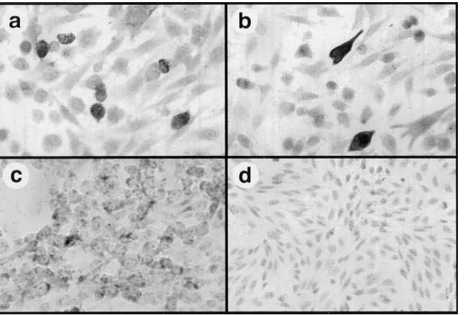
[image:4.612.65.551.497.641.2]rectly demonstrate SAg protein production in in vitro trans-fected cells. Strong positive staining for immunoreactive SEB and SEA proteins was observed intracellularly in 48-h trans-fected CHO cells (Fig. 3 a and b), compared to mock-trans-fected cells (Fig. 3 d). Superantigen expression could be detected beginning as early as 12 h after transfection (not shown). Al-though transiently transfected cells produced substantial amounts of intracellular SAg protein, stably transfected CHO cells expressed much lower levels (Fig. 3 c). Thus, prolonged exposure to high intracellular SAg concentrations may be le-thal to some cells, such that only low levels of intracellular SAg expression can be tolerated for prolonged periods.
Quantitation of intracellular and extracellular SEB produc-tion by transfected CHO cells. An antigen ELISA was used to quantitate SEB protein production by transfected CHO cells. Serial dilutions of supernatants and lysates from 48-h trans-fected CHO cells (5 3 105 cells) were assayed in triplicate,
us-ing an SEB-specific antigen ELISA. Cells were transfected with either empty vector, SEB, or SEA. The concentration of
SEB was determined by comparison to a standard curve gen-erated with recombinant SEB. Results are representative of three separate experiments. Supernatants contained 13.0 pg/ml SEB, whereas lysates contained 3.0 pg/ml SEB (Table I). By immunocytochemistry, 10% of cells (5 3 104 cells) expressed
detectable SEB protein (see Fig. 3). Thus, it was estimated that an individual high-expressing cell produced z 0.12 fg of
intracellular SEB and 0.52 fg of extracellular SEB, assuming that all the intracellular SAg protein was completely liberated by freeze thawing. Depending on the efficiency of gene trans-fer, these data suggest that in vivo transfected cells could therefore serve as a significant source of local SAg activity in vivo, inasmuch as T cells can respond to concentrations of SEB or SEA as low as 1.0–10.0 fg/ml (20).
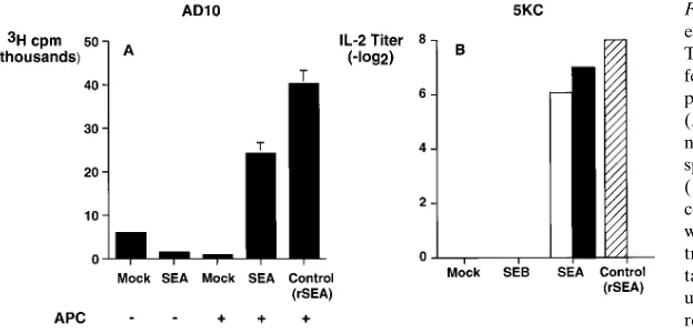
[image:5.612.58.359.92.251.2]Supernatants from SEA-transfected cells stimulate T cell proliferation in a Vb-specific manner. Superantigens differ from conventional lymphocyte mitogens in that they only bind and stimulate T cells expressing certain TCR Vb chains, whereas mitogens such as concanavalin A stimulate T cells irrespective
Figure 2. SEB and SEA are secreted by stably trans-fected cells. Stably transtrans-fected CHO cells were used to detect secretion of biologically active SAg pro-teins. Stably transfected CHO cells were seeded into nine separate wells of a 24-well plate, adhered over-night, then washed with PBS, and 1.0 ml fresh me-dium was added. Supernatants were then harvested from one well at each time point, and frozen before assay for SAg biologic activity. Supernatants were assayed for T cell stimulatory activity, using normal human PBMC, as described in Fig. 1 and Methods. The PBMC stimulatory activity present in serially harvested supernatants from mock-transfected CHO (open squares) versus SEB (A) (closed triangles) or SEA-transfected CHO cells (B) was quantitated. For both SEB- and SEA-transfected CHO cell lines, T cell stimulatory activity in supernatants of the transfectants increased with time of culture, consis-tent with SAg secretion by stably transfected cells.
[image:5.612.59.386.517.739.2]of which TCR Vb they express. In mice, for example, SEA ac-tivates T cells that express Vb 1, 3, 10, 11, 12, and 17 TCRs, whereas SEB activates T cells that express the TCR Vb 7 and 8.1-8.3 chains (3). The TCR Vb specificity of SAgs was there-fore used to demonstrate that the lymphocyte stimulatory ac-tivity present in supernatants of SAg-transfected CHO cells was not due to a nonspecific mitogenic effect. Irradiated, SEA-transfected B16 cells stimulated proliferation of AD10 cells (a Vb31, CD41 T cell clone) in the presence of syngeneic splenic antigen-presenting cells (APC) (Fig. 4 A). In the ab-sence of APCs, or in the preab-sence of mock-transfected B16 cells, the AD10 cells did not proliferate (Fig. 4 A).
Supernatants and lysates from SEA-transfected CHO cells also stimulated IL-2 production by the Vb31 T cell hybridoma 5KC (Fig. 4 B). Supernatants or lysates from SEB-transfected or mock-transfected CHO cells did not stimulate IL-2 produc-tion (Fig. 4 B). Thus, the T cell stimulatory properties of pro-teins produced by mammalian cells transfected with bacterial SAgs were consistent with those of the recombinant SAg mol-ecules, indicating that the SAg produced by transfected mam-malian cells was functional and TCR Vb specific.
Intramuscular injection of SAg genes induces a marked mononuclear cell inflammatory response. Plasmid DNAs in-jected directly into muscle tissue are efficiently expressed, leading to immune responses to proteins encoded by the
in-jected genes (35). Therefore, we inin-jected SAg plasmids into quadriceps muscles of B10.BR mice, and assessed the intramus-cular inflammatory response histologically, as an indirect mea-sure of in vivo gene expression. Injection of either empty vector plasmid DNA or an expression vector encoding the ovalbumin cDNA elicited only minimal inflammatory re-sponses in injected muscle tissues 1 wk after injection (Fig. 5, a
and b). By contrast, injection of plasmid DNA encoding either SEA, SEB, or TSST-1 induced a marked mononuclear cell in-flammatory response (Fig. 5, d–f). The mononuclear cell infil-trate was present between muscle fiber bundles, and also in loose connective tissues adjacent to muscle groups. The in-flammatory infiltrate was not uniform within muscle tissues, but was instead distributed in a multifocal pattern. This inflam-matory infiltrate distribution was consistent with the pattern of gene expression observed in muscle tissues after injection of a b-galactosidase reporter gene (data not shown). The inflam-matory response induced by injection of SEA and TSST-1 DNA was generally stronger than that induced by SEB DNA injection. Intramuscular injection of 1.0 mg recombinant SEA in PBS elicited only a mild inflammatory response, similar to that induced by injection of empty vector DNA (Fig. 5 c). Thus, prolonged SAg expression achieved by gene transfer may be necessary to induce strong local inflammatory re-sponses in tissues such as skeletal muscle, whereas SAg pro-teins injected into such tissues do not persist long enough to in-duce significant inflammatory cell infiltration.
Sequential examination of SEA-injected muscle specimens revealed that mononuclear cell infiltration began by 48 h after DNA injection, reached a maximum at 5–6 d, then began to subside thereafter (data not shown). By 2 wk, the inflamma-tory response had diminished considerably, and consisted largely of monocytes and scattered foci of T cells. Thus, the lo-cal inflammatory effects of SAg gene expression were tran-sient, most likely due to transient in vivo gene expression. The intramuscular inflammatory cell infiltrate was comprised pri-marily of two cell populations, based on immunohistochemical staining of muscle tissues (data not shown). Approximately 50% of the mononuclear cells were T cells (both CD41 and CD81), and the other 50% were Mac-1 positive monocytes and macrophages. Occasional polymorphonuclear cells
(neu-Table I. Quantitation of SEB Production by In Vitro Transfected CHO Cells
Gene transfected Assayed SEB (pg/ml)*
SEB Supernatant 13.0
SEB Lysate 3.0
SEA Supernatant 0
SEA Lysate 0
Empty vector Supernatant 0
Empty vector Lysate 0
[image:6.612.56.300.83.181.2]*Values are mean SEB concentration from triplicate wells of trans-fected CHO cells.
Figure 4. (A) SEA-transfected cells stimulate prolif-eration of Vb31 T cells. To determine whether the T cell stimulatory activity generated by SAg-trans-fected cells was TCR Vb-specific, we assessed the proliferative responses of a Vb31 T cell clone (AD10) to coculture with SEA-transfected B16 mela-noma cells. As a source of syngeneic APC, irradiated splenocytes from B10.BR mice were added to APC (1) wells, while APC (2) wells received only B16 cells and AD10 cells. Recombinant SEA (100 ng/ml) was added to AD10 cells plus APCs as a positive con-trol. After 2 d of incubation, proliferation was quanti-tated by [3H]thymidine incorporation, and the mean
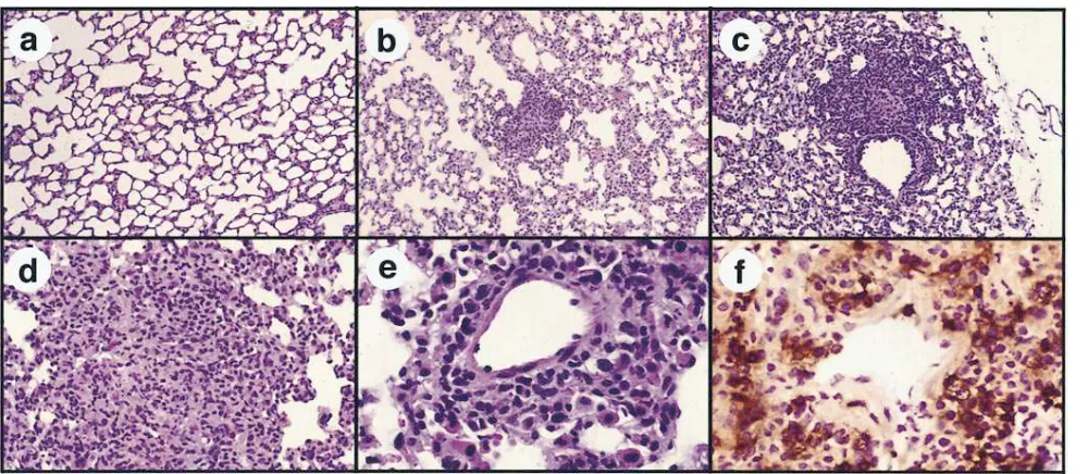
[image:6.612.61.373.540.690.2]Figure 5. Intramuscular injection of SAg plasmid DNA induces a strong mononuclear cell inflammatory response. Quadriceps muscles of B10.BR mice were injected once with 50 mg plasmid DNA encoding SEB, SEA, TSST-1, chicken ovalbumin, or empty vector DNA. The plasmid DNA was diluted to 0.5 mg/ml in PBS before injection. Other mice were injected once intramuscularly with 1.0 mg recombinant SEA protein. 1 wk later (a–e) or 4 d later (f), mice were killed and the quadriceps muscles were harvested, fixed in formalin, sectioned, and stained with hema-toxylin and eosin. Intramuscular injection of empty vector plasmid DNA (a) or a plasmid DNA encoding ovalbumin induced a minimal inflam-matory response (b), as did 1.0 mg recombinant SEA protein (c). By contrast, a marked mononuclear cell infiltrate developed in muscle tissues from mice injected with plasmid DNAs encoding either TSST (d), SEB (e), or SEA (f). In occasional sections, particularly at the 4-d time point, mononuclear cells emigrating from intramuscular blood vessels into perivascular tissues were observed (f). By 2 wk after injection, the inflam-matory response had diminished considerably (data not shown). Similar results have been obtained in numerous SAg DNA-injected mice. 3170 for a–e and 3350 for f.
Figure 7. Intravenous injection of SEA DNA complexed to cationic lipids induces pulmonary perivascular inflammation. Mice (two per group) were injected once intravenously with 120 mg plasmid DNA complexed to a cationic lipid formulation (32). Lung tissues were harvested 6 d after injection and stained for routine histologic evaluation. Multifocal perivascular accumulations of mononuclear cells were observed in multiple sections of lung tissues from SEA DNA-injected mice (b). By contrast, inflammatory changes were not observed in lung tissues of mice injected with empty vector DNA (a). At a higher magnification, the perivascular infiltrate was found to be comprised primarily of mononuclear cells (c, d, and e) and in some cases formed typical granulomas (d). Immunostaining of lung sections with an anti-CD4 antibody revealed numerous CD41 T cells in the perivascular infiltrate (f), whereas CD41 T cells were not increased in lung tissues of empty vector DNA–injected mice (not shown). Numerous Mac-11 cells were also present in the perivascular infiltrates, along with some CD81 T cells (not shown). 385 (a and b),
[image:7.612.58.558.422.640.2]trophils) were also observed in the cellular infiltrate. The T cells probably arrived in muscle tissues by emigration from blood vessels, as suggested by the presence of numerous mononuclear cells adjacent to intramuscular blood vessels at early time points after injection (Fig. 5 f). We have also ob-served local proliferation of T cells in SAg DNA-injected mus-cle tissues (Dow, S.W., unpublished data). Thus, tissue expres-sion of SAg genes serves as a stimulus for both T cell and monocyte immigration and local proliferation.
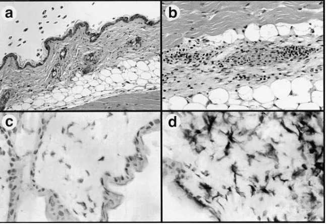
Cutaneous inflammatory responses induced by intradermal injection of SAg DNA. Previous studies have determined that plasmid DNAs can also be expressed efficiently after intrader-mal injection (36). We therefore assessed the response of cuta-neous tissues to intradermal injection of plasmid DNA-encod-ing SEA. Injection of SEA DNA, but not empty vector DNA, induced a mononuclear inflammatory response in dermal tis-sues (Fig. 6, a and b), though the magnitude of the response was less than in muscle tissues. In addition, a pronounced infil-trate of CD41 T cells was observed in epidermal tissues of SEA DNA-injected mice, but not in empty vector–injected mice (Fig. 6 c and d). Infiltration of Mac-11 cells was also ob-served in epidermal and dermal tissues (data not shown). The CD81 T cell response was less in skin than in muscle tissues (data not shown).
Intravenous injection of SAg DNA induces intrapulmonary inflammation. Previous studies have demonstrated intrapul-monary expression of reporter genes after intravenous injec-tion of plasmid DNA complexed to liposomes (32, 37, 38). We therefore investigated the pulmonary inflammatory response to intrapulmonary expression of SAg genes. Lung tissues from mice injected intravenously with SEA plasmid DNA com-plexed to cationic lipids contained numerous foci of mononu-clear cells in a perivascular orientation (Fig. 7, b–e), consistent with endothelial cell SEA gene expression. In most sections, the cellular composition of these foci resembled granulomas (e.g., Fig. 7 d). By immunohistochemistry, the foci were found to be comprised primarily of CD41 T cells (Fig. 7 f) and macrophages (not shown). Injection of empty vector DNA and lipids induced only minimal pulmonary inflammation (Fig. 7
a). Despite the presence of multifocal pulmonary infiltrates in SEA DNA-injected mice, the mice did not exhibit obvious res-piratory distress, and repeated intravenous SEA DNA injec-tions did not induce detectable illness (data not shown). Thus, intrapulmonary expression of SAg genes elicited a strong but apparently self-limiting mononuclear cell perivascular inflam-matory response.
Mice injected with SAg DNA by any of three routes did not develop adverse effects, either locally in the muscles (lameness) or lungs (respiratory distress) or systemically (an-orexia, weight loss). Mice observed for up to 6 mo after intra-muscular SAg DNA injection remained healthy (data not shown). Thus, SAg gene expression in a variety of tissues in-duced local mononuclear cell inflammatory responses without inducing overt toxicity.
Discussion
Numerous studies have established that bacterial SAgs are po-tent, TCR-specific T cell activators (1–7). These properties of SAgs have been extremely valuable tools for investigating mechanisms underlying T cell activation, anergy, tolerance in-duction, and peripheral deletion. Superantigens are also at-tractive agents for use in immunotherapy. For certain applica-tions, the ability to achieve sustained, locally expressed SAg activity has distinct advantages over systemic administration of SAg proteins, both in terms of more specific immune modula-tion and reduced toxicity. We therefore used a genetic therapy approach to address two aims: (a) to evaluate functional SAg expression in mammalian cells in vitro and (b) to assess the tis-sue inflammatory response to SAg gene expression in several different tissues in vivo.
[image:8.612.59.384.60.282.2]The data presented here demonstrate that three different bacterial SAg genes (SEA, SEB, and TSST-1) were capable of expressing biologically active proteins in a variety of different mammalian cells when placed under the control of a strong eu-karyotic promoter. Superantigen proteins present in both supernatants and lysates of transfected cells stimulated prolif-eration of human and mouse lymphocytes. In addition, SAgs
expressed by mammalian cells retained their specificity for TCR Vb chains, indicating that any posttranslational modifica-tions of the SAg protein made by mammalian cells did not al-ter either SAg function or specificity.
Mammalian cells produced transient high concentrations of SAg proteins intracellularly, as demonstrated by intense stain-ing of intracellular deposits of SEB and SEA protein in trans-fected cells (Fig. 3). High concentrations of intracellular SAgs, however, may induce cellular cytotoxicity, as evidenced by the inability to detect the same high levels of SAg gene expression in stably transfected cell lines. A previous study also demon-strated expression of the TSST-1 gene intracellularly in yeast (39). We have also expressed several other staphylococcal SAg genes in mammalian cells (Dow, S., unpublished data). These results suggest that most bacterial SAgs can be expressed in mammalian cells using eukaryotic expression vectors.
Bacterial superantigens were constituitively secreted by in vitro transfected cells at levels more than sufficient for T cell activation (20). The mechanism underlying the movement of SAg proteins across intact cell membranes is currently un-known, but could represent either active transport or passive diffusion along a concentration gradient. Studies with mutated SAgs may determine those portions of the SAg molecules re-sponsible for transcellular movement.
Histologic evaluation of three different SAg DNA–injected tissues indicated that localized expression of SAg genes served as a potent stimulus for mononuclear cell infiltration. Local, sustained SAg expression by SAg gene transfer was necessary to induce the prolonged mononuclear cell inflammatory re-sponse. For example, direct intramuscular injection of recom-binant SEA induced only a minimal inflammatory reaction, compared to intramuscular expression of SEA DNA (Fig. 5). Intradermal injection or percutaneous application of SEB protein was reported recently to induce localized cutaneous in-flammatory responses in mice (40). The response consisted ini-tially of Langerhan’s cell activation, vasodilation, and neu-trophil accumulation, followed in 48 h by mononuclear cell infiltration, which then resolved over 4–5 d (40). A similar re-sponse was observed in cutaneous tissues of mice in our study, except that the duration of the response was prolonged com-pared to the inflammatory response that was reported to have been induced by injection of SAg protein.
The perivascular location of the inflammatory responses observed in lung tissues of SEA DNA–injected mice is consis-tent with endothelial cell transfection and local gene expres-sion. Lesions were not observed histologically in other tissues of SEA DNA–injected mice (kidney, brain, spleen, heart), ex-cept for mild periportal mononuclear cell infiltrates in hepatic tissues (data not shown). These data are consistent with the re-sults of previous reporter gene studies using DOTIM-choles-terol lipids, which demonstrated preferential gene expression in pulmonary tissues (32). Pulmonary inflammatory lesions were also not observed in mice injected intravenously with SEA protein (data not shown), further emphasizing the impor-tance of local SAg gene expression in mediating the inflamma-tory response.
The cellular response to SAg gene expression was similar in all three tissues examined. Approximately 50% of the cellu-lar infiltrate consisted of T cells, with CD41 T cells being more numerous than CD81 T cells, especially in skin and lung tissues. Particularly in the epidermis of SEA DNA–injected skin, the infiltrate consisted almost entirely of CD41 cells
(Fig. 6). Mac-11 cells (monocytes) were also abundant in SAg-induced inflammatory foci, and were particularly numer-ous in muscle tissues. In all three tissues examined, the lesion induced by SAg gene expression was characteristic of a de-layed-type hypersensitivity response. Thus, local SAg expres-sion may be useful for inducing TH1-type T cell responses in tissues, using either direct DNA injection techniques or lipid-mediated gene targeting.
The use of SAg gene expression to induce localized inflam-matory responses has obvious application to the treatment of cancer. It was reported previously that intratumoral injections of plasmid DNA encoding an allogeneic MHC molecule, which also induces local inflammatory reactions, could slow tumor growth experimentally (41). We have found that repeated di-rect injections of SEB DNA plus cytokine DNA into sponta-neous malignant melanomas in dogs induce a pronounced in-flammatory cell infiltrate, followed in many cases by tumor regression, development of systemic antitumor immune re-sponses, and prolonged survival (Dow, S.W., Elmslie, R., and Potter, T.A., manuscript submitted for publication). From a safety standpoint, direct injection of plasmid DNA avoids problems associated with viral vectors, including the dangers of transfection of germ line cells or acquisition of viral replica-tion competence. In addireplica-tion, the extreme potency of SAgs makes it possible to induce biologic effects with even low in vivo transfection efficiencies.
In summary, we have shown that bacterial SAgs can be ex-pressed efficiently by mammalian cells in vitro and in vivo. In vivo expression of SAg genes in several different tissues in-duced a prominent T cell infiltrate, and did not induce toxicity or irreversible local pathology. Thus, direct injection and local expression of bacterial SAg genes may be useful for a variety of immunotherapeutic applications.
Acknowledgments
We thank Kathy Morgan for excellent secretarial assistance, and An-drew Willson for excellent technical assistance. We also thank Drs. Cori Gorman and Lisa Roche at the Megabios Corporation for providing the lipid–DNA complexes used for intravenous injection studies.
This work was supported in part by Public Health Service grant AI952-05 and by a grant from the Colorado Cancer League. T. Potter is a scholar of the Leukemia Society of America.
References
1. Peavy, D.L., W.H. Adler, and R.T. Smith. 1970. The mitogenic effects of endotoxin and staphylococcal enterotoxin B on mouse spleen cells and human
peripheral blood lymphocytes. J. Immunol. 105:1453–1458.
2. Langford, M.P., G.J. Stanton, and H.M. Johnson. 1978. Infect. Immun.
22:62–68.
3. Herman, A., J.W. Kappler, P. Marrack, and A.M. Pullen. 1991.
Superan-tigens: mechanism of T-cell stimulation and role in immune responses. Annu.
Rev. Immunol. 9:745–772.
4. Ochi, A., K. Yuh, K. Migita, and Y. Kawabe. Effects of Staphylococcal
toxins on T cell activity in vivo. 1992. Chem. Immunol. 55:115–136.
5. Kotzin, B.L., D.Y.M. Leung, J. Kappler, and P. Marrack. 1993.
Superan-tigens and their potential role in human disease. Adv. Immunol. 54:99–165.
6. Marrack, P., and J. Kappler. 1990. The staphylococcal superantigens and
their relatives. Science (Wash. DC). 248:1066.
7. Micusan, V.V., and J. Thibodeau. 1993. Superantigens of microbial
ori-gin. Semin. Immunol. 5:3–11.
8. Jardetzky, T.S., J.H. Brown, J.C. Gorga, L. Stern, R.G. Urban, Y. Chi, C. Stauffacher, J.L. Strominger, and D.C. Wiley. 1994. Three dimensional struc-ture of a human class II histocompatibility molecule complexed with
superanti-gen. Nature (Lond.). 368:711–718.
1990. Residues of the variable region of the T cell receptor B-chain that interact
with S. aureus superantigens. Nature (Lond.). 346:471–473.
10. Gasciogne, N.R., and K.T. Ames. 1991. Direct binding of secreted T-cell receptor to superantigen associated with class II major histocompatibility
com-plex protein. Proc. Natl. Acad. Sci. USA. 88:613–616.
11. White, J., A. Herman, A. M. Pullen, R. Kubo, J. Kappler, and P.
Mar-rack. 1989. The Vb-specific superantigen staphylococcal enterotoxin B:
stimu-lation of mature T cells and clonal deletion in neonatal mice. Cell. 56:27–35.
12. Fleischer, B., and H. Schrezenmeier. 1988. T cell stimulation by staphy-lococcal enterotoxins. Clonally variable response and requirement for major
histocompatibility complex class II molecules on accessory target cells. J. Exp.
Med. 167:1697–1707.
13. Fraser, J.D. 1989. High affinity binding of staphylococcal enterotoxins
A and B to HLA-DR. Nature (Lond.). 339:221–223.
14. Scholl, P.R., A. Diez, R. Karr, R.P. Sekaly, J. Trowsdale, and R.S. Geha. 1990. Effect of isotypes and allelic polymorphism on the binding of
staphylo-coccal enterotoxins to MHC class II molecules. J. Immunol. 144:226–230.
15. Herman, A., G. Croteau, R.P. Sekaly, J. Kappler, and P. Marrack. 1990. HLA-DR alleles differ in their ability to present staphylococcal superantigens
to T cells. J. Exp. Med. 172:709–717.
16. Janeway, C.A., J. Yagi, P.J. Conrad, M.E. Katz, B. Jones, S. Vroegop, and S. Buxser. 1989. T-cell responses to Mls and to bacterial proteins that
mimic its behavior. Immunol. Rev. 107:61–88.
17. Choi, Y., B. Kotzin, L. Herron, J. Callahan, P. Marrack, and J. Kappler.
1989. Interaction of Staphylococcus aureus toxin “superantigen” with human T
cells. Proc. Natl. Acad. Sci. USA. 86:8941–8945.
18. Herman, A., N. Labrecque, J. Thibodeau, P. Marrack, and J. Kappler. 1991. Identification of the staphylococcal enterotoxin A superantigen binding
site in the b-1 domain of the human histocompatibility antigen HLA-DR. Proc.
Natl. Acad. Sci. USA. 88:9954–9958.
19. Panina-Bordignon, P., X.T. Fu, A. Lanzavecchia, and R.W. Karr. 1992. Identification of HLA-DR alpha chain residues critical for binding of the toxic
shock syndrome toxin superantigen. J. Exp. Med. 176:1779–1784.
20. Carlsson, R., H. Fischer, and H.O. Sjogren. 1988. Binding of staphylo-coccal enterotoxin A to accessory cells is a requirement for its ability to activate
human T cells. J. Immunol. 140:2484–2488.
21. Rott, O., H. Wekerle, and B. Fleischer. 1992. Protection from
experi-mental allergic encephalitis by application of a bacterial superantigen. Int.
Im-munol. 4:347–353.
22. Kawamura, T., M. Nagata, T. Utsugi, and J. Yoon. 1993. Prevention of
autoimmune type I diabetes by CD41 suppressor T cells in
superantigen-treated non-obese diabetic mice. J. Immunol. 151:4362–4370.
23. Gelfand, E.W., J. Saloga, and G. Lack. 1995. Modification of immediate
hypersensitivity responses by staphylococcal enterotoxin B. J. Clin. Immunol.
15:37S–41S.
24. Schwab, J.H., R.R. Brown, S.K. Anderle, and P.M. Schlievert. 1993.
Su-perantigen can reactivate bacterial cell wall-induced arthritis. J. Immunol. 150:
4151–4159.
25. Schiffenbauer, J., H.M. Johnson, E.J. Butfiloski, L. Wegrzyn, and J.M. Soos. 1993. Staphylococcal enterotoxins can reactivate experimental allergic
encephalomyelitis. Proc. Natl. Acad. Sci. USA. 90:8543–8546.
26. Newell, K.A., J.D.I. Ellenhorn, D.S. Bruce, and J.A. Bluestone. 1991. In
vivo T-cell activation by staphylococcal enterotoxin B prevents outgrowth of a
malignant tumor. Proc. Natl. Acad. Sci. USA. 88:1074–1078.
27. Ochi, A., K. Migita, J. Xu, and K. Simonovitch. 1993. In vivo tumor
im-munotherapy by a bacterial superantigen. J. Immunol. 151:3180–3186.
28. Dohlsten, M. L., P. Abrahamsen, P. Bjork, P.A. Lando, G. Hedlund, G. Forsberg, T. Brodin, N.J.R. Gascoigne, C. Forberg, P. Lind, and T. Kalland. 1994. Monoclonal antibody-superantigen fusion proteins: tumor specific agents
for T-cell-based tumor therapy. Proc. Natl. Acad. Sci. USA. 91:8945–8949.
29. Murray, D.L., D.H. Ohlendorf, and P.M. Schlievert. 1995.
Staphylococ-cal and streptococStaphylococ-cal superantigens: their role in human diseases. ASM News.
61:229–235.
30. Azevedo, J.C.S. 1989. Animal models for toxic shock syndrome: an
overview. Rev. Infect. Dis. 11:S205–S209.
31. Miethke, T., C. Wahl, K. Heeg, B. Echtenacher, P.H. Krammer, and H. Wagner. 1992. T cell mediated lethal shock triggered in mice by the
superanti-gen staphylococcal enterotoxin B: critical role of tumor necrosis factor. J. Exp.
Med. 175:91–98.
32. Solodin, I., C.S. Brown, M.S. Bruno, C. Chow, E. Jang, R.J. Debs, and
T.D. Heath. 1995. A novel series of imidazloinium compounds for in vitro and
in vivo gene delivery. Biochemistry. 34:13537–13544.
33. Zlotnik, A., R.P. Shimonkevitz, M.L. Gefter, J. Kappler, and P. Mar-rack. 1983. Characterization of the gamma interferon mediated induction of
an-tigen presenting activity of P388D1 cells. J. Immunol. 131:2814–2820.
34. Hamad, A.R.A., A. Herman, P. Marrack, and J.W. Kappler. 1994. Mono-clonal antibodies defining functional sites on the toxin superantigen
staphylo-coccal enterotoxin B. J. Exp. Med. 180:615–621.
35. Ulmer, J.B., J.J. Donnelly, S.E. Parker, G.H. Rhodes, P.L. Felgner, V.J. Dwarki, S.H. Gromkowski, R.R. Deck, C.M. DeWitt, A. Friedman, et al. 1993. Heterologous protection against influenza by injection of DNA encoding a viral
protein. Science (Wash. DC). 259:1745–1749.
36. Raz, E., D.A. Carson, S.E. Parker, T.B. Parr, A.M. Abai, G. Aichiniger, S.H. Gromkowski, M. Singh, D. Lew, M.A. Yankauckas, et al. 1994. Intrader-mal gene immunization: the possible role of DNA uptake in the induction of
cellular immunity to viruses. Proc. Natl. Acad. Sci. USA. 91:9519–9523.
37. Thierry, A.R., Y. Lunardi-Iskandar, J.L. Bryant, P. Rabinovich, R.C. Gallo, and L.C. Mahan. 1995. Systemic gene therapy: biodistribution and
long-term expression of a transgene in mice. Proc. Natl. Acad. Sci. USA. 92:9742–
9746.
38. Zhu, N., D. Liggitt, Y. Liu, and R. Debs. 1993. Systemic gene expression
after intravenous DNA delivery into adult mice. Science (Wash. DC). 261:209–211.
39. Deresiewicz, R.L., J.A. Falxenburg, M. Chan, R.W. Finberg, and D.L.
Kasper. 1994. Intracellular expression of toxic shock syndrome toxin 1 in
Sac-charomyces cerevisiae. Infect. Immun. 62:2202–2207.
40. Saloga, J., D.Y.M. Leung, C. Raerdon, R.C. Giorno, W. Born, and E.W. Gelfand. 1996. Cutaneous exposure to the superantigen staphylococcal
entero-toxin B elicits a T-cell dependent inflammatory response. J. Invest. Dermatol.
106:982–988.
41. Plautz, G.E., Z. Yang, B. Wu, X. Goa, L. Huang, and G. Nabel. 1993.
Immunotherapy of malignancy by in vivo gene transfer into tumors. Proc. Natl.