High-flux mitochondrial cholesterol trafficking,
a specialized function of the adrenal cortex
Colin Jefcoate
J Clin Invest.
2002;
110(7)
:881-890.
https://doi.org/10.1172/JCI16771
.
The adrenal cortex is a tissue of excess in terms of both cholesterol metabolism and
cholesterol exchange with the circulation. Exceptionally high levels of lipoprotein receptors
in this highly vascularized tissue provide ready access to dietary cholesterol, allowing the
adrenocortical cells to maintain impressive stores of cytoplasmic cholesterol ester (CE)
droplets. Tightly packed among the CE droplets are specialized mitochondria, carrying in
their inner membranes high levels of the cytochrome P450scc (CYP11A1). This enzyme
carries out the so-called side chain cleavage reaction, consuming cholesterol to produce
pregnenolone, the precursor of cortisol and all other steroids. Glucocorticoid synthesis is
tightly regulated at the level of cholesterol metabolism, which responds to ACTH stimulation
over a period of minutes and ceases equally quickly when this hormone is removed.
Remarkably, this dynamic process is modulated under most circumstances not by control of
the intrinsic enzymatic activity of P450scc, but rather by substrate availability. For this
reason, cholesterol transport within the mitochondrion has emerged as the key control point
for steroidogenesis. The adrenal cortex is not alone in requiring efficient and controlled
delivery of cholesterol into mitochondria. Other steroidogenic cells, including several cell
types in the ovary, the Leydig cells of the testis, and a subset of hippocampal neurons (1),
also employ P450scc to produce pregnenolone and a variety of downstream steroid
hormones or neurosteroids. […]
Perspective
Find the latest version:
The adrenal cortex is a tissue of excess in terms of both cholesterol metabolism and cholesterol exchange with the circulation. Exceptionally high levels of lipoprotein receptors in this highly vascularized tissue provide ready access to dietary cholesterol, allowing the adreno-cortical cells to maintain impressive stores of cytoplas-mic cholesterol ester (CE) droplets. Tightly packed among the CE droplets are specialized mitochondria, carrying in their inner membranes high levels of the cytochrome P450scc (CYP11A1).
This enzyme carries out the so-called side chain cleav-age reaction, consuming cholesterol to produce preg-nenolone, the precursor of cortisol and all other steroids. Glucocorticoid synthesis is tightly regulated at the level of cholesterol metabolism, which responds to ACTH stimulation over a period of minutes and ceases equally quickly when this hormone is removed. Remarkably, this dynamic process is modulated under most circumstances not by control of the intrinsic enzymatic activity of P450scc, but rather by substrate availability. For this reason, cholesterol transport with-in the mitochondrion has emerged as the key control point for steroidogenesis.
The adrenal cortex is not alone in requiring efficient and controlled delivery of cholesterol into mitochon-dria. Other steroidogenic cells, including several cell types in the ovary, the Leydig cells of the testis, and a subset of hippocampal neurons (1), also employ P450scc to produce pregnenolone and a variety of downstream steroid hormones or neurosteroids. In ver-tebrates ranging from birds and fish (2) to mammals, these various cell types all express a short-lived
mito-chondrial import factor now called the steroidogenic acute regulatory protein (StAR), which mediates this process. Here, I examine the often confusing literature on StAR’s mechanism of action, particularly in light of recent work establishing the importance of other play-ers, and I present a model for StAR’s interaction with cholesterol and with some of these other proteins. I also discuss the insights into mitochondrial function that have come from the analysis of patients with con-genital adrenal hyperplasia (CAH), who lack this factor. Finally, I consider the multi-tiered regulation of StAR and related proteins in adrenocortical cells and other steroidogenic cell types.
A StAR is born
Cells of the glomerulosa, fasciculata, and reticularis zones of the cortex express distinct cholesterol-pro-cessing enzymes and act in concert to produce various steroids. In the central fasciculata zone (where the bulk of cortisol is produced) and elsewhere, steroidogenesis is greatly stimulated by exposure to ACTH, which acts primarily by elevating cAMP and protein kinase A (PKA) activity. In 1971, Garren et al. (3) proposed that the effects of ACTH and cAMP are mediated by one or more labile proteins that turn over with a half-time of about 3 minutes. In early spectroscopic studies of P450scc in isolated mitochondria (4), it became clear that cholesterol binding by this cytochrome is enhanced by ACTH treatment. Because this change parallels an increase in a kinetically defined pool of rap-idly metabolized cholesterol, these data indicated that the key action of ACTH is to make cholesterol available to the cytochrome (4). Further findings, indicating that P450scc is active prior to hormone treatment and that ACTH stimulates steroidogenesis only when the mito-chondria are physically intact, were consistent with this idea and suggested that delivery of cholesterol across the mitochondrial membranes was limited and could be increased by ACTH treatment.
Later work (5) tied these threads together by showing that the labile protein factor predicted by Garren et al. (3) is required for cholesterol transfer from the outer mitochondrial membrane (OMM) to the inner mito-chondrial membrane (IMM), where P450scc resides. Inhibition of protein synthesis in the presence of
High-flux mitochondrial cholesterol trafficking, a specialized
function of the adrenal cortex
Colin Jefcoate
Department of Pharmacology, University of Wisconsin Medical School, Madison, Wisconsin, USA
J. Clin. Invest.110:881–890 (2002). doi:10.1172/JCI200216771.
PERSPECTIVE
Biology and biochemistry of cholesterol | Ira Tabas, Series Editor
Address correspondence to:Colin Jefcoate, Department of Phar-macology, University of Wisconsin Medical School,
1300 University Avenue, Madison, Wisconsin 53706, USA. Phone: (608) 263-3975; Fax: (608) 262-1257;
E-mail: [email protected].
Conflict of interest:No conflict of interest has been declared.
ACTH does not affect delivery of cholesterol to mito-chondria but results in accumulation of cholesterol in OMM and a corresponding loss of IMM cholesterol. Cholesterol-P450scc complexes then cease to form, and pregnenolone synthesis is suppressed. The cytochrome itself is not affected by this treatment, since even mild mitochondrial disruption, which relieves the barrier to intermembrane transfer of cholesterol, restores steroidogenesis. Cholesterol transport between the mitochondrial membranes thus became recognized as a limiting, hormonally regulated process (4). This view received further support from work with relatively sol-uble hydroxycholesterol analogs, which can be metab-olized to steroids without any activation process. Because such compounds are not constrained by mem-brane barriers, their turnover to pregnenolone is intrin-sically rapid and cannot be further enhanced by ACTH. These findings suggested that intermembrane trans-fer of cholesterol, the key control point for steroid biosynthesis, requires new synthesis of one or more proteins that become phosphorylated in response to cAMP. In 1983 and in subsequent papers, Orme-John-son and colleagues identified a set of rapidly degraded 30-kDa mitochondrial phosphoproteins, collectively identified as phosphorylated p30 StAR (pp30) (6). These proteins are derived from a p37 precursor, which is phosphorylated in response to cAMP and is processed in the mitochondria to the pp30 form. Like the putative labile factor required for steroidogenesis, pp30 disappears rapidly after addition of the transla-tional inhibitor cycloheximide (CHX). To explain the very rapid loss of activity in the presence of CHX, Orme-Johnson and colleagues proposed that the p37 protein is only effectively phosphorylated during trans-lation, prior to entry into the inner mitochondria where pp30 is then formed (6). This model predicted that any loss of pp30 through intramitochondrial dephosphorylation could only be restored through a new cycle of synthesis. Later work does not support a role for dephosphorylation of pp30 in the rapid inacti-vation of cholesterol metabolism when protein syn-thesis is blocked. Nevertheless, other aspects of p37’s itinerary and fate appear to match Orme-Johnson and colleagues’ early predictions quite closely.
The StARgene, cloned in 1994 by Clark et al. (7), encodes the p37 precursor protein identified by Orme-Johnson and colleagues. The properties of this unusu-al protein account for many of the anomunusu-alous charac-teristics of mitochondrial cholesterol metabolism. As predicted, StAR increases cholesterol metabolism and carries an N-terminal mitochondrial targeting sequence. StAR is readily converted to a form that lacks the 68 N-terminal amino acids and that corre-sponds to the previously reported p30 protein ACTH via cAMP and PKA generates pp30. Consistent with earlier biochemical work that had shown pp30 to be regulated by calcium (8) as well as cAMP, StAR also contains consensus sequences for calmodulin-depend-ent kinases and PKA. As discussed below, the subse-quent discovery that human CAH (a disease in which glucocorticoid production is greatly diminished)
results from loss-of-function mutations in the StAR
gene (9) established the essential role of this protein for normal adrenal steroidogenesis. This pathology is paralleled in StAR-null mice (10).
Pre- and post-StAR events are hormonally regulated
Cholesterol metabolism in normal adrenal cortex cells is acutely regulated by ACTH stimulation, rising appre-ciably within 3 minutes of treatment and peaking with-in 10–15 mwith-inutes (3). Defects with-in either PKA or G protewith-in coupling, as seen in mutant mouse adrenal cell sub-lines, block this response by preventing cAMP forma-tion (11). Other signaling pathways playing key roles in adrenal responses to ACTH include lipoxygenase acti-vation (12) and, at least in adrenal fasciculata cells, stim-ulation mediated by receptors for IGF1, retinoids, and thyroid hormone; several cytokines, conversely, can suppress production of steroid hormones by these cells (13). For the most part, I will focus here on the mecha-nisms of acute adrenal fasciculata responses to cAMP and its analogs, which are generally shared with testic-ular and ovarian cells. It is interesting to note, however, that StAR regulation in another adrenal steroidogenic cell type, the glomerulosa cell, responds via alternative pathways involving Ca2+stimulation (8).
While the unique CHX-sensitivity of adrenal choles-terol metabolism has drawn attention to mitochondr-ial transfer processes, the ACTH regulation of adrenal cells is equally dependent on the ability of cAMP to stimulate the rapid flow of cholesterol between the plasma membrane, cytoplasmic pools, and mitochon-dria. Typically, extramitochondrial transfer is limiting, thus preventing a build-up of OMM cholesterol (14), but such a build-up can occur when mitochondrial transfer is selectively compromised, for example fol-lowing CHX treatment.
In rodent adrenal cells, the stimulation of cholesterol transfer to inner mitochondrial P450scc peaks within 10 minutes of ACTH treatment, prior to a substantial rise in StAR mRNA and protein (15). Thus, StAR rap-idly ceases to be rate-limiting for steroidogenesis in these cells. On the other hand, in cultured human adre-nal cells, as in testis MA10 cells, this increase in choles-terol metabolism is much slower (16, 17) and for sever-al hours parsever-allels StAR synthesis. However, even in these cells, StAR levels can continue to rise after a peak cholesterol metabolism is attained, indicating that some other component must become rate limiting.
The earlier assumption that intrinsic P450scc activi-ty is unaffected by ACTH rested largely on the high, hormone-insensitive turnover of hydroxycholesterol. This finding now requires some reconsideration in light of evidence that P450scc interactions with cho-lesterol, but not with hydroxycholesterols, are strongly affected by the lipid environment of the IMM. Choles-terol interacts strongly with the fatty acid chains of many phospholipids and is thereby constrained from interacting with P450scc. Cholesterol binding to P450scc in lipid vesicles is greatly potentiated when the local membrane is rendered more fluid by the addition of FFAs (18). Cardiolipin, which enhances the active-site binding of cholesterol to cytochrome P450scc, like-wise promotes steroid formation (19). Hydroxycholes-terols bind P450scc approximately ten times more strongly than does cholesterol, and they are not affect-ed by the presence of cardiolipin. Hence, the absence of a hormonal effect on hydroxycholesterol metabolism may still obscure a real effect of ACTH on the relative-ly weak binding of cholesterol to the cytochrome.
Intramitochondrial movement of cytochrome P450scc to incoming cholesterol may also contribute to steroid production. Unlike other microsomal cytochromes, P450scc lacks an N-terminal transbrane sequence and readily dissociates from the mem-brane (20). This enzyme is free to exchange between membranes, and it does so more rapidly than choles-terol, as judged by in vitro experiments using phos-pholipid vesicles (18). As Parker, Chung, Orly, and col-leagues recently found, adrenal mitochondria of cells from mice deficient in P450scc fail to form the typical highly vesicular structure characteristic of the active adrenal cortex (21), suggesting a unique and impor-tant role for P450scc in mitochondrial membrane dynamics. Vesiculation of the IMM follows ACTH stimulation of hormonally suppressed adrenal glands and takes about 12 hours, consistent with the time for appearance of P450scc (22).
Cholesterol metabolism by P450scc is sensitive not only to the lipid environment within the mitochondria, but also to the steady supply of oxygen (the co-substrate of the side chain cleavage reaction) and lipoprotein cho-lesterol (23), both of which reach the adrenal gland through the bloodstream. Blood flow, in turn, depends on ACTH, which stimulates endothelin production (24). Because of the rapidity of cholesterol turnover by P450scc, the pools of available and cytochrome-associ-ated cholesterol in adrenal mitochondria are usually low (25), although it appears to build up when blood flow to the gland is restricted. Acute stimulation of blood flow by ACTH may therefore contribute to corti-sol synthesis by enhancing extracellular access of both cholesterol and oxygen to P450scc.
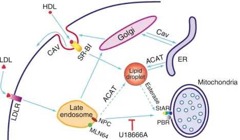
In addition, among its many effects on cholesterol metabolism in the adrenal gland, cAMP elevates the expression of the LDL receptor (26) and SR-BI (27), pro-motes cholesterol delivery to the mitochondria from the plasma membrane (28) and possibly from the late endo-somes (29), and stimulates cholesterol esterase activity while inhibiting acyl-CoA:cholesterol acyltransferase
(ACAT) (30). This combination of events favors the release of cholesterol from stored CEs and its net accu-mulation in mitochondria (3, 31). The late endosomes have been implicated as critical to delivery from LDL as evidenced by the inhibitory effect of the steroidamine U18666A. NPC-1 and the StAR-related protein MLN64 are likely mediators of this step. The various routes by which endogenous and exogenous cholesterol can reach the mitochondrion are shown in Figure 1.
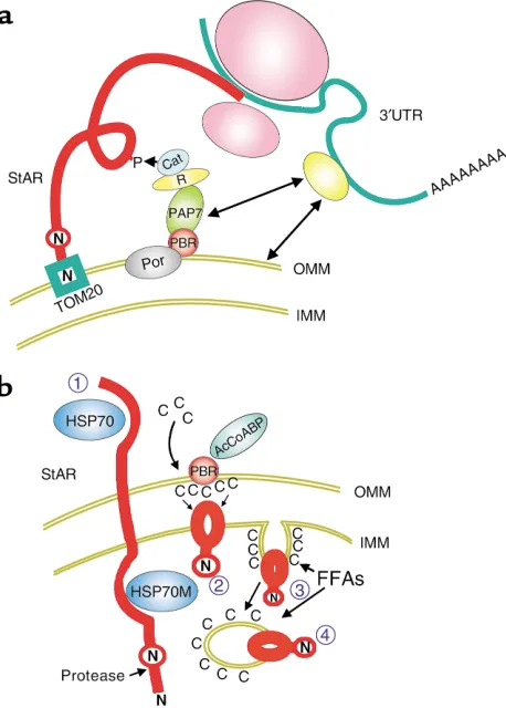
StAR can function at the OMM
[image:4.576.308.542.458.595.2]The site at which StAR acts to facilitate cholesterol metabolism by P450scc has been a matter of consider-able debate and confusion. The 68–amino acid N-ter-minal sequence of p37 StAR directs the protein into the inner mitochondria, where specialized proteases gener-ate the p30 form of the protein. Hormonal stimulation results in PKA-dependent phosphorylation of p37, gen-erating pp30 StAR (6, 7, 32–33), which accumulates within the mitochondrial matrix. Controversy has aris-en because there is clear evidaris-ence from well-defined model systems that p37 StAR, interacting exclusively with the OMM, can mediate cholesterol transfer to IMM P450scc (34–36). Paradoxically, there is equally clear evidence that inner mitochondrial proteolysis in functioning adrenal cells is essential to cholesterol flux-es (33) and that high cholflux-esterol transfer ratflux-es can be sustained aftercomplete processing of pp37 to pp30. These seemingly incompatible findings probably reflect crucial differences between the experimental models, specifically the action of components limited to adrenal cells that permit pp30 to function within the IMM. In addition, it may be that different mechanisms of cho-lesterol transport predominate in different cell types.
Figure 1
The model studies have relied on two types of exper-iments, which differ in important aspects with respect to regulation of cholesterol transport. First, recombi-nant StAR has been transfected into COS-1 cells that have been made functional for steroidogenesis through transfection of a chimeric P450scc/reductase molecule. COS-1 cells are not normally steroidogenic and pre-sumably lack many of the specialized components of the adrenal cholesterol processing system. Indeed, the transfected COS-1 mitochondria appear completely different from adrenal mitochondria and sustain only low rates of cholesterol metabolism in spite of high StAR expression (34). The second model system involves addition of extracts containing recombinant StAR to steroidogenic mitochondria. Here, the mito-chondria usually come from MA10 cells that have not been stimulated by cAMP. These mitochondria have low basal activity for cholesterol metabolism and, again, probably lack key components, some of which will be discussed below.
In COS-1 cells, StAR can enhance cholesterol metabolism independent of the N-terminal targeting sequence (34) that directs it to the OMM receptor TOM20 (36, 37) and then through the IMM, which initiates protein import into this organelle. Indeed, a COS-1 cell extract containing p30, which lacks this targeting sequence, is almost as effective as extracts expressing p37 in mediating cholesterol transfer into isolated MA10 mitochondria (37). These findings suggest that StAR can act on the OMM (34–36).
Indeed, experiments with synthetic phospholipid membranes confirm that p30 and p37 are each capa-ble of mediating transfer of cholesterol between mito-chondrial membranes (38).
The effectiveness of outer membrane activity by StAR has been recently substantiated by an elegant series of experiments in which StAR was linked to components of the mitochondrial protein import system (36). These experiments showed that chimeric StAR, linked to the outer membrane TOM20 but not to intermembrane or inner mitochondrial channel (TIM9 or TIM18) pro-teins, effectively activates cholesterol metabolism in isolated basal MA10 mitochondria. Thus, StAR clearly can mediate cholesterol transfer when linked to the outer membrane of MA10 mitochondria. It should be noted that these experiments do not exclude a role for in the IMM, since, for example, these isolated MA10 cell mitochondria might fail to support StAR activity at this site. As discussed below, the proper regulation of IMM FFAs may be one key factor missing in COS cells and isolated MA10 mitochondria.
StAR processing is preferred for high cholesterol fluxes
[image:5.576.61.290.56.376.2]There are several reasons to question the relevance of the OMM as the major site of StAR action in adrenal cells and, perhaps, in certain other cell types. Some of these concerns center on the properties of COS-1 cells. Thus, unlike in specialized steroidogenic cell types, cholesterol turnover in COS-1 cells is relatively
Figure 2
insensitive to changes in the level of StAR expression (C. Jefcoate, unpublished data). This finding may explain why transfected p30, which is broadly distrib-uted in the COS-1 cells, can be as effective as mito-chondrially localized p37 StAR. COS-1 cells also process p37 to p30 much more slowly than do adre-nal cells, thus providing more opportunity for an OMM mechanism to operate (7).
In addition, several well-established features of adre-nal StAR biosynthesis appear inconsistent with the extramitochondrial StAR hypothesis. First, p37 turns over so quickly that peak cholesterol transfer occurs at a point when p37 can scarcely be detected in steroido-genic cells, particularly MA10 cells (33). Second, intramitochondrial processing is essential to StAR activity. Thus, the mitochondrial proteinase inhibitor orthophenanthrolene and the protein uptake inhibitor CCCP each block pp30 formation and pre-vent cAMP activation of cholesterol metabolism. While it is difficult to exclude other effects of these inhibitors, it is striking that they have no effect on cholesterol metabolism after sufficient pp30 has been generated (33). These experiments also show that once even modest amounts of new pp30 are available in the IMM, activated cholesterol metabolism becomes com-pletely independent not only of processing, but also of both cAMP and protein synthesis.
While p37 certainly has activity when retained at the OMM, pp30, which forms very rapidly in the IMM, may therefore be the major contributor to enhanced cholesterol uptake to P450scc. There is reason to believe that pp30 StAR, acting within the mitochon-drion, is additionally needed to expedite cholesterol entry into the IMM, where multiple proteins involved in respiration and ATP efflux reside. In mitochondria of typical, nonsteroidogenic cells, cholesterol is largely excluded from the IMM, suggesting that this lipid can be detrimental to respiratory functions (39). For instance, incubation of hepatocyte mitochondria with albumin-bound cholesterol can cause a tenfold increas-es in the OMM cholincreas-esterol and even induce the depo-sition of intermembrane cholesterol droplets while fail-ing to increase IMM cholesterol. In adrenal cells, conversely, cholesterol can equilibrate between the OMM and IMM following ACTH treatment in the presence of a P450scc inhibitor (5, 14). ACTH-stimu-lated expansion of the IMM and mobilization of P450scc may represent specialized responses of these mitochondria, allowing cholesterol pools to be physi-cally segregated from the electron transport chain and other normal IMM proteins.
The unusually extensive and vesiculated IMM in adrenal cells, much of which does not have direct access to the OMM, poses a particular problem for efficient cholesterol delivery to P450scc. Primary intermem-brane cholesterol transfer may only occur to regions of the IMM that juxtapose the OMM. As argued below, pp30 StAR may be key to a secondary step in which substrate cholesterol is delivered to P450scc at loca-tions in the inner mitochondrion that are well removed from these peripheral IMM sites.
Intramitochondrial activation, deactivation, and disposal of StAR
While p37 StAR can clearly mediate cholesterol uptake into COS-1 cell mitochondria, its ability to do so in steroidogenic cells in vivo is likely constrained by the small amount of p37 associated with the OMM at steady state and by the distinctive biochemical and morphological features of mitochondria in these spe-cialized cells. An alternative hypothesis (presented in Figure 2) holds that StAR functions as pp30 via inter-action with the inner surface of the IMM. This mecha-nism is consistent with well-established aspects of StAR activation and disposal in active adrenal cells.
Newly synthesized StAR is essential for cholesterol transfer even when large amounts of multiple forms of variously modified StAR are present in the mitochon-dria prior to stimulation of the adrenal cells (33). The conversion of newly formed p37 to the p30 form is highly efficient and reaches a steady state between for-mation and processing within 5 minutes, consistent with very rapid turnover of p37 (t1/2approximately 1–2
minutes). cAMP has no measurable effect on these rates, but maximum stimulation ensures near-com-plete conversion, presumably by promoting the extramitochondrial phosphorylation of p37. The inhi-bition of ACTH-stimulated cholesterol metabolism by protein synthesis inhibitors, even when there is abun-dant IMM pp30, is attributed to the rapid loss of activ-ity of this protein following synthesis. Each molecule of newly synthesized pp30 StAR is estimated to medi-ate the transfer of 400 cholesterol molecules per minute (33). However, most of the pp30 that persists in the mitochondrion, including the bulk of the older molecules, may not be properly localized to contribute to intermembrane cholesterol transfer. Based on immunogold labeling experiments, StAR is found pre-dominantly around the matrix vesicles in normal adre-nal glands (40, 41). At this site, StAR seems unlikely to affect this process. Conversely, newly synthesized pro-tein may be present preferentially in IMM segments that are proximal to the OMM.
Orly has recently provided evidence that inner mito-chondrial p30 turns over slowly via a two-step process (42). Pulse-labeling experiments show that p30 is blocked within 2 hours after transfer to the IMM by selective protease inhibitors that subsequently become ineffective. Orly suggests that the inhibitor-sensitive StAR pool and the protease are located close to the site where p30 first enters the IMM. StAR at this site likely represents the IMM pool that is active in facilitating uptake from the OMM.
Deficiency of StAR function in relation to StAR structure
mis-sense and short deletion alleles affecting the C-termi-nal region are expressed but inactive in stimulating cholesterol metabolism. The positions of these muta-tions fit well with key structural sites predicted by the recent crystal structure of the related MLN64 (43), which shares with StAR a conserved structure termed a StAR-related lipid transfer (START) domain. The key feature of the structure, which presumably is shared by StAR, is a central channel, open at both ends, that can fit a cholesterol molecule. This presumptive cholesterol channel has positive and negatively charged amino acids at one end, including D169, which is mutated in some CAH patients.
Recent work using fluorescence transfer shows that the cholesterol derivative NBD-cholesterol binds with high affinity (Kd= 32 nM) (44) and that StAR activity
alters the lipid mobility of the surrounding membrane, as detected with fluorescent probes. START domains are also found in phosphatidylcholine transfer protein and diverse other proteins that bind phospholipids (see Maxfield, this Perspective series, ref. 45). However, two key residues that are shared with MLN64 (Asp144 and Asn148) are absent in other family members. The rela-tively polar StAR protein associates very strongly with mitochondrial membranes, suggesting possible phos-pholipid interactions.
Disruption of the StARgene in mice produces a simi-lar phenotype to CAH (10). ACTH-stimulated glucocor-ticoid output is lowered about 20-fold. Loss of feedback inhibition by glucocorticoids in the pituitary causes ACTH release to rise with a consequent stimulation of lipoprotein receptor–mediated uptake of CEs and hyper-plasia. The enormous lipid accumulation is ultimately toxic to the gland. Fortunately, the corresponding clini-cal effects can be reversed by early treatment of children with glucocorticoids, and this treatment is also effective in StAR-deficient mice. In males of this strain, testos-terone production is low and large amounts of lipid accumulate in the testis, where hyperplasia also ensues due to the elevated pituitary response. The ovaries are normal at birth but also exhibit interstitial lipid accu-mulation and slow maturation. Residual, StAR-inde-pendent cholesterol metabolism in the adrenal glands is clearly measurable and seems to be sufficient for the slower metabolism to estradiol in the ovaries.
The peripheral benzodiazepine receptor is essential for StAR activity
Like StAR, the peripheral benzodiazepine receptor (PBR) is strongly implicated in mitochondrial choles-terol uptake. PBR is located in the outer membrane of most mitochondria and is elevated in steroidogenic cells. Developing a model for steroidogenesis that inte-grates the information about both of these proteins has proved a significant challenge.
PBR associates with a member of the porin family of outer membrane channel proteins. When PBR is acti-vated by drug agonists or by the adrenal protein acyl-CoA–binding protein (also called DB1 and endozepine), it alters the porin’s permeability and thereby affects a variety of mitochondrial functions. PBR agonists also
stimulate cholesterol metabolism in adrenal and testis cells, while antagonists prevent the induction of mito-chondrial cholesterol transport and inhibit cAMP-stim-ulated steroidogenesis (46). Deletion of PBR severely compromises cholesterol metabolism, which can be restored by transfection of cells with PBR cDNAs. Ago-nist binding causes relocation of the outer membrane cholesterol to PBR, which binds the lipid via a defined sequence distinct from a START domain. The key role for PBR in cholesterol metabolism in adrenal cortex cells is substantiated by the observation that ginkgolide treatment suppresses both cholesterol metabolism and PBR expression without affecting StAR expression (47). Also suggestive is the finding that adrenal cholesterol metabolism in the neonatal rat almost disappears between days 5 and 15 in parallel with a loss of PBR (48); StAR shows only modest changes in expression over this developmental period.
inabili-ty of the mitochondrion to replenish StAR as it is lost from proximal IMM sites could account for the block-ade of ACTH-stimulated cholesterol transfer seen within minutes following CHX treatment.
Stimulation of StAR activity by mitochondrial FFAs
Recent work indicates that StAR is critically sensitive to the presence of FFAs. A novel mechanism has come to light whereby long-chain fatty acids released from CE hydrolysis can accumulate within the mitochondria of steroidogenic cells and stimulate cholesterol metab-olism. This pathway, which is probably not found in other cell types, could help explain the inconsistent functional data that have plagued this field.
The Podesta laboratory has recently established that transfer of fatty acyl-CoA to the inner mitochondria is essential for optimal StAR activity. The mitochon-drial thioesterase (MTE; also called ARTISt), which hydrolyzes acyl-CoA to release FFAs, carries a mito-chondrial targeting sequence and is found in vivo in the inner mitochondrion (54). This enzyme is pre-sumed to release arachidonic acid (AA) and other FFAs inside this organelle. Because linoleic and oleic acids are just as effective as AA at overcoming a block in steroid formation imposed by MTE inhibition (C. Jefcoate and E.J. Podesta, unpublished data), it appears that StAR activation requires fatty acids per se, rather than prostaglandins, leukotrienes, or other AA oxidation products.
Fatty acyl-CoAs derived from the hydrolysis of cyto-plasmic CEs bind to the acyl-CoA–binding protein, which can bind and may thereby activate PBR (46). This interaction favors the accumulation of fatty acids near StAR, perhaps by promoting fatty acyl CoA transfer into the inner mitochondria. While the mitochondrial fate of these fatty acids is clearly critical to the operation of the cholesterol uptake pathway, the mechanism by which fatty acids promote steroidogenesis has not been established. Possibly, the increase in membrane fluidity in the presence of these fatty acids favors the interaction of cholesterol with StAR or P450scc (18, 44). Podesta has pointed out that involvement of fatty acyl CoA and MTE would be compatible with the central role of PBR in cholesterol transport to the IMM (54).
Shaping up for StAR
An often overlooked component of the acute adrenal response is the extraordinarily fast cell contraction that is essential for the hormonal activation of cholesterol metabolism. Adrenal cells and testis in culture com-pletely round up as a result of cytoskeletal structural changes within a few minutes following ACTH stimu-lation (55). This change in cell shape and the attendant changes in cytoskeletal organization play an important role in the acute stimulation of cholesterol metabolism and the induction of StAR activity by ACTH and cAMP. Cytoskeletal disruption, which prevents this response, blocks cholesterol transfer to mitochondria (56).
Notably, the cytoskeleton-associated protein pax-illin, a component of the focal adhesion complex, undergoes cAMP-dependent dephosphorylation with
a half-time of about 5 minutes in parallel with the cell rounding in culture (55). PTPase inhibitors that block paxillin dephosphorylation (54) also substantially inhibit the acute stimulation of cholesterol metabo-lism. The same effects of ACTH on paxillin dephos-phorylation are seen in adrenal glands in vivo, although the relationship to steroidogenesis in the tis-sue remains to be determined. This change is generat-ed via cAMP/PKA activation of a phosphotyrosine phosphatase, SHP2 (55, 64), whose time course is sim-ilar to that of paxillin dephosphorylation.
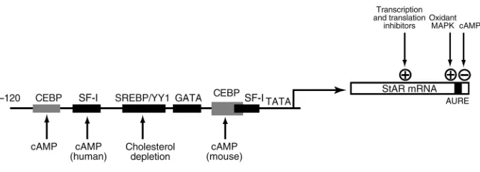
Regulation of StAR expression
Two species of StAR mRNA (1.6 and 3.5 kb) are stimu-lated by cAMP in rodent steroidogenic cells (58). In human but not in rodent cells, the short form seems to predominate both before and after stimulation (57). The StARgene has seven exons (58, 59), with exon 7 con-taining four sites for polyadenylation; these sites give rise to alternative mRNA forms. These predominant mRNA species differ only in their site of polyadenyla-tion, but they can be differentially regulated, since the longer 3′-untranslated region contains AU-rich ele-ments that are recognized specifically by cytoplasmic proteins (59). Stabilization of labile mRNA by such fac-tors provides a much faster response than is possible by transcriptional induction, a strategy that is used by early response genes and cytokines to upregulate gene expres-sion within 15 minutes (60, 61).
StAR expression is also regulated by the transcription factor SF-1, for which several promoter elements have been identified (13). Expression of StAR and other steroidogenic factors is lost in SF-1–deficient mice. The nuclear regulatory protein DAX1 inhibits StAR promot-er activity by binding in the region of the proximal SF-1 site (62). SF-1 is not directly activated by PKA phospho-rylation but can be activated by mitogen-activated pro-tein kinase (MAPK) (63). CEBPβalso plays an important role in cAMP regulation of StAR (13, 64).
StAR: not the last word
While we now have an excellent understanding of CAH, relatively little is known about the role of StAR during the normal acute regulation in human adrenal cells. Available human cell models respond very slowly to cAMP (16) relative to the adrenal gland in vivo, and even in rodent models, most studies have been carried out under maximum stimulatory conditions. More infor-mation is needed on adrenal cell responses to low levels of ACTH, as well as to various cytokines and thyroid and retinoid hormones (67–69). Much also needs to be determined about the signaling that links these activa-tors and suppressors to the key regulaactiva-tors, particularly components of the MAPK pathways (70). Many of these effects are highly tissue- and species-selective and may be sensitive to diet and circadian control.
High cholesterol fluxes through adrenal cortex cells are subject to regulation at multiple points and by diverse signaling processes affecting cytoplasmic trans-fer and transtrans-fer into the mitochondria. These process-es need to be balanced in order to maintain mitochon-drial access to cholesterol while at the same time avoiding an adverse accumulation of cholesterol in the mitochondria. Cholesterol uptake and transfer to the mitochondria are hormonally regulated at multiple lev-els, including lipoprotein receptors, CE hydrolysis, and endosome transport. Mitochondrial transfer from outer to inner membrane, while requiring StAR as an essential component, also shows a diversity of regula-tors and mechanisms.
This diversity of regulation is not surprising in view of the central role of glucocorticoid synthesis in inte-grating acute metabolic and immune responses and the adrenal gland’s key function in organizing respons-es to rapid changrespons-es in environment. While I have focused above on the novel mitochondrial processes involving StAR, it is critical to recognize that these probably only become rate-limiting under unusual cir-cumstances. These conditions have been generated by specific manipulations, including the genetic depletion of StAR and the complete inhibition of protein syn-thesis. MTE and PBR (46, 54) appear to be just as
essen-tial as StAR for steroidogenesis, but the consequences of aberrant adrenal expression or pharmacological manipulation of these proteins are not well under-stood. Nevertheless, it seems clear that the mitochon-drial process is both integrated and flexible, since it can respond to changes in fatty acid metabolism through MTE, to altered mitochondrial energy fluxes through PBR, and to interruptions in protein synthesis through the rapid functional turnover of StAR.
The question of whether StAR functions as p37 in the OMM or as pp30 in the IMM is not fully resolved, but it should be noted that these are not exclusive mecha-nisms and may either work together or be selectively preferred according to the circumstances. The OMM mechanism has been rigorously established, while the evidence for the IMM mechanism is dependent on strong circumstantial data. The mechanism that pre-dominates in a given cell likely depends on biochemi-cal context. OMM transfer could be favored when pro-cessing is slow, when the IMM is not unusually extended, or when MTE is absent. One or more of these conditions seem to apply in COS-1 cells and in isolat-ed mitochondria from unstimulatisolat-ed steroidogenic cells; as argued above, they probably do not apply in highly active adrenal or even testis cells. Which of these mechanisms act in the ovary and the brain (tissues with lower demand for steroid synthesis) is not yet clear.
Essentially all mechanistic experiments to date have focused on comparisons of the fully activated cells, but much remains to be learned about how these various processes function under different stimulation condi-tions, which may alter the balance between the various components. There may also be tissue-specific advan-tages of changes in the favored mechanism. For example, cholesterol transfer driven by an extramitochondrial p37 StAR can be regulated from the cytosol more readily than that driven by the intramitochondrial pp30. The latter, on the other hand, may introduce a greater sensi-tivity to mitochondrial energetics and ion balance.
[image:9.576.106.445.58.180.2]Several methods have been developed for dissecting these processes. In the future, it will be important to focus on more specific methods of dissection under
Figure 3
physiological conditions; for example, antisense and RNA interference to selectively deplete the various components. Unfortunately, the adrenal cortex remains a very enigmatic tissue, in part because cul-tures do not maintain the full physiological function. Even adrenal primary cultures lose a very large amount of the cholesterol flux seen in adrenal glands. Still, adrenal cells continue to be an important system for studying cholesterol transfer. The integration and cell specificity of these processes remain major challenges for future research.
1. Pollack, S.E., et al. 1997. Localization of the steroidogenic acute regula-tory protein in human tissue. J. Clin. Endocrinol. Metabol.82:4243–4251. 2. Bauer, M.P., Bridgham, J.T., Langenau, D.M., Johnson, A.L., and Goetz, F.W. 2000. Conservation of steroidogenic acute regulatory (StAR) pro-tein structure and expression in vertebrates. Mol. Cell. Endocrinol.
168:119–125.
3. Garren, L.D., Gill, G.N., Masui, H., and Walton, G.M. 1971. On the mech-anism of action of ACTH. Recent Prog. Horm. Res.27:433–478. 4. Jefcoate, C.R., et al. 1973. The detection of different states of the P-450
cytochromes in adrenal mitochondria: changes induced by ACTH. Ann. N.Y. Acad. Sci. 212:243–261.
5. Privalle, C.T., McNamara, B.C., Dhariwal, M.C., and Jefcoate, C.R. 1987. ACTH control of cholesterol side-chain cleavage at adrenal mitochon-drial cytochrome P450scc. Regulation of intramitochonmitochon-drial cholesterol transfer. Mol. Cell. Endocrinol. 53:87–101.
6. Epstein, L.F., and Orme-Johnson, N.R. 1991. Regulation of steroid hor-mone biosynthesis. Identification of precursors of a phosphoprotein tar-geted to the mitochondrion in stimulated rat adrenal cortex cells. J. Biol. Chem. 266:19739–19745.
7. Clark, B.J., Wells, J., King, S.R., and Stocco, D.M. 1994. The purification, cloning, and expression of a novel luteinizing hormone-induced mito-chondrial protein in MA-10 mouse Leydig tumor cells. Characterization of the steroidogenic acute regulatory protein (StAR). J. Biol. Chem.
269:28314–28322.
8. Elliott, M.E., Goodfriend, T.L., and Jefcoate, C.R. 1993. Bovine adrenal glomerulosa and fasciculata cells exhibit 28.5-kilodalton proteins sen-sitive to angiotensin, other agonists, and atrial natriuretic peptide.
Endocrinology.133:1669–1677.
9. Bose, H.S., Sugawara, T., Strauss, J.F.R., and Miller, W.L. 1996. The pathophysiology and genetics of congenital lipoid adrenal hyperplasia. International Congenital Lipoid Adrenal Hyperplasia Consortium.
N. Engl. J. Med. 335:141870–141878.
10. Hasegawa, T., et al. 2000. Developmental roles of the steroidogenic acute regulatory protein (StAR) as revealed by StAR knockout mice. Mol. Endocrinol.14:1462–1471.
11. Olson, M.F., Krolczyk, A.J., Gorman, K.B., Steinberg, R.A., and Schim-mer, B.P. 1993. Molecular basis for the 3′,5′-cyclic adenosine monophos-phate resistance of kin mutant Y1 adrenocortical tumor cells. Mol. Endocrinol. 7:477–487.
12. Yamazaki, T., Higuchi, K., Kominami, S., and Takemori, S. 1996. 15-Lipoxygenase metabolite(s) of arachidonic acid mediates adrenocorti-cotropin action in bovine adrenal steroidogenesis. Endocrinology.
137:2670–2675.
13. Christenson, L.K., and Strauss, J.F., III. 2000. Steroidogenic acute regu-latory protein (StAR) and the intramitochondrial translocation of cho-lesterol. Biochem. Biophys. Acta.1529:175–187.
14. DiBartolomeis, M.J., Williams, C., and Jefcoate, C.R. 1986. Inhibition of ACTH action on cultured bovine adrenal cortical cells by 2,3,7,8-tetra-chlorodibenzo-p-dioxin through a redistribution of cholesterol. J. Biol. Chem.261:4432–4437.
15. Jefcoate, C.R., Artemenko, I.P., and Zhao, D. 2000. Relationship of StAR expression to mitochondrial cholesterol transfer and metabolism.
Endocr. Res. 26:663–680.
16. Liu, P.H., Kahri, A.I., and Voutilainen, R. 1996. Expression of the steroidogenic acute regulatory protein mRNA in adrenal tumors and cultured adrenal cells. J. Endocrinol. 150:43–50.
17. Igarashi, Y., and Kimura, T. 1986. Adrenic acid content in rat adrenal mitochondrial phosphatidylethanolamine and its relation to ACTH-mediated stimulation of cholesterol side chain cleavage reaction. J. Biol.
Chem.261:14118–14124.
18. Dhariwal, M.S., and Jefcoate, C.R. 1989. Cholesterol metabolism by puri-fied cytochrome P-450scc is highly stimulated by octyl glucoside and stearic acid exclusively in large unilamellar phospholipid vesicles. Bio-chemistry.28:8397–8402.
19. Lambeth, J.D., Kitchen, S.E., Farooqui, A.A., Tuckey, R., and Kamin, H.
1982. Cytochrome P-450scc-substrate interactions. Studies of binding and catalytic activity using hydroxycholesterols. J. Biol. Chem. 257:1876–1884.
20. Hanukoglu, I., Spitsberg, V., Bumpus, J.A., Dus, K.M., and Jefcoate, C.R. 1981. Adrenal mitochondrial cytochrome P-450scc. Cholesterol and adrenodoxin interactions at equilibrium and during turnover. J. Biol. Chem. 256:4321–4328.
21. Ishii, T., et al. 2002. The roles of circulating high-density lipoproteins and trophic hormones in the phenotype of knock-out mice lacking steroidogenic acute regulatory protein. Mol. Endocrinol.In press. 22. Bornstein, S.R., Ehrhart-Bornstein, M., Guse-Behling, H., and
Scherbaum, W.A. 1992. Structure and dynamics of adrenal mitochon-dria following stimulation with cotricotropin releasing hormone. Anat. Rec.234:255–262.
23. Stevens, V.L., Aw, T.Y., Jones, D.P., and Lambeth, J.D. 1984. Oxygen dependence of adrenal cortex cholesterol side chain cleavage. J. Biol. Chem. 259:1174–1179.
24. Gaillard, I., et al. 2000. ACTH-regulated expression of vascular endothe-lial growth factor in the adult bovine adrenal cortex: a possible role in the maintenance of the microvasculature. J. Cell. Physiol.185:226–234. 25. Jefcoate, C.R., and Orme-Johnson, W.H. 1975. Cytochrome P-450 of adrenal mitochondria. In vitro and in vivo changes in spin states.J. Biol. Chem.250:4671–4677.
26. Kovanen, P.T., Goldstein, J.L., Chappell, D.A., and Brown, M.S. 1980. Regulation of low density lipoprotein receptors by adrenocorticotropin in the adrenal gland of mice and rats in vivo. J. Biol. Chem. 255:5591–5598.
27. Temel, R.E., et al. 1997. Scavenger receptor class B, type I (SR-BI) is the major route for the delivery of high density lipoprotein cholesterol to the steroidogenic pathway in cultured mouse adrenocortical cells. Proc. Natl. Acad. Sci. USA.94:13600–13605.
28. Gocze, P.M., and Freeman, D.A. 1992. A cholesterol ester hydrolase inhibitor blocks cholesterol translocation into the mitochondria of MA-10 Leydig tumor cells. Endocrinology.131:2972–2978.
29. Watari, H., et al. 2000. NPC-1 containing compartment of human gran-ulosa cells: a role in intracellular trafficking of cholesterol supporting steroidogenesis. Exp. Cell Res.255:56–66.
30. Buhman, K.F., Accad, M., and Farese, R.V., Jr. 2000. Mammalian acyl-CoA:cholesterol acyltransferases. Biochem. Biophys. Acta.1529:142–154. 31. Jamal, Z., Suffolk, R.A., Boyd, G.S., and Suckling, K.E. 1985. Metabolism of cholesteryl ester in monolayers of bovine adrenal cortical cells. Effect of an inhibitor of acyl-CoA:cholesterol acyltransferase. Biochim. Biophys. Acta. 834:230–237.
32. Stocco, D.M., and Clark, B.J. 1996. Regulation of the acute production of steroids in steroidogenic cells. Endocrinol. Rev. 17:221–224. 33. Artemenko, I.P., Zhao, D., Hales, D.B., Hales, K.H., and Jefcoate, C.R.
2001. Mitochondrial processing of newly synthesized StAR, but not total StAR, mediates cholesterol transfer to cytochrome P450 side chain cleav-age enzyme in adrenal cells. J. Biol. Chem.276:46583–46596.
34. Arakane, F., et al. 1996. Steroidogenic acute regulatory protein (StAR) retains activity in the absence of its mitochondrial import sequence: implications for the mechanism of StAR action. Proc. Natl. Acad. Sci. USA.
93:13731–13736.
35. King, S.R., et al. 1995. Steroid production after in vitro transcription, translation, and mitochondrial processing of protein products of com-plementary deoxyribonucleic acid for steroidogenic acute regulatory protein. Endocrinology. 136:5165–5176.
36. Bose, H.S., Lingappa, V.R., and Miller, W.L. 2002. Rapid regulation of steroidogenesis by mitochondrial protein import. Nature.417:87–91. 37. Koehler, C.M. 2000. Protein translocation pathways of the
mitochon-drion. FEBS Lett.476:27–31.
38. Kallen, C.B., et al. 1998. Steroidogenic acute regulatory protein (StAR) is a sterol transfer protein. J. Biol. Chem.273:26285–26288.
39. Echegoyen, S., et al. 1993. Cholesterol increase in mitochondria: its effect on inner-membrane functions, submitochondrial localization and ultra-structural morphology. Biochem. J.289:703–708.
40. Lehoux, J.G., et al. 1999. The in vivo effects of ACTH and sodium on the formation of different species of StAR in rat adrenal. Endocrinology.
140:5154–5164.
41. Ronen-Fuhrmann, T., et al. 1998. Spatio-temporal expression patterns of steroidogenic acute regulatory protein (StAR) during follicular devel-opment in the rat ovary. Endocrinology.139:303–315.
42. Orly, J. 2002. Birth and Death of StAR — Easy come, easy go. Endocr. Res.
In press.
43. Tsujishita, Y., and Hurley, H. 2000. Structure and lipid transport mech-anism of a StAR-related domain. Nat. Struct. Biol. 7:408–414. 44. Petrescu, A.D., Gallegos, A.M., Okamura, Y., Strauss, J.F., III, and
Schroeder, F. 2001. Steroidogenic acute regulatory protein binds cho-lesterol and modulates mitochondrial membrane sterol domain dynam-ics. J. Biol. Chem.276:36970–36982.
J. Clin. Invest.110:891–898. doi:10.1172/JCI200216500.
46. Papadopoulos, V. 1998. Structure and function of the peripheral ben-zodiazepine receptor in steroidogenic cells. Proc. Soc. Exp. Biol. Med. 217:130–142.
47. Amri, H., Dreiu, K., and Papadopoulos, V. 1997. Ex vivo regulation of adrenal cortical cell steroid and protein synthesis, in response to adreno-corticotropic hormone stimulation, by the Ginkgo bilobaextract Egb 761 and isolated ginkgolide B. Endocrinology. 138:5415–5426.
48. Zilz, A., Castello, R., Papadopoulos, V., and Widmaier, E.P. 1999. Devel-opmental expression of the peripheral-type benzodiazepine receptor and the advent of steroidogenesis in rat adrenal glands. Endocrinology.
140:859–864
49. West, L.A., et al. 2001. Steroidogenic acute regulatory protein and peripheral-type benzodiazepine receptor associated at the mitochondr-ial membrane. Endocrinology.142:502–505.
50. Hauet, T., Liu, J., Li, H., Papadopoulos, V. 2002 Partners in cholesterol transport. Endocr. Res. In press.
51. Li, H., et al. 2001. Identification, localization, and function of PAP7: a peripheral-type benzodiazepine receptor and PKA (RI[alpha])-associat-ed protein. Mol. Endocrinol. 15:2211–2228.
52. Christensen, K., Bose, H.S., Harris, F.M., Miller, W.L., and Bell, J.D. 2001. Binding of StAR to synthetic membranes suggests an active molten glob-ule. J. Biol. Chem. 276:17044–17051.
53. Murphy, P.J., Kanelakis, K.C., Galigniana, M.D., Morishima, Y., and Pratt, W.B. 2001. Stoichiometry, abundance, and functional significance of the hsp90/hsp70-based multiprotein chaperone machinery in reticulocyte lysate. J. Biol. Chem.276:30092–30098.
54. Maloberti, P., et al. 2000. Regulation of arachidonic acid release in steroidogenesis: role of a new acyl-CoA thioesterase (ARTISt). Endocr. Res.
26:653–662.
55. Rocchi, S., Gaillard, I., Obberghen, E.V., Chambaz, E.M., and Vilgrain, I. 2000. Adrenocorticotrophic hormone stimulates phosphotyrosine phos-phatase SHP2 in bovine adrenocortical cells: phosphorylation and acti-vation by camp-dependent protein kinase. Biochem. J.352:483–490. 56. Hall, P.F. 1995. The roles of microfilaments and intermediate filaments
in the regulation of steroid synthesis. J. Steroid Biochem. Mol. Biol.
55:601–605.
57. Paz, C., Cornejo Maciel, F., Poderoso, C., Gorostizaga, A., and Podesta, E.J. 2000. An ACTH-activated protein tyrosine phosphatase (PTP) is modulated by PKA-mediated phosphorylation. Endocr. Res.26:609–614. 58. Ariyoshi, N., Kim, Y.C., Artemenko, I., Bhattacharyya, K., and Jefcoate,
C.R. 1998. Characterization of rat StAR gene that encodes the predom-inant 3.5-kilobase mRNA. J. Biol. Chem. 273:7610–7619.
59. Sugawara, T., Lin, D., and Holt, J.D. 1995. Structure of the human steroidogenic acute regulatory protein (StAR) gene: StAR stimulates mitochondrial cholesterol 27-hydroxylase activity. Biochemistry. 34:12506–12512.
60. Capowski, E.E., Esnault, S., Bhattacharya, S., and Malter, J.S. 2001. Y box-binding factor promotes eosinophil survival by stabilizing granulocyte-macrophage colony-stimulating factor mRNA. J. Immunol.
167:5970–5976.
61. Steitz, J.A., and Brennan, C.M. 2001. HuR and mRNA stability. Cell. Mol. Life Sci. 58:266–277.
62. Ito, M., Yu, R., and Jameson, J.L. 1997. DAX-1 inhibits SF-1-mediated transactivation via a carboxy-terminal domain that is deleted in adrenal hyoplasia congenita. Mol. Cell. Biol.17:1476–1483.
63. Hammer, G.D., et al. 1999. Phosphorylation of the nuclear receptor SF-1 modulates cofactor recruitment: integration of hormone signaling in reproduction and stress. Mol. Cell.3:521–526.
64. Christenson, L.K., Osborne, T.F., McAllister, J.M., and Strauss, J.F., III. 2001. Conditional response of the human steroidogenic acute regulato-ry protein gene promoter to sterol regulatoregulato-ry element binding protein-1a. Endocrinology.142:28–36.
65. Wang, X., Walsh, L.P., Reinhart, A.J., and Stocco, D.M. 2000. The role of arachidonic acid in steroidogenesis and steroidogenic acute regulatory (StAR) gene and protein expression. J. Biol. Chem. 275:20204–20209. 66. Horton, J.D., Goldstein, J.L., and Brown, M.S. 2002. SREBPs: activators
of the complete program of cholesterol and fatty acid synthesis in the liver. J. Clin. Invest.109:1125–1131. doi:10.1172/JCI200215593. 67. Brand, C., Nury, D., Chambaz, E.M., Feige, J.J., and Bailly, S. 2000.
Tran-scriptional regulation of the gene encoding the StAR protein in the human adrenocortical cell line, H295R, by cAMP and TGFbeta1. Endocr. Res.26:1045–1053.
68. Manna, P.R., et al. 2001. Assessment of mechanisms of thyroid hormone action in mouse Leydig cells: regulation of the steroidogenic acute reg-ulatory protein, steroidogenesis, and luteinizing hormone receptor func-tion. Endocrinology.142:319–331.
69. Hales, K.H., et al. 2000. Diametric effects of bacterial endotoxin lipopolysaccharide on adrenal and Leydig cell steroidogenic acute regu-latory protein. Endocrinology.141:4000–4012.