Journal of Chemical and Pharmaceutical Research, 2019, 11(9):24-36
Research Article
CODEN(USA): JCPRC5
ISSN: 0975-7384
24
Characterization of Photoprotective and Pharmaceutically Important
Compounds from Cyanobacteria and Algal Assemblages from Historical
Kunds of Varanasi, India
Sonal Mishra, Abha Pandey and Rajeshwar P Sinha
*Laboratory of Photobiology and Molecular Microbiology, Centre of Advanced Study in Botany, Banaras Hindu
University, Varanasi, India
_________________________________________________________________
ABSTRACT
Various cyanobacterial (Microcystis aeruginosa, Anabaena sp., Merismopedia sp., Aulosira fertilissima,
Oscillatoria sp. and Gloeocapsa sp.) and algal samples were collected from four historical Kunds of Varanasi,
India, and were investigated for the presence of photoprotective and pharmaceutically important compound such as
mycosporine-like amino acids (MAAs). High-performance liquid chromatography (HPLC) and UV-Vis
spectrophotometry analyses reveal the presence of five different MAAs, identified as porphyra-334 (λmax=334 nm),
palythine (λmax=320 nm), palythenic acid (λmax=337 nm), mycosporine-glycine (λmax=310 nm) and
mycosporine-methylamine-serine (λmax=327 nm) having retention times (RT) of 3.62, 1.65, 4.15, 2.55 and 1.51 min respectively.
These MAAs were further characterised by using Fourier Transform Infrared (FTIR) analysis. To the best of our
knowledge this is the first report of its kind from different historical Kunds of Varanasi. Photoprotective MAAs may
help cyanobacteria and other algal assemblage to protect themselves from deleterious radiations and in turn to
colonize the historical Kunds. These MAAs containing organisms could be a valuable source of photoprotective
compounds of pharmaceutical importance in developing natural sunscreens.
Keywords: Kunds; Mycosporine-like amino acids (MAAs); High-performance liquid chromatography (HPLC);
Fourier Transform Infrared (FTIR) spectroscopy; Ultraviolet radiation
_____________________________________________________________________________
INTRODUCTION
Cyanobacteria are treated as extensive biomass producers having an important role in retaining the trophic energy
dynamics of an ecosystem [1,2]. They are a valuable source of several treasure natural product of economic and
ecological importance. In the past few decades, increase in UV-A (315-400) and UV-B (280-315) radiations on the
25
forms including cyanobacteria and other algae [4]. These organisms have certain defence mechanisms against
harmful UV radiations which are predominantly associated with the biosynthesis of photoprotective compounds
such like mycosporine-like amino acids (MAAs) and scytonemin [5-7].
MAAs and their allied group of organic compounds termed mycosporines are low molecular weight (<400 Da) and
colourless water soluble compounds. These are composed of either aminocyclohexenone or an
aminocyclohexinimine chromophore conjugated with the nitrogen substituent of an amino acid or its imino alcohol.
There are around 40 MAAs known so far with an absorption maxima ranging from 309-365 nm [8-13]. In addition
to the members of cyanophyceae, MAAs have also been reported in some Chlorophyceae [14], Bacillariophyceae
[15] and Chrysophyceae [16]. MAAs have high molar extinction coefficients (ε=28,100-50,000 M-1 cm-1) and the
capacity to prevent UV-induced formation of thymine dimers [2,17]. MAAs are supposed to have antioxidant
properties due to their ability to disperse absorbed energy as heat into their surroundings without producing reactive
oxygen species (ROS) [18]. The link between UV radiation and nascent life is an inseparable evidence, strengthen
with the corroboration for biogenicity of ∼3.5 Ga old euphotic stromatolitic formations [19-25] of seemingly
photosynthetically active, still UV-C bathed, microbial colonies of cyanobacteria like organisms [26,27]. n er
extreme -ra iation ol est stromatolite fossil ( 3 a from an Eoarchean shallow marine environment, is an
oldest evidence for life flourish on Earth [28]. Aquatic organisms are also affected by temperature and pH. At high
temperature and low pH organisms tend to degrade before the synthesis of photoprotective compounds. In this way
photoprotective compounds are affected indirectly at different temperature and pH. In the present study, various
cyanobacterial and algal organisms collected from four historical Kunds of Varanasi, India, were examined for their
ability to synthesise MAAs. Furthermore, these MAAs were characterised by using various techniques.
EXPERIMENTAL SECTION
Sampling Sites and Collection
Our sampling sites were located in Varanasi (25°28' N, 82°96' E), Uttar Pradesh, India. Collection of samples was
done from four different ancient Kunds (water bodies) such as Pisachmochan Kund, Lolark Kund, Kri Kund and
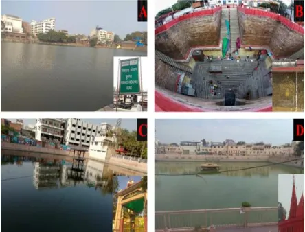
Durga Kund (Figures 1 and 2). All of these ancient and religious Kunds have their own importance and belief.
Samples were filtered by using planktonic net of 10 µm mesh size (Plankton Sampling Kit, Fieldmaster®, Wildco)
and were collected from January 2019 to February 2019 in gallon. These water samples were rich in cyanobacteria
and other algal organisms. We collected the samples again in the month of June 2019 to check the effects of high
intensity solar radiation on the organisms. Sampling sites were taped by using digital camera (Figure 2). UV-A and
26
Figure 1. Sampling sites showing the location of different historical Kunds of Varanasi, India
Figure 2. Historical Kunds (sampling sites) of Varanasi, India. (A) Pisachmochan Kund, (B) Lolark Kund, (C) Kri Kund and (D) Durga Kund
Identification of Microorganisms
The collected samples were centrifuged at 6000 rpm for 15 min and pellets were repeatedly washed with sterile
distilled water. Thereafter, the pellets were examined for the presence of cyanobacteria and other algal organisms by
using light microscope (OLYMPUS, Model number: CX21i-TR). Photographs were taken by using Dewinter-2011
scientific digital camera and analysed with Dewinter software.
Extraction of MAAs
Samples (Water) of different Kunds were centrifuged (6000 rpm for 15 min) and the pellets were dissolved in 2 ml
of 100% (v/v) methanol (HPLC-grade) and placed overnight at fter extraction ali uots ere centrifuge
(6000 rpm for 10 min) and methanolic extracts were subjected to spectroscopic analysis between 200 -700 nm in a
UV-Vis double beam spectrophotometer (U-2600, Shimadzu, Japan). The raw spectra were transferred to computer
and peaks were analyzed by UV-probe software (Version-2, Japan). Thereafter, methanol was evaporated at 45ºC
and redissolved in 80% water and 20% chloroform to separate water soluble compounds from organic compounds. It
was centrifuged at 10,000 rpm for 10 min and upper layer supernatant was taken (water soluble MAAs). Before
being subjected to HPLC analyses samples ere filtere through 0 2 μm pore-sized sterilized microcentrifuge
[image:3.612.195.417.259.428.2]27
HPLC analysis of MAAs
HPLC analysis of partially purified MAAs were carried out by using HPLC system equipped with a Photodiode
Array (PDA) Detector, Waters 2998, 515 HPLC pump, Nova-Pak ® C18 reverse phase guard column (4.6 × 150
mm inside diameter). A 20µL sample was injected into the HPLC column with the help of an auto-injector through a
Waters 717 plus Autosampler. Wavelength was set at 330 nm in detector and scanning of wavelength from 250 to
400 nm by PDA to see the separated peaks. A 0.02% (v/v) acetic acid was used as mobile phase which eluted
isocratically at a flow rate of 1 mL min-1. Identification of MAAs was done by its characteristic absorption maxima
and retention time. Eluted samples were collected with the help of Fraction Collector III. These purified fractions of
MAAs were further used for the characterization and identification.
Characterization of MAAs by Fourier Transform Infrared (FTIR) spectroscopy
HPLC purified fractions of MAAs were lyophilized to perform FTIR by fused with oven-dried potassium bromide
(stored in desiccator) in 1:100 ratio, transparent disk was prepared using hydraulic press and inserted in a Perkin
Elmer FTIR/FIR Spectrometer Frontier version 10 (Perkin Elmer, Waltham, MA, USA) to record the spectra.
RESULTS
Environmental Parameters
Nearly all Kunds had stagnant water (pond) system which were dominated by greenish and blue-green mats of
cyanobacteria and algae and were exposed to the solar radiation for longer period of time. These mats were capable
of producing photoprotective compounds such as MAAs. At study sites, relative humidity was found to be in the
range of 75-97%, tropospheric ozone in the range of 60.98-131.67 µg/m³, A in the range of 6-9 KJ/m² and
UV-B in the range of 11.297-33.928 KJ/m². Environmental conditions such as temperature (18-28°C) and pH
[image:4.612.186.425.473.687.2](6.02-8.31) (Figures 3 and 4) were favorable for the growth of cyanobacterial and algal populations in most of the Kunds.
28
Figure 4. pH at different historical Kunds of Varanasi, India
Screening and Identification of Microorganisms
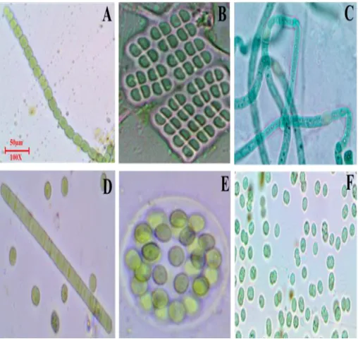
Six species of cyanobacteria and several other algal groups were recorded from the four ancient and religious Kunds
of Varanasi, India (Figures 5 and 6). Kunds were dominated by the cyanobacteria such as Anabaena sp., Oscillatoria
sp., Aulosira fertilissima and Gloeocapsa sp. Other notable algal groups present were Chlorella sp.,
Chlamydomonas sp. Ankistrodesmus sp., Scenedesmus sp. and Nitzschia sp. All the Kunds had stagnant water
(pond) system due to which bloom-forming cyanobacteria such as Microcystis aeruginosa and Merismopedia were
also present but in small number. Organisms identified belong to the class Cyanophyceae, Chlorophyceae,
Chrysophyceae and Bacillariophyceae (Table 1).
Figure 5. Cyanobacterial strains identified from different historical Kunds of Varanasi, India. (A) Anabaena sp., (B) Merismopedia sp.,
[image:5.612.178.432.426.666.2]29
Figure 6. Algal strains identified from different Kunds of Varanasi, India. (A) Pediastrum sp., (B) Scenedesmus sp., (C) Ankistrodesmus
[image:6.612.132.482.305.707.2]sp., (D) Chlamydomonas sp., (E) Chlorella sp., (F)Dictyosphaerium sp., (G) Nitzschia sp. and(H) Desmodesmus opoliensis
Table 1. Cyanobacteria and other algal colonization (+ shows the presence and – shows the absence of organisms) in the different historical Kunds of Varanasi, India.
Kunds (Water bodies)
Organisms Pisachmochan kund Lolark Kund Kri Kund Durga Kund Cyanophyceae
Anabaena sp. + + - +
Oscillatoria sp. + + + -
Aulosira fertilissima + + + -
Merismopedia sp. - - + -
Gloeocapsa sp. + + - +
Microcystis sp. - + - +
Chlorophyceae
Chlamydomonas sp. + + - +
Chlorella sp. + + + +
Scenedesmus sp. - + + -
Ankistrodesmus sp. + - - +
Dictyosphaerium sp. - + + -
Desmodesmus opoliensis - - + +
Pediastrum sp. + + - -
Chrysophyceae
Mallomonas sp. - + - +
Bacillariophyceae
(Diatom)
Nitzschia sp. + + + -
Fragilaria sp. - - + +
Aulacoseira sp. - + - -
30
UV-Vis Spectroscopic analysis
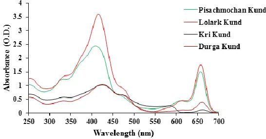
The absorption spectra of methanolic extracts of all samples showed a peak in the range of 310-337 nm which
suggested the presence of MAAs in these organisms. In addition to the peaks for MAAs, methanolic extracts of all
these samples also showed the peaks for chlorophyll a (420 and 660 nm), carotenoids (474 nm) and
[image:7.612.169.442.179.322.2]phycobiliproteins (620 nm) (Figure 7).
Figure 7. Absorption spectra of methanolic extracts of cyanobacterial and algal assemblages from historical Kunds of Varanasi, India
HPLC Analysis of MAAs
HPLC chromatograms showed various peaks with different retention time (RT) corresponding to respective MAAs
(Figure 8A-8D). Identification of different MAAs was done on the basis of retention time and absorption maxima
(Table 2). Results indicate that MAAs porphyra-334 (λmax-334 nm; RT-3.62 min); palythine (λmax-320 nm; RT-1.63
min); palythenic acid (λmax-337 nm; RT-4.15 min); mycosporine-glycine (λmax-310 nm; RT-2.55 min) and
mycosporine-methylamine-serine (λmax-327 nm; RT-1.51 min) were present in the samples. Porphyra-334 was
present in almost all the samples except Kri Kund. Again sampling was done in the period of June 2019
(temperature 37°C). Thereafter centrifugation was done to take supernatant (water) and checked for the presence of
photoprotective compound without adding any solvent. After all these, presence of photoprotective compound
mycosporine-glycine at absorption maxima 310 nm and retention time 2.55 min (Figure 9) was confirmed by HPLC.
This result shows that at high intensity of UV-radiation, organisms degrade (after synthesis of photoprotective
compound) and release their synthesized photoprotective compounds in water bodies. Concentration of these
compounds was low due to mixing in water but these photoprotective compounds protect other organisms from
degradation.
Concentration of all MAAs which was found in various Kunds has been shown in Figure 10. Altogether five
different types of Kund specific MAAs were present in our samples. Palythenic acid was present only in
Pisachmochan kund. Porphyra-334 was present in all Kunds except Kri Kund and its content was highest in Durga
Kund. Palythine was present in Pisachmochan, Lolark, Kri and Durga Kund, in which higher concentration was
observed in Kri Kund. Mycosporine-glycine and Mycosporine-methylamine-serine were present only in Lolark
Kund (Figure 10). These concentrations of MAAs depend on several conditions such as exposure to the UV
31
Kunds. Figure 11 represents the percentage distribution of MAAs in these samples. The results indicate that
porphyra-334 was contributing maximum (58 %) in Durga Kund and minimum (27 %) in Lolark Kund to the total
MAAs. Palythine was contributing maximum (100 %) in Kri Kund and minimum (20 %) in Lolark Kund. Palythenic
acid was contributing 50% in Pisachmochan Kund. Mycosporine-glycine and mycosporine-methylamine-serine
[image:8.612.113.503.158.457.2]were contributing 20 % and 33%, respectively in Lolark Kund.
Figure 8. HPLC chromatograms of the purified MAAs and their corresponding absorption spectra. (A) Pisachmochan Kund, (B) Lolark
Kund, (C) Kri Kund and (D) Durga Kund
Figure 9. HPLC chromatogram and its corresponding absorption spectrum of purified fraction of water sample after centrifugation have
[image:8.612.105.505.502.648.2]32
Table 2. Photoprotective compounds from historical Kunds of Varanasi, India, with their corresponding absorption maxima and
retention times.
Kunds
Mycosporine-like amino acids (MAAS) λmax (nm) Retention time (min) MAAs
334 3.78 Porphyra-334
Pisachmochan Kund 320 1.65 Palythine
337 4.15 Palythenic acid
334 3.62 Porphyra-334
Lolark Kund 320 1.61 Palythine
310 2.55 Mycosporine-glycine
327 1.51 Mycosporine-methylamine-serine
Kri Kund 320 1.63 Palythine
Durga Kund 334 3.52 Porphyra-334
[image:9.612.102.503.118.639.2]320 1.68 Palythine
Figure 10. Concentration of MAAs (nmol/gdw) in different historical Kunds of Varanasi, India. (PR: Porphyra-334, PT: Palythine, PA:
Palythenic acid, MG: Mycosporine-glycine, MMAS: Mycosporine-methylamine-serine). The error bar represents standard deviation of
33
Figure 11. Percentage distribution of MAAs (PR: Porphyra-334, PT: Palythine, PA: Palythenic acid, MG: Mycosporine-glycine, MMAS: Mycosporine-methylamine-serine) at various Kunds)
Effect of Temperature and pH on MAAs in Natural Environment
Temperature and pH does not directly affect MAAs but they affect them indirectly by affecting cyanobacteria. High
temperature (above 32°C) and low pH (less than 6.5) are not favourable for organisms. At this high temperature and
low pH, solubility of oxygen decreases and heavy metals in water become more soluble to increase toxicity and
therefore the death of organisms. This effect of temperature and pH in natural environment are same as in laboratory
conditions. Organisms grow luxuriantly at the temperature ranging from 19-28°C (Figure 3) and pH ranging from
6.72-8.31 (Figure 4). In general the optimum growth temperature for cyanobacteria is higher than that for most of
the algae.
Characterization of MAAs by Fourier Transform Infrared Spectroscopy (FTIR)
Collected fractions of FTIR containing KBr pellets revealed the presence of four prominent bands (shown by
arrows) in all the four samples (Figure 12). Band of 3353 cm−1 may be assigned to OH group, 2947 cm−1 for side
chain vibrations consisting of CH stretching and indicating the presence of NH2⁺ and band of 2836 cm−1 indicating
the presence of OH stretching in COOH (carboxylic group) and band of 1650 cm−1 may be assigned for the presence
34
Figure 12.Fourier Transform Infrared (FTIR) spectroscopy showing four prominent spectral bands (marked by arrows) revealing the presence of MAAs
DISCUSSION AND CONCLUSION
Cyanobacteria and other algal groups are worthwhile and beneficial source of natural products with potential
biological activities. Members of Chlorophyceae such as Chlamydomonas sp., Chlorella sp., Scenedesmus sp. and
Ankistrodesmus sp. are known to produce MAAs viz., porphyra-334, mycosporine-glycine and shinorine [29].
Chlorella sp. has been reported to produce porphyra-334, palythine, mycosporine-glycine and shinorine by their
symbiotic association with ciliates [30]. Small mass of 44 Da (CO2) and 18 Da (H2O) misplaying repeatedly discern
commonly in porphyra-334 [31,32]. MAAs such as MAA-322 (322 nm) and MAA-324 (324 nm) have also been
reported to be produced by the green alga Chlorella [33]. Scenedesmus sp. is known to produce MAAs such as
porphyra-334, palythine, shinorine, asterina-330 and MAA-302 [34]. Production of asterina-330 and shinorine has
been reported earlier in Ankistrodesmus sp. [34]. The Bacillariophycean member Nitzschia sp. has also been
reported to produce MAAs [35]. These different types of MAAs were identified on the basis of varying absorption
maxima and their respective retention times by HPLC followed by FTIR spectroscopy. Organisms which are able to
synthesize or accumulate MAAs are supposed to provide protection to the internal organelles and components from
harmful UV radiations [36]. In comparison to other MAAs, palythine and asterina-330 have been reported in a
limited number of microalgae or macroalgae [37-39].
Our study suggests that changing temperature and pH are responsible for the optimum growth and assemblage of
cyanobacteria and other algal groups [40]. Cyanobacterial growth rate increases at higher temperature (up to 25°C)
due to change in climate and therefore there is dominance of cyanobacteria in temperate water bodies [41-43]. They
are the main source of photoprotective compound MAAs, which have several other pharmaceutical importances
such as antiaging, anticancerous, anti-inflammatory and anti-immunosuppressive. Erythema, edema and sunburn are
acute UV-damages which have been shown to reduce in the presence of MAAs [44]. DNA lesions have also been
reported to be eliminated in the presence of MAAs [45]. MAAs has the ability to inhibit the overexpression of
biomarker of skin inflammation and in this way may protect from psoriasis, a chronic condition caused by overcome
35
mycosporine-glycine and its precursor gadusol act as antioxidants to avert cellular damage arising from
UVR-induced ROS [47]. In conclusion, cyanobacteria together with algal assemblages are useful in producing natural
photoprotective compounds even under various stress conditions and therefore could be a valuable source for the
development of natural sunscreens and antioxidants.
ACKNOWLEDGEMENTS
S. Mishra is thankful to the In charge, CIL, Department of Botany, Banaras Hindu University, Varanasi, for
providing necessary facilities. A. Pandey (09/013(0619)/2016-EMR-I) is thankful to CSIR, New Delhi, India, for the
financial support in the form of SRF.
Author Contributions
RPS: conceived and designed the experiments, analyzed and interpreted the data and edited the MS; SM: Performed
the experiments and drafted the paper; AP: Helped in performing the experiments and critically revised the MS. All
authors read and approved the final MS.
Conflict of Interest Disclosure
The authors declare that there is no conflict of interest.
REFERENCES
1) RMM Abed; S Dobretsov; K Sudesh. J Appl Microbiol.2009, 106, 1-12.
2) RP Rastogi; RP Sinha. Biotechnol Adv.2009, 27, 521-539.
3) CE Williamson; RG Zepp; RM Lucas; S Madronich; AT Austin; CL Ballare; M Norval; B Sulzberger, AF
Bais; RL McKenzie; SA Robinson, Nat Clim Chang.2014, 4, 434-441.
4) D-P Häder; CE Williamson; SA Wangberg; M Rautio; KC Rose; K Gao; EW Helbling; RP Sinha; R
Worrest, Photochem Photobiol Sci.2015, 14, 108-126.
5) F Garcia-Pichel; RW Castenholz, Appl Environ Microbiol.1993, 59, 163-169.
6) RP Rastogi; Richa; RP Sinha; SP Singh; D-P Häder, J Ind Microbiol Biotechnol.2010, 37, 537-558.
7) RP Rastogi; A Incharoensakdi, FEMS Microbiol Ecol. 2014, 87, 244-256.
8) WC Dunlap; BE Chalker, Coral Reefs. 1986, 5, 155-159.
9) JM Shick; WC Dunlap. Annu Rev Plant Physiol. 2002, 64, 223-262.
10) JI Carreto; MO Carignan; G Daleo; SG De Marco. J Plankton Res.1990, 12, 909-921.
11) NN Rosic; S Dove. Appl Environ Microbiol. 2011, 77, 8478-8486.
12) N Wada; T Sakamoto; S Matsugo. Antioxidants. 2015, 4, 603-646.
13) T Řezanka; M Temina; Tolsikov; M Dembitsky. Folia Microbiol. 2004, 49, 339-352.
14) MF Shih; JY Cherng. Eur J Dermatol. 2008, 18, 303-307.
15) L Riegger; D Robinson. Mar Ecol Prog Ser. 1997, 160, 13-25
16) CA Llewellyn; RL Airs. Mar drugs. 2010, 8(4), 1273-1291.
17) T Misonou; J Saitoh; S Oshiba; Y Tokitomo; M Maegawa; Y Inoue; H Hori; T Sakurai. Mar Biotechnol.
2003, 5, 194-200.
18) FR Conde; MS Churio; CM Previtali. J Photochem Photobiol B: Biol. 2000, 56, 139-144.
36
20) SM Awramik; JW Schopf; MR Walter. Precambr Res. 1983, 20, 357-374.
21) JW Schopf; BM Packer. Science. 1987, 237, 70-73.
22) JW Schopf. Science. 1993, 260, 640-646.
23) JW Schopf; AB Kudryavtsev; DG Agresti; TJ Wdowiak; AD Czaja. Nature. 2002, 416, 73-76.
24) MM Tice; DR Lowe, Nature. 2004, 431(7008), 549-552.
25) MJ Van Kranendonk; P Philippot; K Lepot; S Bodorkos; F Pirajno. Precambr Res. 2008, 167, 93-124.
26) F Westall; CEJ de Ronde; G Southam; N Grassineau; M Colas; C Cockell; H Lammer, Trans Royal Soc B:
Boil Sci.2006, 361, 1857-1875.
27) F Westall. Science, 2009, 323, 471-472.
28) AP Nutman; VC Bennett; CRL Friend; MJ Van Kranendonk; A Chivas. Nature. 2016, 537, 535-538.
29) SS Suh; J Hwang; M Park; H Seo; HS Kim; J Lee; S Moh; TK Lee. Mar drugs. 2014, 12(10), 5174-5187.
30) B Sonntag; M Summerer; R Sommaruga. Freshw Biol. 2007, 52, 1476-1485.
31) KHM Cardozo; VM Carvalho; E Pinto; Colepicolo P. Rapid Commun Mass Spectrom, 2006, 20, 253-258
32) MO Carignan; KHM Cardozo; D Oliveira-Silva; P Colepicolo; JI Carreto. J Photochem Photobiol B: Biol.
2009, 94, 191-200
33) U Karsten; T Friedl; R Schumann; K Hoyer; S Lembcke. J Phycol. 2005, 41(3), 557-566.
34) F Xiong; J Kopecky; L Nedbal. Aquat Bot.1999, 63(1), 37-49.
35) EW Helbling; BE Chalker; WC Dunlap; O Holm-Hansen; VE Villafañe. J Exp Mar Biol Ecol. 1996, 204,
85-101.
36) SP Singh; D-P Häder; RP Sinha. Ageing Res Rev. 2010, 9, 79-90
37) RP Sinha; SP Singh; D-P Häder, J Photochem Photobiol B: Biol. 2007, 89, 29-35.
38) VK Kannaujiya; Richa; RP Sinha, Front Environ Sci. 2014, 2, 1-6.
39) A Pandey; S Pandey; Rajneesh; J Pathak; H Ahmed; V Singh; SP Singh; RP Sinha, Int J Appl Sci
Biotechnol. 2017, 5(1), 12-21.
40) HW Paerl, Life,2014, 4(4), 988-1012.
41) HW Paerl; J Huisman, Env. Microbiol. Rep., 2009, 1(1), 27-37.
42) KD Joehnk; JEF Huisman; J Sharples; BEN Sommeijer; PM Visser; JM Stroom. Glob Change Biol. 2008,
14(3), 495-512.
43) TW Davis; DL Berry; GL Boyer; CJ Gobler. Harmful Algae. 2009, 8(5), 715-725.
44) Richa; RP Sinha. Mar Biol.2013, 14, 509-534.
45) RP Rastogi, UV-B induced DNA damage and repair in cyanobacteria, PhD Thesis, Banaras Hindu
University, Varanasi, India, 2010.
46) KP Lawrence; PF Long; AR Young, Curr Med Chem. 2018, 24(40),5512-5527.