0095-1137/04/$08.00⫹0 DOI: 10.1128/JCM.42.12.5462–5466.2004
Copyright © 2004, American Society for Microbiology. All Rights Reserved.
Development of a Rapid PCR Method Using the Insertion Sequence
IS
1203
for Genotyping Shiga Toxin-Producing
Escherichia coli
O157
Masahiro Suzuki,* Masakado Matsumoto, Mami Hata, Masao Takahashi, and Kenji Sakae
Department of Microbiology, Aichi Prefectural Institute of Public Health, Nagoya, Aichi, Japan
Received 7 May 2004/Returned for modification 14 July 2004/Accepted 13 August 2004
We developed a rapid PCR method utilizing the diversity of the insertion site IS1203for genotyping Shiga
toxin-producingEscherichia coli(STEC) O157 (IS1203PCR typing). DNA fragments digested by PvuII, which
cut IS1203at one site, were ligated with themselves and detected by PCR with outward-facing primer pairs for
IS1203. To minimize nonspecific bands, nested PCR was also performed. Two fingerprinting patterns produced
from the upstream or downstream regions of IS1203were obtained within 1 or 2 days. By combining the two
patterns, 79 STEC O157 isolates were classified into 39 types, which were then classified into 36 subtypes by
pulsed-field gel electrophoresis (PFGE). The discriminatory power of IS1203PCR typing (Dⴝ0.974) is similar
to that of PFGE (Dⴝ0.981). This method can be used for rapid and simplified genotyping.
Shiga toxin-producingEscherichia coli(STEC) O157 is as-sociated with outbreaks and sporadic cases of diarrhea, hem-orrhagic colitis, and hemolytic-uremic syndrome. Genotyping is a useful way to identify strains with the same genetic back-ground. Pulsed-field gel electrophoresis (PFGE) is a standard and effective method for genotyping STEC O157 (11, 16). Many studies have reported that STEC O157 can be classified into numerous genotypes by PFGE (1, 5, 7). However, PFGE is time-consuming and technically demanding and requires a special electrophoresis system.
The genomes of most bacteria contain distinct classes of insertion sequences (IS). IS typing involves utilizing the diver-sity of insertion sites. For some bacterial species, IS typing can be utilized for genotyping methods other than PFGE. For
Mycobacterium tuberculosis, restriction fragment length poly-morphism (RFLP) analysis using the sequence IS6110 is a standard genotyping method (12, 18). This method is time-consuming because it is a standard Southern blotting-based technique. Several studies have investigated genotyping meth-ods using PCR with insertion sequence primers. Otal et al. (13) reported on an IS6110-based PCR method using single out-ward-facing primers for IS6110that is comparable to standard-ized IS6110RFLP analysis methods. IS typing by PCR has also been developed for STEC O157. Thompson et al. (17) re-ported that the band patterns yielded by IS3typing forE. coli
using outward-facing primers are not as variable as those pro-duced by PFGE.
STEC O157 contains the IS1203sequence in addition to IS3. IS1203was reported by Paton and Paton (14) for theE. coli
O111:H⫺ strain PH. IS1203 is a 1,312-nucleotide sequence with imperfect 26-bp terminal inverted repeats and is closely related to the IS629 sequence of Shigella sonnei (10) and IS3411ofE. coli(4). At least 12 copies of IS1203have been found in the genome of theE. coliO111:H⫺strain PH (14). IS1203sequences were found in the genome sequence data of STEC O157 EDL933 (accession no. NC_002655) (12 copies)
and of the Sakai strain (accession no. NC_002695) (19 copies). IS1203was also found in thestx1andstx2genes of STEC O157
(8, 15) and in phage carryingstx2(6).
In this study, we developed a genotyping method using IS1203(referred to here as IS1203PCR typing) which is sim-ilar to the IS6110–inverse-PCR method developed by Otal et al. (13). We attempted to create a rapid and reproducible PCR genotyping protocol using ligation and nested PCR.
MATERIALS AND METHODS
Strains.Seventy-nine STEC O157 strains derived from patients (n⫽64) and cattle (n⫽15) were used. They were isolated in Aichi Prefecture, Japan, from 1999 to 2002 except for one strain, which was derived from the Sakai outbreak in 1996 (2, 9). The origins of strains obtained from patients were as follows: 29 strains from 9 outbreak cases, 1 strain from the Sakai outbreak, and 34 strains from sporadic cases.
PFGE.PFGE was performed as described elsewhere (1), with minor modifi-cations. In brief, bacterial cells cultured in Luria-Bertani (LB) broth at 37°C overnight with shaking were directly embedded in low-melting-temperature aga-rose (Nippon Gene, Tokyo, Japan). After appropriate preparations for restric-tion endonuclease digesrestric-tion were made, the DNA in each plug was digested with 30 U of XbaI (Roche Diagnostics, Mannheim, Germany) at 37°C overnight. PFGE was performed on a 1% agarose gel by using a Gene Navigator system (Pharmacia Biotech AB, Uppsala, Sweden) in 0.5⫻Tris-borate-EDTA buffer at 200 V. For whole-genome separation, linearly ramped switching times from 1 to 40 s were applied for 22 h.DNA concatemers (Bio-Rad Laboratories, Her-cules, Calif.) were used as size markers, and the gels were visualized by ethidium bromide staining and photographed under a transilluminator. The patterns were analyzed with FingerPrinting II software (version 4.2; Bio-Rad Laboratories). A dendrogram was drawn from this data by the unweighted pair group method with arithmetic averages (UPGMA), and similarity coefficients were calculated by the Jaccard method. PFGE patterns were sorted by FingerPrinting II and visually compared by using the criteria of Tenover et al. (16). Major strain types were designated by letters, with numbers added for subtypes.
IS1203PCR typing.A schematic drawing of IS1203PCR typing is shown in Fig. 1. Strains were cultured in LB broth at 37°C overnight with shaking, and chromosomal DNA was purified by using a DNeasy tissue kit (QIAGEN, Hilden, Germany) according to the manufacturer’s instructions. Five hundred nano-grams of purified DNA was digested by 10 U of the PvuII restriction enzyme (Roche Diagnostics) for 1 h at 37°C. The digested DNA was purified by phenol extraction and ethanol precipitation and was then dissolved in 500l of distilled water. As IS1203was digested by PvuII at one site (Fig. 1a), each of the digested fragments contained an upstream or a downstream region of IS1203. To detect fragments containing IS1203, digested DNA fragments were self-ligated by using a Rapid DNA Ligation kit (Roche Diagnostics). Ligation was performed in a total volume of 10l containing 2 to 4 ng of digested DNA for 30 min at 20°C.
* Corresponding author. Mailing address: 7-6 Nagare Tsuji-machi Kita-ku, Nagoya 462-8576, Aichi, Japan. Phone: 81-52-910-5669. Fax: 81-52-913-3641. E-mail: [email protected].
5462
on May 15, 2020 by guest
http://jcm.asm.org/
The self-ligated DNA was detected by PCR using primer pair 1203-82 (5⬘-CAC GTT CCA GCT CTT TCA G-3⬘) and 1203-11 (5⬘-GAC TGG CTG AAG AGA GAG AT-3⬘) for fragments containing the upstream region of IS1203(upstream typing), and primer pair 1203-62 (5⬘-TAA ACG CCA CAT AGA CGA AGC C-3⬘) and 1203-31 (5⬘-GTA TGA CAA CGC GAT GGC T-3⬘) for fragments containing the downstream region (downstream typing). PCR was performed in a volume of 25l containing 2.5l of ligated DNA, EX-Taq buffer, deoxynucleo-side triphosphates, 0.2M each primer, and 1 U of EX-Taq (Takara Bio, Ohtsu, Japan). PCR cycles consisted of 94°C for 1 min, 55°C for 45 s, and 72°C for 1 min, and PCR was performed for 20 cycles by using a GeneAmp PCR system 9600 (Applied Biosystems Japan, Tokyo, Japan).
To minimize nonspecific bands, nested PCR was performed by using primer pair 1203-72 (5⬘-GCG TAC AGC CAA TCT TTG G-3⬘) and 1203-21 (5⬘-GTT ATG GGA CTT GCC GGT GTT-3⬘) for upstream typing and primer pair 1203-52 (5⬘-TGC CAT GTG CTG ACG TAA G-3⬘) and 1203-41 (5⬘-GAA AAA CCG TGC AGA AGT GG-3⬘) for downstream typing. PCR was performed as described above except that 1l of the product from the first PCR procedure was used as the DNA template. Five microliters of nested PCR products was sepa-rated by electrophoresis in a 2.0% DNA agar gel (Marine BioProducts, Delta, British Columbia, Canada) at a constant voltage of 50 V for 2.5 h by using a Mupid-2 Mini Electrophoresis unit (Advancebio, Tokyo, Japan). Gels were vi-sualized by ethidium bromide staining and photographed under a UV transillu-minator. A 100-bp molecular size marker ladder (Nippon Gene) was used as a reference. The patterns were analyzed with FingerPrinting II software (version 4.2) as a composite data set of upstream and downstream typing. A dendrogram was drawn from these data by use of UPGMA, with the weight between the upstream and downstream typing set at 1:1.
IS1203PCR type names were assigned as follows. Clusters with more than 30% similarity were given Greek letters, from␣to; subclusters with more than 60% similarity were designated by Roman numerals within the same cluster; and IS1203PCR patterns with 1 or 2 differences in the bands had Arabic numerals added. Clusters or subclusters without subdivisions were not given a lower class name. Seven randomly selected strains, including two strains with the same PFGE pattern, were used to test for reproducibility. The reproducibility tests were performed under the same conditions three times.
Statistical analysis. The numerical index of discrimination (D index) for IS1203PCR typing and PFGE, defined as the average probability that different genotyping was assigned to two unrelated strains in a population of a given genus, was calculated by the method of Hunter and Gaston (3).
RESULTS
PFGE analysis.Seventy-nine strains were classified into 24
types (A to Y; to avoid confusion with the number zero, the letter O was not assigned) by PFGE. Strains within the same type had similarity coefficients of ⬎70% (Fig. 2). Five types were further subdivided into 17 subtypes: A1 to A7, B1 and B2, J1 and J2, U1 to U4, and X1 and X2. From the results of PFGE subtyping, 79 strains were classified into 36 PFGE subtypes. Strains obtained from an outbreak were classified as belonging to the same PFGE type, and theDindex was 0.981 for PFGE.
IS1203PCR typing.We repeated IS1203PCR typing three
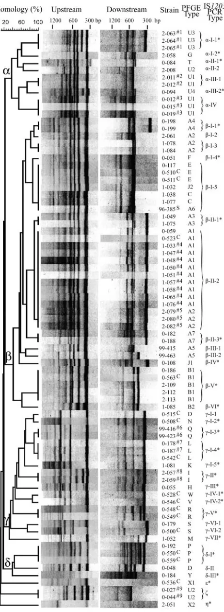
times for seven strains (Fig. 3). For these tests, the same electrophoresis patterns were obtained in the 1.2-kbp region. Bands above 1.2 kbp for downstream typing of strains 2–051 and 0–536 were not stable in the repeated test.
Three to nine bands under 1.2 kbp were detected by up-stream typing for each strain, and the strains were classified into 29 upstream types. Thirty of 79 strains were classified into 18 types with one-to-one correspondence to PFGE, and the other 49 strains, classified into 11 upstream types, were divided into 18 subtypes by PFGE. TheDindex was 0.921 for upstream typing.
One to nine bands under 1.2 kbp were detected by
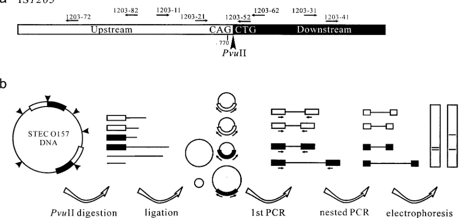
down-FIG. 1. Schematic diagram of IS1203PCR typing. (a) IS1203sequences were divided into two fragments, the upstream (open box) and downstream (solid box) fragments, by PvuII digestion between nucleotides 1032 and 1033 (arrowhead). Arrows indicate the primer regions for upstream typing (1203-82 and 1203-11 for first PCR, and 1203-72 and 1203-21 for nested PCR) and downstream typing (1203-62 and 1203-31 for first PCR, and 1203-52 and 1203-41 for nested PCR). (b) Detection of PvuII-digested fragments containing IS1203. STEC O157 genomic DNA contains more than 10 copies of IS1203(two copies of IS1203are shown in the figure for simplicity). Open box, IS1203 upstream region; solid box, downstream region. STEC O157 genomic DNA was digested at the PvuII recognition sites (arrowheads). Digested fragments were then self-ligated. Ligated fragments containing IS1203were amplified by PCR in the direction of the arrows. Fragments containing upstream or downstream regions of IS1203were reacted. Nested PCR was performed for accuracy, and two electrophoresis patterns (upstream and downstream patterns) were obtained from one strain.
on May 15, 2020 by guest
http://jcm.asm.org/
[image:2.585.59.527.74.296.2]stream typing, but two strains exhibited no band. Seventy-seven strains which exhibited more than one band were classified into 27 downstream types. Seventeen of these 77 strains were clas-sified into 10 types with one-to-one correspondence to PFGE. The other 60 strains, which were classified into 17 downstream types, were divided into 24 subtypes by PFGE. TheDindex was 0.952 for downstream typing.
Based on the composite data set that combined the finger-printing patterns of upstream and downstream typing, 79 strains were classified into 39 types (Fig. 4). IS1203PCR types were divided into four major clusters (clusters␣to␦) and three independent types (ε,, and). Forty-one strains exhibited 25 patterns with one-to-one correspondence to PFGE (Fig. 4). Another 17 strains were classified into 12 types by IS1203PCR typing and into 6 types by PFGE (Table 1). Some strains with the same PFGE type had IS1203PCR types in different clus-ters: two strains with PFGE type U2 were classified as␣-II-2 and , respectively, and two strains with PFGE type D were classified as␥-I-1 and␦-II, respectively. Finally, 21 strains were classified into two types by IS1203PCR typing and into six subtypes by PFGE (Table 2). Seven strains of one IS1203
PCR type (-I-5) were classified into four major PFGE types. Strains collected from the same outbreak had the same IS1203
PCR type. TheDindex was 0.974 for IS1203PCR typing with composite data sets.
DISCUSSION
This is the first report of PCR typing of STEC O157 strains by use of IS1203. To easily carry out IS typing, several
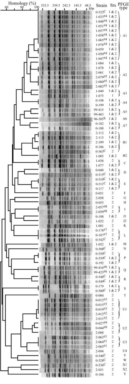
[image:3.585.49.274.69.735.2]re-FIG. 2. A dendrogram based on PFGE patterns was drawn by using UPGMA. PFGE patterns are also shown. Shiga toxin-producing types and PFGE types are shown in the Stx and PFGE type columns. Su-perscript numbers or letters following strain designations indicate or-igins, as follows: #1 through #9, strains isolated from outbreaks; S, strain isolated from the Sakai outbreak; C, strains isolated from cattle stool.
FIG. 3. Electrophoresis patterns of IS1203PCR typing repeated three times. Electrophoresis patterns under 1.2 kbp were the same for all strains. Large fragments had a tendency to be unstable (down-stream typing, strains 2–051 and 0–536).
on May 15, 2020 by guest
http://jcm.asm.org/
[image:3.585.301.541.69.245.2]searchers have developed IS-typing PCR methods. Most of these use single primers for the IS3sequence ofE. coli(17) or the IS6110sequence ofM. tuberculosis(13).
In this study, we developed an IS-typing PCR method that utilizes and is similar to the IS6110–inverse-PCR method de-veloped by Otal et al. (13), which involves IS typing using digestion with the restriction endonuclease PstI, ligation, and PCR. Although Otal et al. expected more bands by IS6110 -inverse PCR, the number of bands was not higher than that obtained by IS6110PCR typing without the ligation step. The fragment size produced in standard RFLP analysis with IS6110
by PstI digestion is greater than 1 kbp (12). We hypothesized that the fragments should be short enough to be amplified by PCR. Prolonging the PCR extension step was not effective for amplifying large fragments (data not shown). The advantages of this protocol are as follows. First, to increase the discrimi-natory power of IS1203PCR typing, PvuII digestion and liga-tion were performed. As IS1203was cut at one site by PvuII, we obtained two fingerprint patterns containing the upstream and downstream regions of IS1203, respectively. This produced characteristic patterns for each strain, and the combination of these patterns increases the discriminatory power of IS1203
PCR typing. In addition, the fragments were short enough to facilitate PCR amplification (the expected length was less than 1 kbp in about one-third of the bands generated from the Sakai strain), and we could obtain stable bands. Second, we acquired reproducible results, because ligation and nested PCR mini-mized nonspecific bands under 1.2 kbp. We observed that some amplicons in the first PCR disappeared from nested-PCR products (data not shown). This suggested that some nonspecific reaction(s) occurred in the first PCR and that such nonspecific bands could be removed by the second PCR. We expected that minimization of nonspecific bands would im-prove reproducibility. Therefore, we performed nested PCR to obtain reproducible results. Although we could obtain ducible results for most strains, further interlaboratory repro-ducibility studies are required.
Concerning the discriminatory power of IS1203PCR typing, PFGE identified 36 different subtypes and IS1203PCR typing
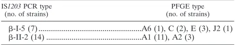
FIG. 4. A dendrogram was drawn by using a composite data set of upstream and downstream patterns obtained from IS1203PCR typing by UPGMA. The electrophoresis patterns of upstream and
[image:4.585.51.282.68.698.2]downstream typing are also shown. The patterns were divided into four major clusters (clusters␣to␦) and three independent types. Pattern designations obtained from composite IS1203 PCR typing data and from PFGE are shown in the respective columns. IS1203PCR types exhibiting one-to-one correspondence with the PFGE subtypes are asterisked. Superscript numbers or letters following strain designations indicate origins, as follows: #1 through #9, strains isolated from out-breaks; S, strain isolated from the Sakai outbreak; C, strains isolated from cattle stool.
TABLE 1. IS1203PCR types with the same PFGE pattern
IS1203PCR type (no. of strains)
PFGE type (no. of strains)
␣-II-2 (1),(2) ... U2 (3) ␣-III-1 (2),␣-IV (3) ... U1 (5) -I-2 (1),-I-3 (2)... A2 (3) -III-1 (1),-III-2 (1)... A5 (2) ␥-I-1 (1),␦-II (1) ... D (2) ␥-VI-1 (1),␥-VI-2 (1) ... S (2)
on May 15, 2020 by guest
http://jcm.asm.org/
identified 39. Thompson et al. (17) developed a subtyping method based on PCR amplification of variable DNA se-quences between the repetitive IS3elements of STEC O157 and reported that PFGE identified 25 different subtypes and IS3-based PCR identified 15. IS1203 PCR typing allows for higher discriminatory power than IS3-based PCR. TheDindex (0.974) for IS1203PCR typing was slightly lower than that for PFGE (0.981) in this study, suggesting that the discriminatory power of our method is slightly lower than that of PFGE. However, theDindex (0.974) indicates that if two strains from a population are randomly sampled, 97.4% of the time they will belong to different types (3). We believe that most strains can be identified by IS1203PCR typing.
IS1203PCR typing is an easy and rapid genotyping method with a sensitivity similar to that of PFGE. This method can be used to screen STEC O157 isolates.
ACKNOWLEDGMENT
This work was supported by grants from the Ministry of Health, Labor, and Welfare of Japan.
REFERENCES
1.Barrett, T. J., H. Lior, J. H. Green, R. Khakhria, J. G. Wells, B. P. Bell, K. D. Greene, J. Lewis, and P. M. Griffin.1994. Laboratory investigation of a multistate food-borne outbreak ofEscherichia coliO157:H7 by using pulsed-field gel electrophoresis and phage typing. J. Clin. Microbiol.32:3013–3017. 2.Hayashi, T., K. Makino, M. Ohnishi, K. Kurokawa, K. Ishii, K. Yokoyama, C. G. Han, E. Ohtsubo, K. Nakayama, T. Murata, M. Tanaka, T. Tobe, T. Iida, H. Takami, T. Honda, C. Sasakawa, N. Ogasawara, T. Yasunaga, S. Kuhara, T. Shiba, M. Hattori, and H. Shinagawa.2001. Complete genome sequence of enterohemorrhagicEscherichia coliO157:H7 and genomic com-parison with a laboratory strain K-12. DNA Res.28:11–22.
3.Hunter, P. R., and M. A. Gaston.1988. Numerical index of the discrimina-tory ability of typing systems: an application of Simpson’s index of diversity. J. Clin. Microbiol.26:2465–2466.
4.Ishiguro, N., and G. Sato.1988. Nucleotide sequence of insertion sequence
IS3411, which flanks the citrate utilization determinant of transposon Tn3411.J. Bacteriol.170:1902–1906.
5.Izumiya, H., J. Terajima, A. Wada, Y. Inagaki, K. I. Itoh, K. Tamura, and H. Watanabe.1997. Molecular typing of enterohemorrhagicEscherichia coli
O157:H7 isolates in Japan by using pulsed-field gel electrophoresis. J. Clin. Microbiol.35:1675–1680.
6.Johansen, B. K., Y. Wasteson, P. E. Granum, and S. Brynestad.2001. Mosaic structure of Shiga-toxin-2-encoding phages isolated fromEscherichia coli
O157:H7 indicates frequent gene exchange between lambdoid phage ge-nomes. Microbiology147:1929–1936.
7.Johnson, J. M., S. D. Weagant, K. C. Jinneman, and J. L. Bryant.1995. Use of pulsed-field gel electrophoresis for epidemiological study ofEscherichia coliO157:H7 during a food-borne outbreak. Appl. Environ. Microbiol.61:
2806–2808.
8.Kusumoto, M., Y. Nishiya, Y. Kawamura, and K. Shinagawa.1999. Identi-fication of an insertion sequence, IS1203variant, in a Shiga toxin 2 gene of
Escherichia coliO157:H7. J. Biosci. Bioeng.87:93–96.
9.Makino, K., K. Ishii, T. Yasunaga, M. Hattori, K. Yokoyama, C. H. Yutsudo, Y. Kubota, Y. Yamaichi, T. Iida, K. Yamamoto, T. Honda, C. G. Han, E. Ohtsubo, M. Kasamatsu, T. Hayashi, S. Kuhara, and H. Shinagawa.1998. Complete nucleotide sequences of 93-kb and 3.3-kb plasmids of an entero-hemorrhagicEscherichia coliO157:H7 derived from Sakai outbreak. DNA Res.5:1–9.
10.Matsutani, S., H. Ohtsubo, Y. Maeda, and E. Ohtsubo.1987. Isolation and characterization of IS elements repeated in the bacterial chromosome. J. Mol. Biol.196:445–455.
11.Olive, D. M., and P. Bean.1999. Principles and applications of methods for DNA-based typing of microbial organisms. J. Clin. Microbiol.37:1661–1669. 12.Otal, I., C. Martin, V. Vincent Levy Frebault, D. Thierry, and B. Gicquel.
1991. Restriction fragment length polymorphism analysis using IS6110as an epidemiological marker in tuberculosis. J. Clin. Microbiol.29:1252–1254. 13.Otal, I., S. Samper, M. P. Asensio, M. A. Vitoria, M. C. Rubio, R. Go´mez
Lus, and C. Martı´n.1997. Use of a PCR method based on IS6110 polymor-phism for typingMycobacterium tuberculosisstrains from BACTEC cultures. J. Clin. Microbiol.35:273–277.
14.Paton, A. W., and J. C. Paton.1994. Characterization of IS1203, an insertion sequence inEscherichia coliO111:H⫺. Gene150:67–70.
15.Suzuki, M., F. Kondo, Y. Ito, M. Matsumoto, M. Hata, H. Oka, M. Taka-hashi, and K. Sakae.2004. Identification of a Shiga-toxin type I variant containing an IS1203-like element, from Shiga-toxin producingEscherichia coliO157:H7. FEMS Microbiol. Lett.234:63–67.
16.Tenover, F. C., R. D. Arbeit, R. V. Goering, P. A. Mickelsen, B. E. Murray, D. H. Persing, and B. Swaminathan.1995. Interpreting chromosomal DNA restriction patterns produced by pulsed-field gel electrophoresis: criteria for bacterial strain typing. J. Clin. Microbiol.33:2233–2239.
17.Thompson, C. J., C. Daly, T. J. Barrett, J. P. Getchell, M. J. Gilchrist, and M. J. Loeffelholz.1998. Insertion element IS3-based PCR method for sub-typingEscherichia coliO157:H7. J. Clin. Microbiol.36:1180–1184. 18.van Embden, J. D., M. D. Cave, J. T. Crawford, J. W. Dale, K. D. Eisenach,
[image:5.585.42.284.79.126.2]B. Gicquel, P. Hermans, C. Martin, R. McAdam, T. M. Shinnick, and P. M. Small.1993. Strain identification of Mycobacterium tuberculosisby DNA fingerprinting: recommendations for a standardized methodology. J. Clin. Microbiol.31:406–409.
TABLE 2. IS1203PCR types with different PFGE patterns
IS1203PCR type (no. of strains)
PFGE type (no. of strains)
-I-5 (7) ...A6 (1), C (2), E (3), J2 (1) -II-2 (14) ...A1 (11), A2 (3)