A Preprocessing Approach For Image Analysis Using
Gamma Correction
S. Asadi Amiri
Department of Computer Engineering,
Shahrood University of Technology, Shahrood, Iran
H. Hassanpour
Department of Computer Engineering,
Shahrood University of Technology, Shahrood, Iran
ABSTRACT
This paper presents a new, simple and robust image enhancement algorithm for image analysis by modifying the gamma value of its individual pixels. Considering the fact that gamma variation for a single image is actually nonlinear, the proposed method locally estimates the gamma values in an image. First, for local gamma correction the image is divided to overlapping windows and then the gamma value of each window is estimated by minimizing the homogeneity of co-occurrence matrix. This feature represents image details, the minimum value of this feature shows maximum details of the image. As the enhanced image shows details better, the method is a useful preprocessing technique for image analysis. In this study, it is shown that the proposed method has performed well in improving the quality of images. Subjective and objective image quality assessments used in this study attest superiority of the proposed method compared to the existing methods in image quality enhancement.
Keywords
Image Enhancement, Gamma Correction, Windowing, Co -Occurrence Matrix, Homogeneity.
1.
INTRODUCTION
Due to technical limitations, many imaging devices may not display the actual appearance of objects. This technical limitation, known as gamma distortion, often disturbs the image [1]. The gamma distortion is not monotonic. It mainly depends to the relative illumination reflection of objects in adjacent pixels. In other words, image distortion depends to the depth, texture, and relative reflection of objects in the image. Since an image contains objects with variety of texture and depth gamma distortions may not be the same on all objects. Hence, it needs an adaptive approach to enhance the image.
Image enhancement is developed to improve the visual important features, or to provide better input for other automated image processing techniques [2]. For example in face recognition systems, the algorithms may fail to recognize faces correctly due to changes in illumination [3,4]. It needs to be noted that image enhancement techniques such as histogram equalizationand homomorphic filtering may not be used to enhance images suffering from gamma distortion. For example, the main objective of histogram equalization is to achieve a uniform distributed histogram by using the cumulative density function of the input image. This may not be a suitable objective, where brightness of some areas (or objects) of the image are satisfactory [5]. In histogram equalization technique, the pixel values are either added or multiped by a value [1]. It mainly cares about histogram of the image not the actual appearance of the image which is the case in gamma correction.
[image:1.595.313.535.322.521.2]Hence, conventional image enhancement techniques, like global brightness, contrast enhancement, histogram equalization, and homomorfic filtering are incapable of providing satisfactory enhancement results for images suffering from gamma distortion. Hence, gamma correction is a necessary preprocessing approach for this distortion. Figure 1 shows an example of superiority of proposed gamma correction to histogram equalization, homomorphic filtering and a method proposed in [4].
(a)Original image (b)Proposed method
(c)Histogram equalization
(d)Homomorphic filtering
(e)Method proposed in [4]
Fig 1: Comparison between the proposed method, histogram equalization, homomorphic filtering and a method proposed in [4] for face recognition. This image is from http://cvc.yale.edu/projects/yalefaces/yalefaces.html.
Medical image enhancement is also necessary for better diagnostic information. For example, one of the most common positioning errors in panoramic radiography is palatoglossal air space above the apices of the root of maxillary teeth [6]. It causes a radiolucency obscuring the apices of maxillary teeth. In the case of this positioning error, the imaging should be repeated. This causes the patient be exposed to radiation again. To avoid the repetition of harmful x-Rays to the patient, it is necessary to improve the panoramic images.
Imaging devices apply the power low transformation on pixel of the image, hence gamma correction is required to enhance the image
In this paper we present a simple technique for estimating the gamma values without any calibration information or knowledge of the imaging device. As mentioned earlier, since an image may contain objects with variety of texture and depth, the gamma distortions may not be the same on all objects. Hence in this method for local gamma correction, an input image is divided to overlapping windows and each window is enhanced individually. To determine the gamma value of each window, first twenty different gamma values (from 0.2 to 2.2 interval o.1) are applied to each window. Then, gray level co-occurrence matrix (GLCM) is calculated for each window with different gamma values (luminance). At last homogeneity feature is extracted from each of these
matrices, and selecting the gamma value for each window that minimizing the homogeneity feature value. We will show that, homogeneity feature measures the image details and the minimum value of this feature shows maximum details of the image.
This technique can be used to enhance both gray images and color images. The HSV color model is adopted in processing color images [5]. In practice, the value (V) is only processed with the proposed method. Then the HVS color model with the modified V is transformed into the RGB color model. In the next section, gamma correction is described. In Section 3 gray level co-occurrence matrix is briefly described. Image quality assessment is introduced in Section 4. The proposed algorithm is presented in Section 5. Section 6 shows the results, and Section 7 contains the conclusions
(a)Original image (b)Proposed gamma correction
(c) Histogram equalization (d) Homomorphic filtering
Fig 2. An example of a panoramic radiograph with positioning error and its proposed gamma corretion, histogram equalization and homomorphic filtering result.
2.
GAMMA CORRECTION
Many devices used for capturing, printing or displaying the images generally apply a transformation, called power-law [1], on each pixel of the image that has a nonlinear effect on luminance:
u
u
g
(1)In the above equation u ϵ [0, 1] denotes the image pixel intensity, γ is a positive constant introducing the gamma value. By this assumption, the value of γ typically can be determined experimentally, by passing a calibration target with a full range of known luminance values through the imaging device. When the value of γ is known, inverting this process is trivial:
11
u u
g (2)
Often such calibration is not available or direct access to the imaging device is not possible. Hence an algorithm is needed to enhance an image for its gamma values without any
knowledge about the imaging device [7]. In addition to this problem, in practice, these nonlinear effects aren’t consistent across all regions of the image. In other words, the value of gamma may change from one region to another. For instance, it is possible that a scene contains a large dynamic illumination range that an imaging device is not able to adequately capture. Thus, especially in very dark or bright regions of the image, some details may become clustered together within a small intensity range [8]. Hence a local enhancement process is needed to adjust the image quality in different regions in a way that the human viewers grasp these details.
[image:2.595.55.541.260.523.2]each value is the one that minimizes those higher order correlations. This method is time consuming and has limited success. Another global gamma correction based on texture analysis has been introduced in [9]. Although this method is not time consumig, but because of global gamma correction this method may not be succeed to enhance some images that need local gamma correction. In [3] a mapping function is considered to correlate gamma values with pixel values. In fact, the algorithm is a nonlinear transformation that makes pixels with low values brighter, whereas pixels with high values become darker. This transformation leaves midtons with less correction or even no correction. This approach is a pixel wise operation that may be successful on reducing the illumination on the scene.Since local information of the pixels is not used, image distortion may occur in natural scene images. A new local gamma correction method based on nearest neighbor algorithm and two feature vectors: pixel intensity histograms and dispersion-versus-location distributions is presented in [10]. Although this method produces satisfying results, but its computational complexity is high and it only works on grayscale images.
3.
THE
GRAY
LEVEL
CO-OCCURRENCE MATRIX
The co-occurrence matrix is often used for feature extraction in texture analysis of an image. The co-occurrence matrix of a gray level image is regarded as a two dimensional matrix. Its size is proportional to the number of gray levels in an image. For instance, the images used in this paper have 256 gray levels; thus, their GLCM is a matrix of size 256×256. In contrast to histogram, GLCM describes the relationship between the values of neighbouring pixels. It measures the probability that a pixel of a particular gray level occurs at a specified direction and a distance from its neighbouring pixels. This can be calculated by the function𝑃(𝑖, 𝑗, 𝑑, 𝜃), where 𝑖 is the gray level at location with coordinate (𝑥, 𝑦), 𝑗 is the gray level of its neighbouring pixel at a distance 𝑑 and a direction 𝜃 from a location (𝑥, 𝑦) [11]. 𝜃 usually ranges from: 0, 45, 90, to 135 [12]. This is mathematically defined by Equation (3):
,
,
,
,
, ,
} , , , , , , { # ) , , , ( 2 2 1 1 2 2 1 1 2 2 1 1 2 2 1 1
y x y x d y x y x j y x f i y x f y x y x d j i P (3)In [13], fourteen different features of GLCM have been defined. These features consist of texture information, but, there may be correlation between them. In this paper homogeneity feature is extracted from co-occurrence matrix𝑃, this feature is defined in the following:
𝐻𝑂𝑀 = 𝑃(𝑖, 𝑗, 𝑑, 𝜃) 1 + 𝑖 − 𝑗 256
𝑗 256
𝑖
(4) Homogeneity returns a value that measures the closeness of the distribution of elements in the GLCM to the GLCM diagonal, and its range is between 0 to 1. In other words, it describes how uniform the texture is.
4.
IMAGE QUALITY ASSESSMENT
The two types of image quality assessment techniques are the subjective method, which involves human beings to evaluate the quality of the images, and the objective method, which numerically computes the image quality. Since human beings are the ultimate receivers in most image processing applications, subjective evaluation is the most reliable way of
assessing the quality of an image. But, it is not usually useful for real world applications because this method is expensive and time consuming [14]. The goal of objective image quality assessment is to design computational models that can predict perceived image quality accurately and automatically. The goal of the objective image quality assessment research is thus to predict the quality of an image as closely as to the subjective assessment. These numerical measures should correlate well with human subjectivity.
MSE and PSNR are the two common objective methods but they do not correlate well with the subjective assessment. They depend on only the difference between the original reference image and the enhanced image, and do not measure whether the enhanced version contains more visual information or not [8].
Thus, a lot of objective image quality assessments have developed in the past few decades to replace them. The structural similarity metric (SSIM) proposed in [15] is correlated with human visual system.
Let x, y be the original and the test images, respectively. SSIM is defined as:
(5)
x
y
l
x
y
c
x
y
s
,
,
,
S is the correlation coefficient between x and y, which measures the degree of linear correlation between x and y. L measures how much the x and y are close in luminance. C measures the similarities between the contrasts of the images.
Where: (6)
N i i N i iy
N
Y
x
N
X
1 11
,
1
(7) (8)
N i i y N i i xy
y
N
x
x
N
1 2 2 1 2 21
1
1
1
(9)
N i i ixy
x
X
y
Y
N
12
1
1
The dynamic range of SIMM is [0, 1]. The best value ,1, is achieved if x=y.
5.
PROPOSED METHOD
As mentioned earlier, the goal of the present research is to estimate the gamma value of an image in a local approach. The basic idea is the fact that homogeneity value in an image not suffering from gamma distortion has a lower value (near to zero). These homogeneity values can be calculated by co-occurrence matrix. The gamma value is then estimated by minimizing these homogeneities.
In the proposed method, for adaptive gamma correction, the image is divided into overlapping windows. A sliding window of size 64×64 is moved across the image from top-left side to bottom-right by thirty pixels in each movement. A value of 64×64 pixels was chosen for images with the size 256×256 as this window size gives the best trade off between the rendering
2 2 2 2of local details and the need for reducing space dimensionality. To find a proper gamma value for each window, we apply a range of inverse gamma values from 0.2 to 2.2 interval 0.1 to each window. Different windows may need different gamma value for a proper enhancement. To find the best gamma value for each window, we compute the co-occurrence matrix of the window to extract the homogeneity feature. Then, the gamma value associated with the minimum homogeneity is considered as the best gamma value for enhancement.
(a) Original image
(b) Gamma corrected image
Fig 3. Blocking effects in gamma correction. This image is from http:// dragon.larc.nasa.gov/retinex/pao/news.
[image:4.595.59.284.200.344.2]In this approach, each window in the image has its own gamma value. Because of overlapping windows, pixels may settle under different windows, hence, different gamma values may apply. We apply only one gamma value on each pixel which is the average of the gamma values in the covering windows. In other words, a matrix M of gamma values with the same size as the image is achieved. To enhance the image, according to Equation (2) the gamma values are applied to each pixel. Figure 3 shows the result. As it is shown in this figure, this approach has unpleasure blocking effects on the image. In this step, to eliminate the blocking effects, first we apply average filter on M containing the gamma values. Then the filtered gamma values are applied to the image for gamma correction. Figure 4 is shown this result. As is clear the blocking effects are eliminated.
Fig 4. Image enhancement by our proposed method without the problem of blocking effect as exist in Figure 3(b).
6.
EXPERIMENTAL RESULTS
[image:4.595.56.285.227.552.2] [image:4.595.313.540.249.395.2](a)Original image (b) Proposed method
(c)Histogram equalization (d)Method proposed in [3]
(e)Method proposed in [7] (f)Method proposed in [9]
[image:5.595.62.529.98.621.2](a)Original image (b)Proposed method
(c)Histogram equalization (d)Method proposed in [3]
(e)Method proposed in [7] (f)Method proposed in [9]
[image:6.595.58.542.67.615.2](a)Original image (b) Proposed method
(c)Histogram equalization (d)Method proposed in [3]
(e)Method proposed in [7] (f)Method proposed in [9]
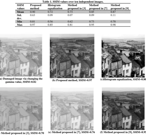
[image:7.595.62.535.70.639.2]Numerical assessment has also been performed to show the performance of the proposed method. We have used SSIM measure as the full reference numerical image quality assessment. Since the reference versions of the images in the quality assessment were not available, ten standard images have been used from Matlab. First, quality of these images has been damaged by random gamma values, then these images have been applied to the five algorithms to be restored. Table 1
[image:8.595.64.532.186.620.2]shows the SSIM values of different approaches in restoring these ten images. As it is shown in Table 1, the proposed algorithm provides a better SSIM value, hence outperforms the other algorithms in enhancing the images. Figure 8 represents a sample of the ten images with their SSIM values. These images show that the scores in Table 1 are in agreement with subjective evaluations of human observers.
Table 1. SSIM values over ten independent images. SSIM
values
Proposed method
Histogram equalization
Method proposed in [3]
Method proposed in [7]
Method proposed in [9]
Mean 0.90 0.72 0.73 0.85 0.87
Std. dev.
0.63 0.09 0.07 0.09 0.11
Min 0.81 0.56 0.62 0.73 0.70
Max 0.97 0.85 0.81 0.95 0.98
(a) Damaged image via changing the
gamma value, SSIM=0.82 (b) Proposed method, SSIM=0.97 (c)Histogram equalization, SSIM=0.80
(d) Method proposed in [3], SSIM=0.70 (e) Method proposed in [7], SSIM=0.76 (f) Method proposed in [9], SSIM=0.95 Fig 8: Comparison between the proposed gamma corretion method, histogram equalization, and the methods proposed in [3,7,9], (objective quality assessment).
7.
CONCLUSIONS
We have introduced a new image enhancement method based on gamma correction that estimates image gamma values without any calibration information or knowledge of the imaging device. The proposed method is a necessary preprocessing stage for most image analysis. Experimental results in this research indicate that the proposed method improves image quality, enhances the dynamic range and details of the image.
8.
REFERENCES
[1] R.C. Gonzalez, R.E. Woods, “Digital Image Processing”, Prentice Hall, Upper Saddle River, NJ 07458, 2002. [2] A. Pizurica, W. Philips, “Estimating the probability of the
[3] Y. Shi, J. Yang, R. Wu, “Reducing Illumination Based on Nonlinear Gamma Correction”, Proc. ICIP, San Antonio, 2007, pp. 529-539.
[4] Ch.N. Fan, F.Y. Zhang, “Homomorphic filtering based illumination normalization method for face recognition”, Pattern Recognition Letters, Vol. 32, 2011, pp. 1468-1479. [5] Q. Chen, X. Xu, Q. Sun, D. Xia, “A solution to the deficiencies of image enhancement”, Signal Processing, Vol. 90, 2010, p.p. 44-56.
[6] P.W. Goaz, S. C. White, “Oral radiology: Principles and Interpretation”, 2009.
[7] H. Farid, “Blind inverse gamma correction”, IEEE Transactions on Image Processing, 2001, Vol. 10, pp. 1428-1433.
[8] S. Lee, “Content-based image enhancement in the compressed domain based on multi-scale α-rooting algorithm”, Pattern Recognition Letters, 2006, Vol. 27, pp. 1054-1066.
[9] S. Asadi Amiri, H. Hassanpour, A.K. Pouyan, “Texture Based Image Enhancement Using Gamma Correction”, Middle-East Journal of Scientific Research, 2010, Vol. 6, pp. 569-574.
[10] M. FarshbafDoustar, H. Hassanpour, “A Locally-Adaptive Approach For Image Gamma Correction”, Proc. Signal Processing and their Applications (ISSPA), 2010, pp. 73-76.
[11] P. Gastaldo, R. Zunino, I. Heynderickx, E. Vicario, “Objective quality assessment of displayed images by using neural networks”, Signal Processing Image Communication, 2005, pp. 643-661.
[12] L.I. Voicu, H.R. Myler, A.R. Weeks, “Practical considerations on color image enhancement using homomorphic filtering”, Journal of Electronic Imaging, Vol. 6, No. 1, (1997), pp. 108-113.
[13] R.M. Haralick, K. Shanmugan, I. Dinstein, “Textural features for image classification”, IEEE Trans. SMC, 1973, Vol. 3, pp. 610-621.
[14] Z. Wang, A.C. Bovik, ”Modern Image Quality Assessment”, Morgan and Claypool Publishing Company, New York, 2006.
![Figure 1 shows an example of superiority of proposed gamma correction to histogram equalization, homomorphic filtering and a method proposed in [4]](https://thumb-us.123doks.com/thumbv2/123dok_us/8126273.795586/1.595.313.535.322.521/superiority-proposed-correction-histogram-equalization-homomorphic-filtering-proposed.webp)

![Fig 5: Comparison between the proposed gamma corretion method, histogram equalization, and the methods proposed in [3,7,9] (subjective quality assessment, Sample 1)](https://thumb-us.123doks.com/thumbv2/123dok_us/8126273.795586/5.595.62.529.98.621/comparison-proposed-corretion-histogram-equalization-proposed-subjective-assessment.webp)
![Fig 6: Comparison between the proposed gamma corretion method, histogram equalization, and the methods proposed in [3,7,9] (subjective quality assessment, Sample 2)](https://thumb-us.123doks.com/thumbv2/123dok_us/8126273.795586/6.595.58.542.67.615/comparison-proposed-corretion-histogram-equalization-proposed-subjective-assessment.webp)
![Fig 7: Comparison between the proposed gamma corretion method, histogram equalization, and the methods proposed in [3,7,9] (subjective quality assessment, Sample 3)](https://thumb-us.123doks.com/thumbv2/123dok_us/8126273.795586/7.595.62.535.70.639/comparison-proposed-corretion-histogram-equalization-proposed-subjective-assessment.webp)