Amplification Tests on a Multipathogen External Quality Assessment
Panel
K. Loens,aA. M. van Loon,bF. Coenjaerts,bY. van Aarle,cH. Goossens,aP. Wallace,dE. J. C. Claas,cand M. Ievena on behalf of the GRACE Study Group
Department of Microbiology, Vaccine and Infectious Diseases Institute, University of Antwerp, Antwerp, Belgiuma; Department of Virology, Medical Microbiology, University Medical Center, Utrecht, The Netherlandsb; Department of Medical Microbiology, Leiden University Medical Center, Leiden, The Netherlandsc; and Quality Control for Molecular Diagnostics, Glasgow, Scotland, United Kingdomd
An external quality assessment (EQA) panel consisting of a total of 48 samples in bronchoalveolar lavage (BAL) fluid or
trans-port medium was prepared in collaboration with Quality Control for Molecular Diagnostics (QCMD) (
www.qcmd.org
). The
panel was used to assess the proficiency of the three laboratories that would be responsible for examining the 6,000 samples to be
collected in the GRACE Network of Excellence (
www.grace-lrti.org
). The main objective was to decide on the best-performing
testing approach for the detection of influenza viruses A and B, parainfluenza virus types 1 to 3, respiratory syncytial virus
(RSV), human metapneumovirus, coronavirus, rhinovirus, adenovirus,
Chlamydophila pneumoniae,
Mycoplasma pneumoniae,
and
Legionella pneumophila
by nucleic acid amplification techniques (NAATs). Two approaches were chosen: (i) laboratories
testing samples using their in-house procedures for extraction and amplification and (ii) laboratories using their in-house
am-plification procedures on centrally extracted samples. Furthermore, three commercially available multiplex NAAT tests—the
ResPlex (Qiagen GmbH, Hilden, Germany), RespiFinder plus (PathoFinder, Maastricht, The Netherlands), and RespiFinder
Smart 21 (PathoFinder) tests—were evaluated by examination of the same EQA panel by the manufacturer. No large differences
among the 3 laboratories were noticed when the performances of the assays developed house in combination with the
in-house extraction procedures were compared. Also, the extraction procedure (central versus local) had little effect on
perfor-mance. However, large differences in amplification efficacy were found between the commercially available tests; acceptable
re-sults were obtained by using the PathoFinder assays.
G
RACE (www.grace-lrti.org) is a Network of Excellence
focus-ing on the complex and controversial field of
community-acquired lower respiratory tract infections (CA-LRTI), which are
among the leading reasons for seeking medical care. The
promis-cuous use of antibiotics for the treatment of CA-LRTI accounts for
a major part of the community burden of antibiotic use and
con-tributes dramatically to the rising prevalence of resistance among
major human pathogens. The overall objective of GRACE is to
combat antimicrobial resistance by integrating centers of research
excellence and exploiting genomics in the investigation of
CA-LRTI.
A multitude of nucleic acid amplification techniques (NAATs)
for the detection of pathogenic organisms in respiratory
speci-mens have been described (5, 8, 10). Currently, a few commercial
assays are available, but the majority of assays applied in clinical
diagnostic laboratories have been developed in-house. Therefore,
there is a need for interlaboratory exchange of clinical samples in
order to compare results and evaluate individual assays,
particu-larly when collaboration takes place in a multicenter network.
Part of the GRACE project is dedicated to the evaluation and
validation of rapid diagnostic tests such as NAATs. One of the
objectives is to select the best-performing strategy for nucleic acid
(NA) extraction, amplification, and detection of pathogenic
or-ganisms involved in lower respiratory tract infections. The
proce-dure selected will then be applied to specimens obtained from
3,000 adult patients presenting with lower respiratory tract
infec-tions at their general practitioners’ offices and 3,000 matched
con-trols.
In the present study, the complete coded external quality
as-sessment (EQA) panel, consisting of 48 samples, was analyzed by
PCR in two out of three diagnostic laboratories participating in
the GRACE network. The third laboratory analyzed only the
sub-panel 3 samples. The three laboratories applied their own
‘‘in-house’’ PCR protocols for extraction, amplification, and
detec-tion. Moreover, laboratory 3 also extracted the nucleic acids by
using a NucliSens EasyMag extraction protocol, after which the
extracted nucleic acids were sent to the other two laboratories for
analysis with their in-house amplification and detection
proto-cols. Thus, in total, two different DNA extraction methods, as well
as different amplification and detection protocols, were evaluated.
In addition, the GRACE EQA panel was also analyzed by three
commercially available tests.
MATERIALS AND METHODS
Panel preparation and panel composition.The EQA panel consisted of a
total of 48 samples that had been included in previous Quality Control for Molecular Diagnostics (QCMD) EQA panels (2, 9, 11–14, 19, 20) and was
Received31 January 2011 Returned for modification26 May 2011
Accepted26 October 2011
Published ahead of print14 December 2011
Address correspondence to K. Loens, [email protected]. Copyright © 2012, American Society for Microbiology. All Rights Reserved.
doi:10.1128/JCM.00200-11
on May 16, 2020 by guest
http://jcm.asm.org/
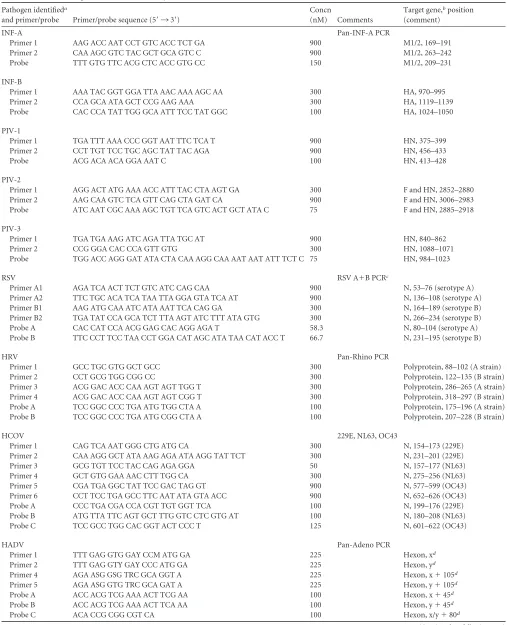
TABLE 1Primers and probes used at laboratory 1
Pathogen identifieda
and primer/probe Primer/probe sequence (5=¡3=)
Concn
(nM) Comments
Target gene,bposition (comment)
INF-A Pan-INF-A PCR
Primer 1 AAG ACC AAT CCT GTC ACC TCT GA 900 M1/2, 169–191
Primer 2 CAA AGC GTC TAC GCT GCA GTC C 900 M1/2, 263–242
Probe TTT GTG TTC ACG CTC ACC GTG CC 150 M1/2, 209–231
INF-B
Primer 1 AAA TAC GGT GGA TTA AAC AAA AGC AA 300 HA, 970–995
Primer 2 CCA GCA ATA GCT CCG AAG AAA 300 HA, 1119–1139
Probe CAC CCA TAT TGG GCA ATT TCC TAT GGC 100 HA, 1024–1050
PIV-1
Primer 1 TGA TTT AAA CCC GGT AAT TTC TCA T 900 HN, 375–399
Primer 2 CCT TGT TCC TGC AGC TAT TAC AGA 900 HN, 456–433
Probe ACG ACA ACA GGA AAT C 100 HN, 413–428
PIV-2
Primer 1 AGG ACT ATG AAA ACC ATT TAC CTA AGT GA 300 F and HN, 2852–2880
Primer 2 AAG CAA GTC TCA GTT CAG CTA GAT CA 900 F and HN, 3006–2983
Probe ATC AAT CGC AAA AGC TGT TCA GTC ACT GCT ATA C 75 F and HN, 2885–2918
PIV-3
Primer 1 TGA TGA AAG ATC AGA TTA TGC AT 900 HN, 840–862
Primer 2 CCG GGA CAC CCA GTT GTG 300 HN, 1088–1071
Probe TGG ACC AGG GAT ATA CTA CAA AGG CAA AAT AAT ATT TCT C 75 HN, 984–1023
RSV RSV A⫹B PCRc
Primer A1 AGA TCA ACT TCT GTC ATC CAG CAA 900 N, 53–76 (serotype A)
Primer A2 TTC TGC ACA TCA TAA TTA GGA GTA TCA AT 900 N, 136–108 (serotype A)
Primer B1 AAG ATG CAA ATC ATA AAT TCA CAG GA 300 N, 164–189 (serotype B)
Primer B2 TGA TAT CCA GCA TCT TTA AGT ATC TTT ATA GTG 300 N, 266–234 (serotype B)
Probe A CAC CAT CCA ACG GAG CAC AGG AGA T 58.3 N, 80–104 (serotype A)
Probe B TTC CCT TCC TAA CCT GGA CAT AGC ATA TAA CAT ACC T 66.7 N, 231–195 (serotype B)
HRV Pan-Rhino PCR
Primer 1 GCC TGC GTG GCT GCC 300 Polyprotein, 88–102 (A strain)
Primer 2 CCT GCG TGG CGG CC 300 Polyprotein, 122–135 (B strain)
Primer 3 ACG GAC ACC CAA AGT AGT TGG T 300 Polyprotein, 286–265 (A strain)
Primer 4 ACG GAC ACC CAA AGT AGT CGG T 300 Polyprotein, 318–297 (B strain)
Probe A TCC GGC CCC TGA ATG TGG CTA A 100 Polyprotein, 175–196 (A strain)
Probe B TCC GGC CCC TGA ATG CGG CTA A 100 Polyprotein, 207–228 (B strain)
HCOV 229E, NL63, OC43
Primer 1 CAG TCA AAT GGG CTG ATG CA 300 N, 154–173 (229E)
Primer 2 CAA AGG GCT ATA AAG AGA ATA AGG TAT TCT 300 N, 231–201 (229E)
Primer 3 GCG TGT TCC TAC CAG AGA GGA 50 N, 157–177 (NL63)
Primer 4 GCT GTG GAA AAC CTT TGG CA 300 N, 275–256 (NL63)
Primer 5 CGA TGA GGC TAT TCC GAC TAG GT 900 N, 577–599 (OC43)
Primer 6 CCT TCC TGA GCC TTC AAT ATA GTA ACC 900 N, 652–626 (OC43)
Probe A CCC TGA CGA CCA CGT TGT GGT TCA 100 N, 199–176 (229E)
Probe B ATG TTA TTC AGT GCT TTG GTC CTC GTG AT 100 N, 180–208 (NL63)
Probe C TCC GCC TGG CAC GGT ACT CCC T 125 N, 601–622 (OC43)
HADV Pan-Adeno PCR
Primer 1 TTT GAG GTG GAY CCM ATG GA 225 Hexon, xd
Primer 2 TTT GAG GTY GAY CCC ATG GA 225 Hexon, yd
Primer 4 AGA ASG GSG TRC GCA GGT A 225 Hexon, x⫹105d
Primer 5 AGA ASG GTG TRC GCA GAT A 225 Hexon, y⫹105d
Probe A ACC ACG TCG AAA ACT TCG AA 100 Hexon, x⫹45d
Probe B ACC ACG TCG AAA ACT TCA AA 100 Hexon, y⫹45d
Probe C ACA CCG CGG CGT CA 100 Hexon, x/y⫹80d
(Continued on following page)
on May 16, 2020 by guest
http://jcm.asm.org/
divided into three subpanels (see Tables 4, 5, and 6). The 21 samples in respiratory virus subpanel 1 contained a virus transport medium spiked with the following viruses in various concentrations: human metapneu-movirus (hMPV) (n⫽4), influenza A virus (INF A) (n⫽5), influenza B virus (INF B) (n⫽1), respiratory syncytial virus (RSV) (n⫽3), parain-fluenza virus type 1 (PIV-1) (n⫽3), PIV-2 (n⫽1), and PIV-3 (n⫽1). Three samples were negative for all viruses. The 13 samples in EQA sub-panel 2, prepared in Dulbecco’s modified Eagle’s medium and fetal calf serum, were spiked with the following viruses in various concentrations: human coronaviruses (HCOV) (n⫽3), human rhinoviruses (HRV) (n⫽ 5), and human adenoviruses (HADV) (n⫽4). One sample was negative for all viruses. EQA subpanel 3 consisted of 14 samples spiked with the following bacteria in various concentrations:Mycoplasma pneumoniae (n⫽4),Chlamydophila pneumoniae(n⫽4), andLegionella pneumophila (n⫽4). Two samples were negative. The following EQA subpanel 3 sam-ples were prepared in bronchoalveolar lavage (BAL) fluid: GRACE-37, 38, 39, 40, 44, 45, GRACE-47, and GRACE-48.
All BAL fluid pools spiked with a respiratory virus or withM. pneu-moniae,C. pneumoniae, orL. pneumophilawere tested in triplicate by mono-PCRs for the presence of that specific organism but not for the presence of the other organisms, unless they were part of the same EQA panel.
Laboratories 1 and 2, as well as Qiagen GmbH (Hilden, Germany) and PathoFinder (Maastricht, The Netherlands), analyzed the complete panel. In laboratory 3, only samples from EQA subpanel 3 were analyzed.
Distribution of the proficiency panels.The panel samples were
ran-domized by QCMD, freeze-dried, labeled, packed, and distributed at am-bient temperature to participants along with a panel receipt form and an instruction manual. Results were reported back to QCMD.
External quality assessment process.The laboratories were given 5
weeks to test the panel samples using their routine molecular diagnostic tests and to return their results to QCMD. Participants were asked to return qualitative data (presence/absence) separately for each pathogen and, if available, (semi)quantitative data, e.g., cycle threshold (CT) values.
RNA and DNA extractions. (i) RNA and DNA extractions at
labora-tory 1.Before the extraction of nucleic acid (NA), QCMD samples were
reconstituted in 1 ml NA-free water and were spiked with internal con-trols—a known amount of phocine herpesvirus (DNA) and a known amount of encephalomyocarditis virus (RNA)—to monitor the efficient extraction of DNA and RNA, respectively, as described previously (4). All
48 samples were tested separately for the pathogens. RNA and DNA ex-traction was performed by using a MagnaPure LC total nucleic acid kit (Roche Diagnostics, Mannheim, Germany) as described by Houben et al. (4). Briefly, 200l of a clinical specimen was mixed with lysis buffer and proteinase K and was subsequently incubated with magnetic particles to allow binding of the nucleic acid. Unbound material was removed by several washing steps. The nucleic acid was then eluted in 200l of elution buffer and was directly used for cDNA synthesis (pathogens carrying an RNA genome) and real-time TaqMan PCR (RNA plus DNA pathogens).
(ii) RNA and DNA extractions at laboratory 2.The freeze-dried
sam-ples were resuspended in 1 ml of NA-free water. Subsequently, 200l of this suspension was subjected to nucleic acid extraction using the Mag-NApure LC total nucleic acid kit, by following the same procedure as that used by laboratory 1. In laboratory 2, however, equine arteritis virus (EAV) was used as an internal control for RNA extractions.
(iii) RNA and DNA extractions at laboratory 3.Nucleic acids were
extracted with the NucliSens EasyMag system (bioMérieux, Grenoble, France) as described previously (7). After extraction, three aliquots were prepared and were frozen at⫺70°C until frozen shipment to laboratories 1 and 2.
Amplification methods. (i) Amplification methods at laboratory 1.
The isolated viral RNA was reverse transcribed using a MultiScribe reverse transcriptase (RT) kit and random hexamers (Applied Biosystems, Foster City, CA), according to the manufacturer’s guidelines, followed by RT inactivation for 5 min at 95°C.
[image:3.585.37.548.75.244.2]Primers and probes were selected using Primer Express software (Perkin-Elmer Applied Biosystems) and were based on highly conserved genomic regions. To cover subgroups, type-specific primers and probes were chosen for INF A and B, as well as for PIV-1 to -3. The forward and reverse primers, as well as the probes used, are given in Tables 1 through 3. Samples were assayed in duplicate in a 25-l reaction mixture containing 5l of cDNA, 12.5l of 2⫻TaqMan universal PCR master mix (Perkin-Elmer Applied Biosystems), and the concentrations of the forward primers, reverse primers, and probes indicated in Table 1. The fluorogenic probes were both labeled with the 5=reporter dye 6-carboxyfluorescein (FAM) and the 3= quencher dye 6-carboxytetramethylrhodamine (TAMRA). Amplification and detection were performed with an ABI Prism 7700 sequence detection system under the following conditions: 2 min of incubation at 50°C to attain optimal AmpErase uracil-N-glycosylase activity, 10 min at 95°C to activate the AmpliTaq Gold DNA polymerase, and 45 cycles of 15 s at 95°C and 1 min at 60°C.
TABLE 1(Continued)
Pathogen identifieda
and primer/probe Primer/probe sequence (5=¡3=)
Concn
(nM) Comments
Target gene,bposition (comment)
M. pneumoniae
Primer 1 GGT CAA TCT GGC GTG GAT CT 50 P1, 3967–3986
Primer 2 TGG TAA CTG CCC CAC AAG C 300 P1, 4032–4014
Probe TCC CCC GTT GAA AAA GTG AGT GGG T 125 P1, 3988–4012
L. pneumophila
Primer 1 GCA ATG TCA ACA GCA ATG GC 300 MIP, 13–32
Primer 2 CGG CAC CAA TGC TAT AAG ACA A 300 MIP, 94–73
Probe CAA CCG ATG CCA CAT CAT TAG CTA CAG ACA 100 MIP, 35–64
C. pneumoniae
Primer 1 AAA CAA TTT GCA TGA AGT CTG AGA A 900 MOMP, 756–732
Primer 2 TCC GCA TTG CTC AGC C 300 MOMP, 631–646
Probe TAA ACT TAA CTG CAT GGA ACC CTT CTT TAC TAG G 75 MOMP, 667–700
aINF, influenza virus; PIV, parainfluenza virus; RSV, respiratory syncytial virus; HRV, human rhinovirus; HCOV, human coronavirus; HADV, human adenovirus. bM1/2, matrix 1 and 2; HA, hemagglutinin; HN, hemagglutinin-neuraminidase; F, fusion; N, nucleoprotein; P1, cytadhesin; MIP, macrophage infectivity potentiator; MOMP,
major outer membrane protein.
cSee reference 18a.
dStart position differs with serotype.
on May 16, 2020 by guest
http://jcm.asm.org/
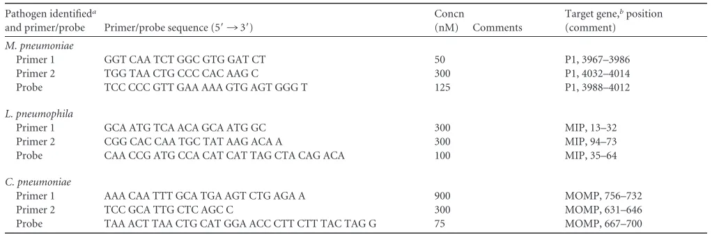
TABLE 2Primers and probes used at laboratory 2b
Pathogen identified and
primer/probe namea Primer or probe Sequence
Fragment length (bp)
Probe typec
Labeld
5= 3=
PIV-1
609PIV1-TQ-YAK Probe CAAACGATGGCTGAAAAAGGGA 164 TQ YAK BHQ-1
1101PIV1s Sense primer AAAAACTTAGGGTTAAAGACAATCCA
1102PIV1as Antisense primer GCCAGATGTRTGTCYTTCCTGCTGGT
PIV-2
621PIV2-TQ-TEXAS RED Probe AATCGCAAAAGCTGTTCAGTCAC 113 TQ TXR BHQ-2
231PIV2s Sense primer CCATTTACCTAAGTGATGGAA
232PIV2as Antisense primer CGTGGCATAATCTTCTTTTT
1065PIV2as Antisense primer TGTGGCATAATCTTCTTTCT
PIV-3
566PIV3-TQ-FAM Probe ACCCAGTCATAACTTACTCAACAGCAAC 154 TQ FAM BHQ-1
1106PIV3s Sense primer CAGGAAGCATTGTRTCATCTGT
1107PIV3as Antisense primer ATAGTGTGTAATGCAGCTYGT
PIV-4
675PIV4-TQ-CY5 Probe GTCTCAAAATTTGTTGATCAAGAYAATACAATT 200 TQ Cy5 BHQ-2
264PIV4 Sense primer CCTGGAGTCCCATCAAAAGT
1071PIV4as Antisense primer GCATCTATACGAACRCCTGCT
INF A
1815FLUA-TQ-FAM Probe CCTCGCTCACTGGGCACGGT 119 TQ FAM BHQ-1
1233FluAs Sense primer CATGGARTGGCTAAAGACAAG
1234FluAas Antisense primer TYTGGACAAAGCGTCTACG
INF B
681FLUB-TQ-TEXAS-RED Probe GCAAACACTGGGCTGCARCT 145 TQ TXR BHQ-2
220FluBs Sense primer GTCCATCAAGCTCCAGTTTT
221FluBas Antisense primer TCTTCTTACAGCTTGCTTGC
RSV
654RSV-TQ-HEX Probe CCATGTGAATTCCCTGCATCAAT 155 TQ HEX BHQ-1
674RSV-TQ-HEX Probe CCTGcGAATTCCCTGCcTCAAT 155 TQ HEX BHQ-1
1070RSVs Sense primer TTTCCACAATATTTAAGTGTtAA
412RSVs Sense primer TTTCCACAATATYTAAGTGTCAA
413RSVas Antisense primer TCATCWCCATACTTTTCTGTTA
hMPV
567MPV-TQ-YAK Probe GCATGYCAYTGGTGTGGGATATT 170 TQ YAK BHQ-1
342MPVs Sense primer CATGCCCACTATAAAAGGTCAG
343MPVas Antisense primer CACCCCAGTCTTTCTTGAAA
1068MPVs Sense primer TATGCCTACCATAAAAGGTCAA
1069MPVas Antisense primer CACCCCAGTCTTTCCTAAAG
EAV
615EAV-TQ-CY5 Probe CGCTGTCAGAACAACATTATTGCCCAC 134 TQ Cy5 BHQ-2
417EAVs Sense primer CATCTCTTGCTTTGCTCCTTAG
418EAVas Antisense primer AGCCGCACCTTCACATTG
HRV
606HRV-TQ-FAM Probe TCCTCCGGCCCCTGAATGYGGCTAA 142 TQ FAM BHQ-1
777HRV_1s Sense primer GACAGGGTGTGAAGAGCC
778HRV_2s Sense primer GACATGGTGTGAAGACCC
779HRV_3S Sense primer GACAAGGTGTGAAGAGCC
780HRV_4s Sense primer GACATGGTGTGAAGACTC
1039HRVs Sense primer GACATGGTGTGAAGATCT
1037HRVas Antisense primer ACACGGACACCCAAAGTAGT
(Continued on following page)
on May 16, 2020 by guest
http://jcm.asm.org/
The viral load was determined by the number of amplification cycles needed for a positive PCR test (CT). ACTvalue of 45 was chosen as a cutoff
for sample positivity. Samples were controlled for the presence of possible inhibitors of the extraction or amplification reaction by the indicated internal controls:CTvalues had to range within clear-cut intervals.
Posi-tive results were confirmed by a second analysis of the same sample. In case of discrepant results, a third analysis was performed.CTvalues are
mean values for duplicate reactions.
(ii) Amplification methods at laboratory 2.Primers and probes were
[image:5.585.46.544.83.568.2]designed using Beacon Designer software (Premier Biosoft International, Palo Alto, CA) and have been described previously (15, 16), except that molecular beacons were replaced by TaqMan hydrolysis probes, using the same target sequences. The assays were performed as four multiplex real-time PCR assays, combining INF A, INF B, and RSV; PIV-1 to -4; HRV, hMPV, and the EAV internal control; and finally the four HCOV 229E, OC43, NL63, and HKU1.
TABLE 2(Continued)
Pathogen identified and
primer/probe namea Primer or probe Sequence
Fragment length (bp)
Probe typec
Labeld
5= 3=
HCOV-NL63
599HCOV-NL63-TQ-TXR Probe CGCATACGCCAACGCTCTTGAACA 143 TQ TXR BHQ-2
750HCOV-NL63s Sense primer GTTCTGATAAGGCACCATATAGG
751HCOV-NL63as Antisense primer TTTAGGAGGCAAATCAACACG
HCOV-229E
598HCOV-229E-TQ-FAM Probe ATGAACCTGAACACCTGAAGCCAATCTATG 137 TQ FAM BHQ-1
741HCOV-229Es Sense primer CATACTATCAACCCATTCAACAAG
742HCOV-229Eas Antisense primer CACGGCAACTGTCATGTATT
HCOV-OC43
587HCOV-43-TQ-YAK Probe TGCCCAAGAATAGCCAGTACCTAGT 110 TQ YAK BHQ1
484HCOV43s Sense primer CATACTCTGACGGTCACAATAATA
485HCOV43as Antisense primer ACCTTAGCAACAGTCATATAAGC
HCOV-HKU1
677HCOV-HKU1-TQ-CY5 Probe TYCGCCTGGTACGATTTTGCCTCA 147 TQ Cy5 BHQ-2
864HCOV-HKU1s Sense primer TCCTACTAYTCAAGAAGCTATCC
865HCOV-HKU1as Antisense primer AATGAACGATTATTGGGTCCAC
HADV
692ADV-XS-FAM Probe AGCCCACCCTKCTTTAT 139 TQ FAM BHQ-1
658ADV4-TQ-YAK Probe GAGTCYACCCTTCTCTATGT YAK BHQ-1
372ADVs Sense primer CATGACTTTTGAGGTGGATC
346ADVas Antisense primer CCGGCCGAGAAGGGTGTGCGCAGGTA
423ADV4s Sense primer CATGAATTTCGAAGTCGACC
424ADV31s Sense primer TATGACATTTGAAGTTGACC
M. pneumoniae
612MYCPN-TQ-YAK Probe CAAAGCCACCCTGATCACCC 151 TQ YAK BHQ-1
224MYCPNs Sense primer ATTCCCGAACAAAATAATG
225MYCPNas Antisense primer GTTTGACAAAGTCCGTGAAG
C. pneumoniae
611CPN-TQ-FAM Probe GGGATCTTCGGACCTTTCGG 154 TQ FAM BHQ-1
214CPN16Ss Sense primer GCGGAAGGGTTAGTAGTACA
215CPN16Sas Antisense primer ATCGCATAAACTCTTCCTCA
Legionellaspp.
539LEGSP-MB-FAM Probe CCGAGCGGTGAGTAACGCGTAGGAATATGGCTCGG 212 MB FAM Dabcyl
156LegSPs Sense primer AGGCTAATCTTAAAGCGCC
157LegSPas Antisense primer CCTGGCTCAGATTGAACG
L. pneumophila
593LEGPN-TQ-YAK Probe GCATTGGTGCCGATTTGGGA 124 TQ YAK BHQ-1
269LGPNs Sense primer TGGTGACTGCAGCTGTTATG
270LGPNas Antisense primer CATTGCTTCCGGATTAACAT
aPIV, parainfluenza virus; INF, influenza virus; RSV, respiratory syncytial virus; hMPV human metapneumovirus; EAV, equine arteritis virus; HRV, human rhinovirus;; HCOV,
human coronavirus; HADV, human adenovirus.
bYAK, Yakima Yellow; TXR, Texas Red; HEX, hexachlorofluorescein; R⫽(AG); Y⫽(CT); K⫽(GT); W⫽(AT). cMB, molecular beacon; TQ, TaqMan.
dBHQ, black hole quencher.
on May 16, 2020 by guest
http://jcm.asm.org/
Real-time PCR was performed in 50l of a reaction mixture consist-ing of 10l of 5⫻one-step RT-PCR buffer (One-Step RT-PCR kit; Qia-gen, Hilden, Germany), 10 mM deoxynucleoside triphosphates (dNTPs), 4.5 mM MgCl2, 0.6M each primer (Table 2), and 0.34M TaqMan
probes, with 5l of the template. The PCR thermal profile consisted of an initial cDNA step of 30 min at 50°C, followed by 15 min at 95°C and 45 cycles of 30 s at 95°C, 30 s at 55°C, and 30 s at 72°C.
For DNA targets, published assays (17) were used; these were per-formed in HotStarTaqmaster mix (Qiagen). Amplification, detection, and data analysis were performed using the iCycler IQ real-time detection system (Bio-Rad, Veenendaal, The Netherlands).
When samples were tested in duplicate reactions, one positive reaction was considered a positive result, since in proficiency testing, samples with concentrations around the limit of detection (LOD) can be detected. In case both reactions were positive, the value in the table is the mean value.
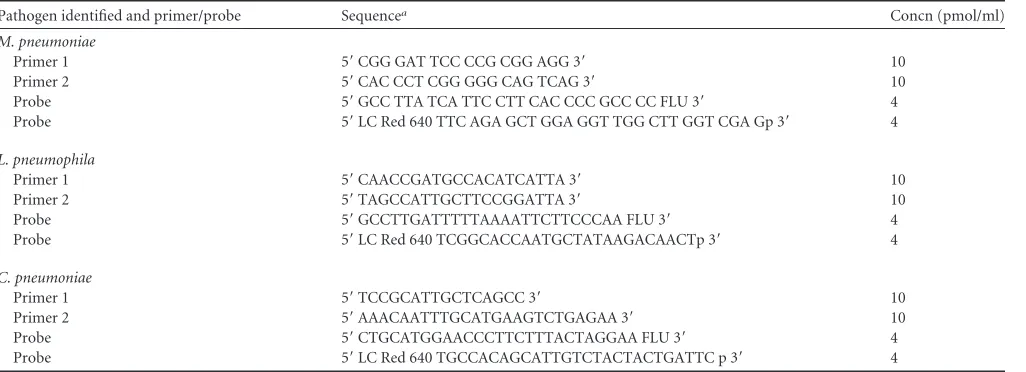
(iii) Amplification methods at laboratory 3. Real-time in-house
mono-PCRs were applied as described previously forM. pneumoniae,C. pneumoniae, andL. pneumophiladetection (6, 18). Primers and probes are presented in Table 3. Positive results were confirmed by a second analysis of the same sample. In case of discrepant results, a third analysis was performed.
Commercially available PCRs.The panels were sent to Qiagen GmbH
to be analyzed by the ResPlex 1 assay and to PathoFinder to be analyzed by the RespiFinder plus and RespiFinder Smart 21 assays according to the manufacturer’s instructions. After receiving the first ResPlex results, the manufacturer made modifications to the kit, producing the ResPlex 2 assay.
The RespiFinder Smart 21 assay is a real-time variant (under develop-ment) of the MultiFinder PCR technology (13) that enables the detection and differentiation of 21 respiratory pathogens: INF A, INF A H1N1, INF B, RSV-A, RSV-B, HADV, HRV, PIV-1 to -4, HCOV 229E, NL63, OC43, and HKU1, hMPV, bocavirus,M. pneumoniae,C. pneumoniae,L. pneu-mophila, andBordetella pertussis.
RESULTS
Results of in-house PCRs for the detection of respiratory
vi-ruses.
The performances of the NAATs for the detection of the
respiratory viruses (subpanels 1 and 2) at laboratories 1 and 2 on
extracts obtained with the routine MagNA pure LC and NucliSens
EasyMag nucleic acid extraction procedures were comparable; no
major differences in sensitivity and specificity were observed.
Us-ing its own protocol for extraction and amplification, laboratory 1
obtained one false-positive result (sample GRACE-02; PIV-2/4)
and three false-negative results, all on samples with very low viral
loads (samples GRACE-02, GRACE-08, and GRACE-33) (Tables
4 and 5). When examining NucliSens EasyMag-extracted samples,
laboratory 1 reported the correct virus in samples GRACE-09,
GRACE-16, and GRACE-18, but each time in combination with
PIV-2/4.
Laboratory 2 reported two and three false-negative results after
applying its own nucleic acid extraction procedure and the
NucliSens EasyMag procedure, respectively, for the 21 samples of
subpanel 1 and one and three false-negative results for the 13
samples of subpanel 2 (Tables 4 and 5). No false-positive results
were reported after NucliSens EasyMag extraction, in contrast to
four false-positive results obtained after laboratory 2 used its own
nucleic acid extraction (Tables 4 and 5).
In general, after NucliSens EasyMag extraction, both
laborato-ries obtained
C
Tvalues equal to or lower than those obtained with
their in-house extraction procedures (Tables 1 to 4).
Results of in-house PCRs for the detection of atypical
patho-gens.
In Table 6, the results of the
M. pneumoniae
,
C. pneumoniae
,
and
L. pneumophila
EQA subpanel are presented. No
false-negative results were reported by laboratories 1 and 3. Laboratory
2 failed to detect 3 positive samples: GRACE-38, containing the
lowest concentration of
M. pneumoniae
, 50 color-changing units
(CCU)/ml, and GRACE-45 and GRACE-47, spiked with 18 and 60
CFU/ml of
L. pneumophila
, respectively. None of the
negative-control samples (negative transport medium [NTM]) were found
positive by the three GRACE laboratories. In addition to the
cor-rect pathogens, laboratories 1 and 3 also detected
M. pneumoniae
in two samples. Laboratory 2 also found
M. pneumoniae
in one of
these samples, indicating that contamination had occurred before
the start of analysis of these samples.
Results by commercially available PCRs: the ResPlex,
Respi-Finder Plus, and RespiRespi-Finder Smart 21 assays.
After
examina-tion of the EQA panel for the presence of hMPV, INF A/B, PIV-1
to -3, and RSV in subpanel 1 (Tables 4 and 7), correct results were
reported for 9/21, 11/21, 17/21, and 15/21 samples by use of the
ResPlex 1, ResPlex 2, RespiFinder plus, and RespiFinder Smart 21
assays, respectively. Sample GRACE-11, containing transport
me-TABLE 3Primers and probes used at laboratory 3Pathogen identified and primer/probe Sequencea Concn (pmol/ml)
M. pneumoniae
Primer 1 5=CGG GAT TCC CCG CGG AGG 3= 10
Primer 2 5=CAC CCT CGG GGG CAG TCAG 3= 10
Probe 5=GCC TTA TCA TTC CTT CAC CCC GCC CC FLU 3= 4
Probe 5=LC Red 640 TTC AGA GCT GGA GGT TGG CTT GGT CGA Gp 3= 4
L. pneumophila
Primer 1 5=CAACCGATGCCACATCATTA 3= 10
Primer 2 5=TAGCCATTGCTTCCGGATTA 3= 10
Probe 5=GCCTTGATTTTTAAAATTCTTCCCAA FLU 3= 4
Probe 5=LC Red 640 TCGGCACCAATGCTATAAGACAACTp 3= 4
C. pneumoniae
Primer 1 5=TCCGCATTGCTCAGCC 3= 10
Primer 2 5=AAACAATTTGCATGAAGTCTGAGAA 3= 10
Probe 5=CTGCATGGAACCCTTCTTTACTAGGAA FLU 3= 4
Probe 5=LC Red 640 TGCCACAGCATTGTCTACTACTGATTC p 3= 4
aFLU, fluorescein.
on May 16, 2020 by guest
http://jcm.asm.org/
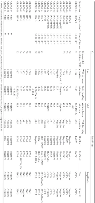
[image:6.585.42.555.77.263.2]TABLE 4 EQA subpanel 1 results Sample no. Sample content a Concn/dilution C T Result b by: Reference lab (real-time PCR) Lab 1 Lab 2 ResPlex 1 ResPlex 2 RespiFinder With in-house extraction With EasyMag extraction With in-house extraction With EasyMag extraction Plus Smart 21 GRACE-14 hMPV-I 1.0 ⫻ 10 ⫺ 4 31 31.38 31.19 32.2; INF A, 40.3 32.3 hMPV hMPV hMPV hMPV GRACE-01 hMPV-I 1.0 ⫻ 10 ⫺ 6 36 37.93 39 45.1 40.5 Negative Negative Negative Negative GRACE-09 hMPV-II 1.0 ⫻ 10 ⫺ 4 33 34.02 34.08 35.3 37.4 hMPV hMPV hMPV hMPV GRACE-07 hMPV-II 1.0 ⫻ 10 ⫺ 5 35 37.85 36.52 41.5 Negative Negative hMPV hMPV hMPV GRACE-02 INF A type H3 1.0 ⫻ 10 ⫺ 7 PIV-2/4, 44.17 Negative Negative Negative Negative Negative Negative Negative GRACE-08 INF A type H3 1.0 ⫻ 10 ⫺ 7 Negative 37.9 39.5 Negative Negative Negative Negative Negative GRACE-15 INF A type H3 1.0 ⫻ 10 ⫺ 6 37.75 35.97 36.3 37.2 Negative Negative INF A INF A GRACE-17 INF A type H1 1.0 ⫻ 10 ⫺ 5 35.38 33.75 34.4 35 Negative INF A INF A INF A GRACE-12 INF A type H1 1.0 ⫻ 10 ⫺ 6 40.39 35.05 Negative 36.7 Negative HCOV, EV INF A Negative GRACE-06 INF B 1.0 ⫻ 10 ⫺ 6 38.9 36.43 39.3 Negative Negative Negative INF B INF B GRACE-21 RSV A 1.0 ⫻ 10 ⫺ 3 24 24.24 27.75 29.5 29.8 RSV, HADV RSV A, HADV RSV A, HADV RSV A, HADV GRACE-19 RSV A 1.0 ⫻ 10 ⫺ 5 30 32.26 30.07 40.7 35.3 HRV HCOV, EV RSV A RSV A GRACE-16 RSV B 5.0 ⫻ 10 ⫺ 4 32 28.88 30.36; PIV-2/ 4, 42.4 33.4 34.4 Negative INF B, HRV RSV B RSV B GRACE-20 PIV-1 27 27.85 27.19 31.1 30.9 PIV-1 PIV-1 PIV-1 PIV-1 GRACE-10 PIV-1 30 30.53 29.87 34 33.8 PIV-1 PIV-1 PIV-1 PIV-1 GRACE-13 PIV-1 34 33.71 34.08 38.1 38 PIV-1 PIV-1 PIV-1 Negative GRACE-03 PIV-2 34 32.7 31.57 33.1 30.9 PIV-2 PIV-2, HCOV, EV PIV-2 PIV-2 GRACE-18 PIV-3 32 34.7 32.85; PIV-2/ 4, 40.87 31.3 32.5 PIV-3 PIV-3 PIV-3 PIV-3 GRACE-04 NTM 0 Negative Negative Negative Negative Negative Negative Negative Negative GRACE-05 NTM 0 Negative Negative Negative Negative Negative Negative Negative Negative GRACE-11 NTM 0 Negative Negative Negative Negative HRV Negative Negative Negative a hMPV, human metapneumovirus; INF, influenza virus; RSV, respiratory syncytial virus; PIV, parainfluenza virus; NTM, negative transport medium. b HCOV, human coronavirus; EV, enterovirus; HADV, human adenovirus; HRV, human rhinovirus.
on May 16, 2020 by guest
http://jcm.asm.org/
[image:7.585.137.446.63.721.2]dium only, was reported to be positive for HRV by the ResPlex
assay. GRACE-19 was spiked with RSV-A but was found to be
positive for HRV by the ResPlex 1 assay and positive for HCOV
and enterovirus by the ResPlex 2 assay. GRACE-16 was spiked
with RSV-B but was found to be positive for INF B and HRV by
the ResPlex 2 assay, whereas GRACE-12 contained INF A but was
reported to be positive for HCOV and enterovirus by the same
assay. Additionally, the ResPlex 2 assay identified 2 extra viruses in
GRACE-03. Sample GRACE-21 was found to be positive for both
HADV and RSV by all 4 commercial tests.
No difference in sensitivity was observed between the
Respi-Finder plus and the RespiRespi-Finder Smart 21 assay in examination of
subpanel 2 for HADV, HCOV, and HRV (Tables 5 and 7): both
assays failed to detect HADV 31, HRV 16, and HRV 90 in samples
GRACE-24, GRACE-25, and GRACE-33, respectively. The latter
sample was also reported as negative by all of the in-house tests.
The ResPlex assay (ResPlex 1) failed to detect the same three
sam-ples. Additionally, five other samples were also reported as
nega-tive by the ResPlex 1 assay. After modification of the assay to the
ResPlex 2 format, sensitivity improved slightly, with five
false-negative results. No false-positive results were obtained with any
of the commercially available assays.
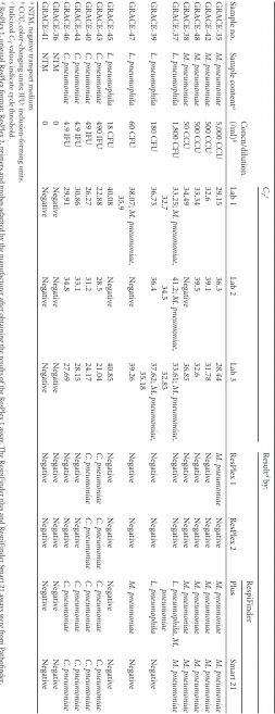
Upon examination of subpanel 3, the RespiFinder plus assay
failed to detect the sample spiked with 18 CFU of
L. pneumophila
(Table 6). All other samples were correctly identified. The
Respi-Finder Smart 21 assay did not detect any of the four samples
spiked with
L. pneumophila
. The manufacturer was contacted on
this issue and improved the assay. GRACE-37 and GRACE-39
were correctly identified as
L. pneumophila
-positive samples after
retesting with a newer version of the RespiFinder Smart 21 assay.
No other false-negative or false-positive results were reported by
use of the RespiFinder assays.
The sensitivities of both ResPlex assays were very low. The
original format (ResPlex 1) yielded only three positive samples, all
with the highest loads of
M. pneumoniae
or
C. pneumoniae
. The
assay failed to detect any
L. pneumophila
-positive samples. After
the assay was adapted (ResPlex 2 results), no improvement was
seen.
DISCUSSION
One of the objectives of this study was to check whether in-house
nucleic acid extraction procedures could be replaced by a central
nucleic acid extraction method with subsequent transport of
ex-tracts to other centers for nucleic acid amplification purposes in
the context of a large study. The RNA and DNA sent to the
par-ticipating laboratories were extracted from the EQA panel at
lab-oratory 3 with the NucliSens EasyMag system, producing nucleic
acid extracts of high quality, as reflected by the results obtained.
After comparison of the results, the different extraction methods
did not reveal significant differences: comparable sensitivities and
specificities were obtained with both in-house nucleic acid
extrac-tion methods and the NucliSens EasyMag extracextrac-tion procedure.
Considering the overall workload and the results obtained, the
method of choice for extraction of nucleic acids from respiratory
samples collected in the GRACE network is the NucliSens
Easy-Mag procedure performed in laboratory 3.
For comparison of the sensitivities and specificities of the
dif-ferent nucleic acid amplification methods, it was decided that
lab-oratory 1 would apply its in-house PCRs for the detection of RSV,
INF A/B, HCOV OC43, NL63, and 229E, and the polyomaviruses
TABLE 5 EQA subpanel 2 results Sample no. Sample content a Concn/dilution CT Result bby: Reference lab (real-time PCR) Lab 1 Lab 2 ResPlex 1 ResPlex 2 RespiFinder With in-house extraction With EasyMag extraction With in-house extraction With EasyMag extraction Plus Smart 21 GRACE-31 HCOV 229E 2.0 ⫻ 10 ⫺ 4 30–32 31.75 33.39 37.0; HADV, 45.0 43.7 HCOV 229E HCOV 229E HCOV 229E HCOV 229E GRACE-30 HCOV OC43 1.0 ⫻ 10 ⫺ 4 34–36 35.35 31.86 42.4 Negative Negative HCOV OC43, EV HCOV OC43 HCOV OC43 GRACE-22 HCOV NL63 2.0 ⫻ 10 ⫺ 6 35–37 32.79 34.62 Negative Negative Negative Negative HCOV NL63 HCOV NL63 GRACE-28 HRV 16 1.0 ⫻ 10 ⫺ 4 29–30 40.25 29.91 34.6; HADV, 40.0 33.8 HRV HRV HRV HRV GRACE-25 HRV 16 1.0 ⫻ 10 ⫺ 6 38–40 39.7 37.36 42.3 39.7 Negative Negative Negative Negative GRACE-34 HRV 72 1.0 ⫻ 10 ⫺ 5 33–34 35.43 32.97 39.1 36.7 Negative Negative HRV HRV GRACE-23 HRV 90 1.0 ⫻ 10 ⫺ 3 29–30 29.58 28.15 31.4; HADV, 44.6 30.3 HRV HRV HRV HRV GRACE-33 HRV 90 1.0 ⫻ 10 ⫺ 6 37–38 Negative Negative Negative Negative Negative Negative Negative Negative GRACE-27 HADV 3 5.0 ⫻ 10 2 34.06 31.34 35.4 33.5 Negative HADV HADV HADV GRACE-29 HADV 4 2.5 ⫻ 10 2 31.04 31.75 37.1 35.8 HADV HADV HADV HADV GRACE-24 HADV 31 1.0 ⫻ 10 2 38.89 41.2 39.8 37 Negative HADV Negative Negative GRACE-32 HADV 1 1.0 ⫻ 10 2 31.83 36.69 38.4 38.7 Negative Negative HADV HADV GRACE-26 NTM 0 Negative Negative Negative Negative Negative Negative Negative Negative aHCOV, human coronavirus; HRV, human rhinovirus; HADV, human adenovirus; NTM, negative transport medium. bEV, enterovirus.
on May 16, 2020 by guest
http://jcm.asm.org/
[image:8.585.65.260.44.725.2]TABLE 6 EQA subpanel 3 results Sample no. Sample content a Concn/dilution (/ml) b C T c Result d by: Lab 1 Lab 2 Lab 3 ResPlex 1 ResPlex 2 RespiFinder Plus Smart 21 GRACE-35 M. pneumoniae 5,000 CCU 29.15 36.3 28.44 M. pneumoniae Negative M. pneumoniae M. pneumoniae GRACE-42 M. pneumoniae 500 CCU 32.6 39.1 31.78 Negative Negative M. pneumoniae M. pneumoniae GRACE-48 M. pneumoniae 500 CCU 33.34 39.5 32.6 Negative Negative M. pneumoniae M. pneumoniae GRACE-38 M. pneumoniae 50 CCU 34.49 Negative 36.85 Negative Negative M. pneumoniae M. pneumoniae GRACE-37 L. pneumophila 1,800 CFU 33.25; M. pneumoniae , 32.7 41.2; M. pneumoniae , 34.5 33.61; M. pneumoniae , 32.85 Negative Negative L. pneumophila , M. pneumoniae M. pneumoniae GRACE-39 L. pneumophila 180 CFU 36.73 36.4 37.62; M. pneumoniae , 35.18 Negative Negative L. pneumophila Negative GRACE-47 L. pneumophila 60 CFU 38.07; M. pneumoniae , 35.9 Negative 39.26 Negative Negative M. pneumoniae Negative GRACE-45 L. pneumophila 18 CFU 40.08 Negative 40.85 Negative Negative Negative Negative GRACE-43 C. pneumoniae 490 IFU 22.88 28.5 21.04 C. pneumoniae C. pneumoniae C. pneumoniae C. pneumoniae GRACE-40 C. pneumoniae 49 IFU 26.27 31.2 24.17 C. pneumoniae C. pneumoniae C. pneumoniae C. pneumoniae GRACE-44 C. pneumoniae 4.9 IFU 30.86 33.1 28.15 Negative Negative C. pneumoniae C. pneumoniae GRACE-46 C. pneumoniae 4.9 IFU 29.91 34.8 27.69 Negative Negative C. pneumoniae C. pneumoniae GRACE-36 NTM 0 Negative Negative Negative Negative Negative Negative Negative GRACE-41 NTM 0 Negative Negative Negative Negative Negative Negative Negative a NTM, negative transport medium. b CCU, color-changing units; IFU: inclusion-forming units. c Italicized C T values indicate cycle threshold. d ResPlex 1, original ResPlex format; ResPlex 2, primers and probes adapted by the manufacturer after obtaining the results of the ResPlex 1 assay. The R espiFinder plus and RespiFinder Smart 21 assays were from Pathofinder, Maastricht, Netherlands; the ResPlex assays were from Qiagen GmbH, Hilden, Germany.
on May 16, 2020 by guest
http://jcm.asm.org/
[image:9.585.166.419.66.723.2]WUPyV and KIPyV. Laboratory 2 would examine samples for
PIV-1 to -4, HRV, hMPV, HAdV, and bocavirus by using its
in-house PCRs, and the in-in-house PCRs of laboratory 3 would be used
for the detection of
M. pneumoniae
,
C. pneumoniae
, and
L.
pneu-mophila
. This decision was not based on
CT
values, since the
Bio-Rad iCycler, used in laboratory 2, usually gives higher
C
Tvalues
than the real-time equipment used in the other two laboratories.
The RespiFinder Smart 21 assay (PathoFinder) is a real-time
multiplex PCR assay under development and is not yet
commer-cially available. It is a further development of the MultiFinder
technology as applied in the RespiFinder plus assay. According to
the manufacturer (personal communication), the analytical
sen-sitivity of the RespiFinder ranges from 5 to 50 copies per reaction
for most targets when commercially available quantitated DNA/
RNA PCR controls (Vircell) are used. Seven samples positive for a
respiratory virus were missed by the assay, two more than with the
RespiFinder plus assay. All these samples contained low viral
loads. All
L. pneumophila
-spiked samples were classified as
nega-tive based on the RespiFinder Smart 21 results, whereas two had
been positive with the RespiFinder plus assay. The manufacturer
was contacted on this issue and improved the assay. The
commer-cially available ResPlex assay (Qiagen GmbH), a multiplex PCR,
was also evaluated in this study but did not perform well. Even
when the company had made some modifications to the kit after
their first results (ResPlex 1), the performance of the assay
im-proved only slightly (ResPlex 2), and it was considered too
insen-sitive for further evaluations. The manufacturer was contacted
and is aware of the sensitivity problems of the ResPlex assay. It
intends to improve the sensitivity of the test. According to the
literature, the analytical sensitivity reported by the supplier of the
ResPlex II assay is about 500 viral genomes per reaction (21).
Serial dilutions of titrated strains were prepared by Wang et al.,
and sensitivities on the order of 3.0 · 10
⫺250% tissue culture
infective dose (TCID
50)/reaction for INF A, 1.0 · 10
⫺3TCID
50/
reaction for INF B, 1.4 · 10
⫺1TCID
50
/reaction for RSV, and 7.0
TCID
50s/reaction for human enterovirus were found (21). Lower
sensitivities for the ResPlex II assay than for multiplex NAATs are
also reported in the literature (1, 3).
All samples used in this GRACE quality control (QC) panel
originated from previous EQA distributions. All pools spiked with
a respiratory virus or with
M. pneumoniae
,
C. pneumoniae
, or
L.
pneumophila
were tested for the presence of that specific organism
but not for the presence of the other organisms, unless they were
part of the same EQA panel. When the commercially available
multiplex assays were applied to the GRACE QC samples, more
than 1 target organism was detected in some GRACE samples. If
the result was confirmed by at least one of the other commercially
available multiplex tests, e.g., GRACE-21 and GRACE-37, the
ad-ditional organism was probably already present in the original
pool, and the result should be considered correct. On the other
hand, the detection of PIV-2/4 in GRACE-09, GRACE-16, and
GRACE-18 (Table 4) and of HADV in GRACE-23, GRACE-28,
and GRACE-31 (Table 5) was probably due to contamination that
occurred during the extraction/amplification procedure. This
conclusion is supported by the fact that the reported
C
Tvalues are
similar.
In conclusion, this study demonstrated the importance of
in-cluding a sufficient number of weakly positive samples and
nega-tive controls in amplification runs to detect possible false-posinega-tive
and false-negative results when the best-performing test must be
selected and when a new assay is to be validated.
ACKNOWLEDGMENT
K. Loens is supported through Priority 1 (Life Sciences, Genomics and Biotechnology for Health) of the European Union’s FP6, contract LSHM-CT-2005-518226, GRACE.
REFERENCES
1.Balada-Llasat JM, LaRue H, Kelly C, Rigali L, Pancholi P.2011. Eval-uation of commercial ResPlex II v2.0, MultiCode-PLx, and xTAG respi-ratory viral panels for the diagnosis of respirespi-ratory viral infections in adults. J. Clin. Virol.50:42– 45.
2.De Mendonca R, MacKay WG.2007. QCMD 2007Legionella pneumo-phila(LPDNA07) proficiency programme. QCMD report. QCMD, Glas-gow, Scotland.
3.Gharabaghi F, Hawan A, Drews SJ, Richardson SE.2011. Evaluation of multiple commercial molecular and conventional diagnostic assays for the detection of respiratory viruses in children. Clin. Microbiol. Infect. doi: 10.1111/j.1469-0691.2011.03529.x.
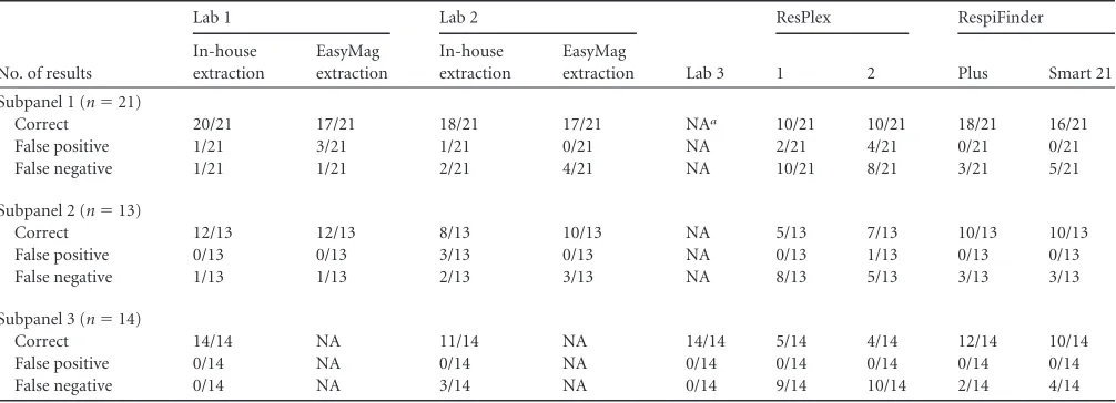
[image:10.585.42.545.77.259.2]4.Houben ML, et al.2010. Disease severity and viral load are correlated in TABLE 7Summary of results
No. of results
Lab 1 Lab 2
Lab 3
ResPlex RespiFinder
In-house extraction
EasyMag extraction
In-house extraction
EasyMag
extraction 1 2 Plus Smart 21
Subpanel 1 (n⫽21)
Correct 20/21 17/21 18/21 17/21 NAa 10/21 10/21 18/21 16/21
False positive 1/21 3/21 1/21 0/21 NA 2/21 4/21 0/21 0/21
False negative 1/21 1/21 2/21 4/21 NA 10/21 8/21 3/21 5/21
Subpanel 2 (n⫽13)
Correct 12/13 12/13 8/13 10/13 NA 5/13 7/13 10/13 10/13
False positive 0/13 0/13 3/13 0/13 NA 0/13 1/13 0/13 0/13
False negative 1/13 1/13 2/13 3/13 NA 8/13 5/13 3/13 3/13
Subpanel 3 (n⫽14)
Correct 14/14 NA 11/14 NA 14/14 5/14 4/14 12/14 10/14
False positive 0/14 NA 0/14 NA 0/14 0/14 0/14 0/14 0/14
False negative 0/14 NA 3/14 NA 0/14 9/14 10/14 2/14 4/14
aNA, not applicable.
on May 16, 2020 by guest
http://jcm.asm.org/
infants with primary respiratory syncytial virus infection in the commu-nity. J. Med. Virol.82:1266 –1271.
5.Kumar S, Hammerschlag MR.2007. Acute respiratory infection due to Chlamydia pneumoniae: current status of diagnostic methods. Clin. Infect. Dis.44:568 –576.
6.Loens K, et al.2008. Evaluation of different nucleic acid amplification techniques for the detection ofM. pneumoniae,C.pneumoniaeand Legio-nella spp. in respiratory specimens from patients with community-acquired pneumonia. J. Microbiol. Methods73:257–262.
7.Loens K, Bergs K, Ursi D, Goossens H, Ieven M.2007. Evaluation of NucliSens easyMAG for automated nucleic acid extraction from various clinical specimens. J. Clin. Microbiol.45:421– 425.
8.Loens K, Goossens H, Ieven M.2010. Acute respiratory infection due to Mycoplasma pneumoniae: current status of diagnostic methods. Eur. J. Clin. Microbiol. Infect. Dis.29:1055–1069.
9.Loens K, et al.2010. A multicenter pilot external quality assessment programme to assess the quality of molecular detection ofChlamydophila pneumoniaeandMycoplasma pneumoniae.J. Microbiol. Methods82:131– 135.
10. Mahony JB.2008. Detection of respiratory viruses by molecular methods. Clin. Microbiol. Rev.21:716 –747.
11. Meijer A, Niesters H, MacKay WG.2007. QCMD 2007 influenzavirus A and B (INFA07) proficiency programme. QCMD, Glasgow, Scotland. 12. Niesters H, MacKay WG, van Loon AM.2007. QCMD 2007 influenza
virus haemagglutinin typing INFHT07 proficiency programme. QCMD, Glasgow, Scotland.
13. Niesters H, van Loon AM, MacKay WG.2007. QCMD 2007 metapneu-movirus and respiratory syncytial virus (MPVRSV07) proficiency pro-gramme. QCMD report. QCMD, Glasgow, Scotland.
14. Pandit A, Claas EC.2007. QCMD 2007 adenovirus (ADVDNA07) pro-ficiency programme. QCMD, Glasgow, Scotland.
15. Scheltinga SA, Templeton KE, Beersma MF, Claas EC.2005. Diagnosis of human metapneumovirus and rhinovirus in patients with respiratory tract infections by an internally controlled multiplex real-time RNA PCR. J. Clin. Virol.33:306 –311.
16. Templeton KE, Scheltinga SA, Beersma MF, Kroes AC, Claas EC.2004. Rapid and sensitive method using multiplex real-time PCR for diagnosis of infections by influenza A and influenza B viruses, respiratory syncytial virus, and parainfluenza viruses 1, 2, 3, and 4. J. Clin. Microbiol.42:1564 – 1569.
17. Templeton KE, et al. 2005. Improved diagnosis of the etiology of community-acquired pneumonia with real-time polymerase chain reac-tion. Clin. Infect. Dis.41:345–351.
18. Ursi D, Dirven K, Loens K, Ieven M, Goossens H.2003. Detection of Mycoplasma pneumoniaein respiratory samples by real-time PCR using an inhibition control. J. Microbiol. Methods55:149 –153.
18a.van de Pol AC, et al. 2010. Molecular quantification of respiratory syncytial virus in respiratory samples: reliable detection during the initial phase of infection. J. Clin. Microbiol.48:3569 –3574.
19. van Loon AM, MacKay WG.2006. QCMD 2006 influenza virus A and B (INFRNA06) proficiency programme. QCMD, Glasgow, Scotland. 20. van Loon AM, Niesters H, MacKay WG.2007. QCMD 2007 rhinovirus
and coronavirus (RVCVRNA07) proficiency programme. QCMD, Glas-gow, Scotland.
21. Wang H, Kong F, Jelfs P, James G, Gilbert GL.2004. Simultaneous detection and identification of common cell culture contaminant and pathogenicMollicutesstrains by reverse line blot hybridization. Appl. En-viron. Microbiol.70:1483–1486.