The Genus
Corynebacterium
and Other Medically Relevant
Coryneform-Like Bacteria
Kathryn Bernarda,b
National Microbiology Laboratory, Public Health Agency of Canada, Winnipeg, Manitoba, Canada,aand University of Manitoba, Department of Medical Microbiology, Winnipeg, Manitoba, Canadab
Catalase-positive Gram-positive bacilli, commonly called “diphtheroids” or “coryneform” bacteria were historically nearly al-ways dismissed as contaminants when recovered from patients, but increasingly have been implicated as the cause of significant infections. These taxa have been underreported, and the taxa were taxonomically confusing. The mechanisms of pathogenesis, especially for newly described taxa, were rarely studied. Antibiotic susceptibility data were relatively scant. In this minireview, clinical relevance, phenotypic and genetic identification methods, matrix-assisted laser desorption ionization–time of flight (MALDI-TOF) evaluations, and antimicrobial susceptibility testing involving species in the genusCorynebacteriumand other medically relevant Gram-positive rods, collectively called coryneforms, are described.
T
his minireview will be limited to “club-shaped” Gram-posi-tive, medically relevant species of the genusCorynebacteriumand of the “irregular” Gram-positive coryneforms mostly but not exclusively from the suborderMicrococcineae. Those coryneforms were selected because they most closely resembleCorynebacterium
speciessensu strictophenotypically and so can be difficult to dif-ferentiate from members of that genus. Coryneforms described here are rare, opportunistic human pathogens, and they are Gram-positive bacilli or coccobacilli. Nearly all are catalase posi-tive, and they express a range of pigments and metabolic pro-cesses.
Clinical relevance.These coryneform bacteria are increasingly being recognized as causing opportunistic disease under specific circumstances, such as in patients who are immunocompromised, have prosthetic devices, or have been in hospitals/nursing homes for long-term periods of time (20). The most significant pathogen of this group remainsCorynebacterium diphtheriae, the primary cause of diphtheria, a disease which has essentially disappeared from developed countries after implementation of universal vac-cination that targets the primary virulence factor, the diphtheria toxin (DT) (52). Comprehensive reviews outline epidemiology, the diseases caused byC. diphtheriae, vaccine recommendations, immune response to the vaccine at population levels, and the function and pathogenesis of DT (20,52), with other manifesta-tions shown inTable 1.
The zoonotic agentsCorynebacterium pseudotuberculosis(the cause of caseous lymphadenitis primarily in sheep and goats) (3), transmitted to humans by contact with diseased animals (38) and
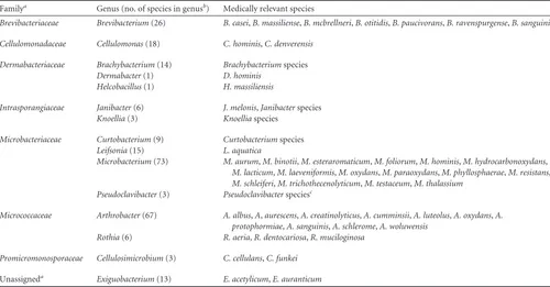
C. ulcerans, historically thought to cause disease in humans after contact with contaminated milk or farm animals, but more re-cently linked to transmission between humans and their compan-ion pets such as cats and dogs (13,51), are also able to produce diphtheria toxin and cause diphtheria-like disease in humans. It is therefore recommended that those in frequent contact with ani-mals (veterinarians, animal care technologists, or farm workers) ensure that they maintain adequate vaccine coverage against diph-theria.Corynebacterium species that have caused disease in hu-mans with some also considered as zoonotic agents are reviewed inTable 1. Infections or case reports involving coryneforms are outlined inTable 2.
Determining clinical relevancy: when to identify to genus and species. Studies have demonstrated that Corynebacterium
species and various coryneforms, particularly those taxa found as part of normal skin flora, are prominent contaminants of clinical materials, although occasionally it is difficult to correctly decide in a timely fashion if recovery of such bacteria implies contamina-tion or has clinical relevance (53). Therefore, it is recommended that clinicians should attempt to identify any of the taxa described in this minireview to the genus and species level if the organism is isolated (i) from normally sterile body sites, e.g., blood culture (with the exception if recovered from only one of multiple speci-mens taken becoming positive), (ii) from adequately collected clinical material where Corynebacterium species or coryne-forms are the predominant organism, and (iii) if recovered from urine specimens, e.g.,C. urealyticum, is the sole bacte-rium encountered with a bacterial count of⬎104/ml or if it is
the predominant organism recovered and the total bacterial count is⬎105/ml (19,53).
Taxonomy and habitat.The genusCorynebacteriumconsists of 88 validly published species, including 3 species in press or published since theBergey’s Manualchapter was compiled (6). Of these, 53 species are occasional or extremely rare causes of infec-tion in humans or are transmitted to humans by zoonotic contact, with the remaining 35 species having been recovered solely from animals or birds, the environment, water, foodstuffs or synthetic materials. Many medically relevant species are thought to be part of human skin flora, includingC. amycolatum,C. jeikeium, as well as some of the other lipophilic species, many of which have been found to be resistant to multiple drug classes and can cause signif-icant and occasionally fatal disease, particularly in immunocom-promised patients (37). Over the past 30 years, all Corynebacte-rium species that had genetic and chemotaxonomic features inconsistent with those currently attributed to this genus have
Published ahead of print25 July 2012
Address correspondence to [email protected].
Copyright © 2012, American Society for Microbiology. All Rights Reserved.
doi:10.1128/JCM.00796-12
on May 16, 2020 by guest
http://jcm.asm.org/
TABLE 1Medically relevantCorynebacteriumspeciesa
Taxonb Infection or site recovered in humans or animalsc
C. accolens(lipophile) Blood, osteomyelitis, abscess C. afermentanssubsp.afermentans(FR) Blood, abscess (MDR) C. afermentanssubsp.lipophilum(FR) (lipophile) Blood, abscess
C. ammoniagenes Feces
C. amycolatum[zoonotic] (FR) Blood, variety of sterile sites, cellulitis, wounds, sepsis endocarditis, peritonitis C. appendicis(lipophile) Abscess
C. argentoratense Throat, respiratory tract, blood culture
C. atypicum Unknown
C. aurimucosum Blood culture, urogenital sites, complications of pregnancy
C. auris Healthy and diseased ears
C. bovis[zoonotic] (lipophile) (FR) Blood culture (human); mastitis (cows) C. canis[zoonotic] Wound (human) after dog bite C. confusum Abscess, bone, blood culture C. coyleae Blood cultures, abscess, sepsis, ulcer C. diphtheriae[zoonotic], biovar mitis, gravis,
belfanti; biovar intermedium (lipophile) (FR)
Diphtheria, throat, respiratory specimens, cutaneous lesions, blood culture, abscesses, sequelae from diphtheria toxin (humans, animals)
C. durum Throat, respiratory specimens, blood cultures C. falsenii[zoonotic] Blood culture, CSF, mouth of eagle C. freiburgense[zoonotic] Wound (human) after dog bite
C. freneyi Wound, abscess, ulcer, sperm, female genital tract, blood
C. glucuronolyticum[zoonotic] Prostitis, urethritis; male urogenital tract, semen, blood, peritoneal fluid, disk fluid (humans); urogenital tract (pigs)
C. hansenii Pus
C. imitans Pharyngeal infection, throat, blood culture
C. jeikeium[zoonotic] (lipophile) (FR) Endocarditis, sepsis, prosthetic device infections; blood cultures, heart valves, bone marrow, bile, other C. kroppenstedtii(lipophile) Granulomatous mastitis, breast abscess; otitis externa
C. kutscheri[zoonotic] Soft tissue, skin infection (human) after rat bite; found in mice and rats C. lipophiloflavum(lipophile) Vaginal secretion
C. macginleyi(lipophile) (FR) Conjunctivitis, ocular infections, bacteremia, endocarditis, aortic abscess, urine, tracheostomy site C. massiliense Hip fluid from orthopedic prosthesis
C. mastitidis-like [zoonotic] (lipophile) Cataract; diabetic retinopathy, ocular sites
C. matruchotii Oral cavity
C. minutissimum(FR) Bacteremia, meningitis, endocarditis, cellulitis, abscesses, peritonitis, pyelonephritis C. mucifaciens Bacteremia, wound, joint fluid, abscess, peritoneal fluid, tissue biopsy, cavitary pneumonia
C. mycetoides Skin ulcer
C. pilbarense Ankle aspirate
C. propinquum Blood cultures, respiratory specimens, endocarditis, osteosynthesis aspirate, pleural effusion C. pseudodiphtheriticum(FR) Respiratory specimens after pneumonia; exudative pharyngitis; blood, wound, keratitis, conjunctivitis,
urine, peritoneal fluid, cervical necrosis, ear, synovial fluid
C. pseudotuberculosis[zoonotic] (FR) Lymphadenitis (human); potential for diphtheria-like disease (humans); caseous lymphadenitis (sheep, goats, other animals)
C. pyruviciproducens Groin abscess, blood culture, synovial fluid C. resistans(lipophile) Blood cultures, bronchial aspirates, cellulitis (MDR) C. riegelii UTI, urosepsis, blood culture
C. simulans Abscess, boil, axillar lymph node, blood, bile C. singulare Semen, blood culture
C. sputi(lipophile) Pneumonia-sputum
C. stationis Blood cultures, stool, also seawater
C. striatum(FR) Bacteremia, pneumonia, bronchitis, endocarditis, osteomyelitis, necrotic fasciitis, abscess, wound (MDR)
C. sundsvallense Blood culture, intrauterine device, draining sinus C. thomsenii Pleural fluid, also from air sample
C. timonense Blood culture, endocarditis C. tuberculostearicum(lipophile) (includes most
CDC group G-2 strains)
Blood culture, lymph node, urethra, skin, urine, peritoneum/peritonitis, urogenital tract, abscess, CSF, Hickman catheter site, synovial fluid
C. tuscaniense Blood culture/endocarditis
C. ulcerans[zoonotic] (FR) Diphtheria-like disease; pharyngitis, sinusitis, tonsillitis, pulmonary nodules, skin ulcers (humans); bovine mastitis; otherwise infections in cats, dogs, monkeys, squirrels, otters, orcas, camels, lions, pigs, and goats
(Continued on following page)
on May 16, 2020 by guest
http://jcm.asm.org/
been reassigned to other genera, or conversely, relevant taxa as-signed to other genera and those withCorynebacterium-like fea-tures, have been added to the genus (6). Centers for Disease Con-trol and Prevention had published identification schemes and methods forCorynebacteriumspecies and coryneforms, based on phenotypic features of strains from their culture collection and gaveCorynebacterium-like taxa provisional “group names” (24,
25). Nearly all have since been assigned to validly named genera and species, but occasionally, case reports or GenBank accession deposits that use these provisional names are published. The pro-visional “group names” relevant to the genusCorynebacterium
include the following (provisional name shown first, and the cur-rent species name shown in pacur-rentheses after the provisional name): CDC group 6 (C. accolens), CDC group ANF-1 (C.
afer-mentans), CDC group ANF-3 (C. propinquum), CDC group D-1 (C. pseudodiphtheriticum), CDC group D-2 (C. urealyticum), CDC group F-2 (C. amycolatum), CDC group G-1 (C. macginleyi), CDC group G-2 (assignable toC. tuberculostearicum), CDC group I-1 (assignable toC. striatum), CDC group I-2 (C. amycolatum), and CDC group J-K (C. jeikeium). Some strains of CDC group F-1 from recognized culture collections with a presence on the Inter-net have not yet been assigned to a species. The provisional “group names” relevant to catalase-positive coryneforms include the fol-lowing: CDC group A-1, A-2 (OerskoviaorCellulosimicrobium
[image:3.585.42.547.79.154.2]spp.), CDC groups A-3, A-4 (Cellulomonas spp.), CDC groups A-4, A-5 (Microbacteriumspp.), CDC group B-1 and B-3 (primar-ilyBrevibacterium casei), and CDC groups 3 and 5 (Dermabacter hominis) (6,8,12,19,20).
TABLE 1(Continued)
Taxonb Infection or site recovered in humans or animalsc
C. urealyticum(lipophile) [zoonotic] (FR) Urinary tract infections especially in patients with underlying genitourinary disorders; also blood cultures, endocarditis, respiratory specimens associated with pneumonia; soft tissue infection; cause of UTIs in cats, dogs, and other animals (MDR)
C. ureicelerivorans(lipophile) Blood culture; ascitic fluid
C. xerosis[zoonotic] (FR) Ear, brain abscess, osteomyelitis; cause of abscess, arthritis, mastitis, abortion, septicaemia, osteomyelitis in goats, pigs, cows, and sheep
aTheCorynebacteriumspecies shown here are infrequently described pathogens with the exception of the frequently reported (FR) pathogens. Data in this table are from references
4,6, and19.
bThe taxon and whether the species is described as being lipophilic or zoonotic are shown. FR, frequently reported, defined as⬎10 cases reported in the literature for the species or
provisional name.
cMDR, described as usually multidrug resistant; CSF, cerebral spinal fluid; UTI, urinary tract infection.
TABLE 2Medically relevant coryneforms
Familya Genus (no. of species in genusb) Medically relevant species
Brevibacteriaceae Brevibacterium(26) B. casei,B. massiliense,B. mcbrellneri,B. otitidis,B. paucivorans,B. ravenspurgense,B. sanguinis
Cellulomonadaceae Cellulomonas(18) C. hominis,C. denverensis
Dermabacteriaceae Brachybacterium(14) Brachybacteriumspecies Dermabacter(1) D. hominis
Helcobacillus(1) H. massiliensis
Intrasporangiaceae Janibacter(6) J. melonis,Janibacterspecies Knoellia(3) Knoelliaspecies
Microbacteriaceae Curtobacterium(9) Curtobacteriumspecies Leifsonia(15) L. aquatica
Microbacterium(73) M. aurum,M. binotii,M. esteraromaticum,M. foliorum,M. hominis,M. hydrocarbonoxydans, M. lacticum,M. laeveniformis,M. oxydans,M. paraoxydans,M. phyllosphaerae,M. resistans, M. schleiferi,M. trichothecenolyticum,M. testaceum,M. thalassium
Pseudoclavibacter(3) Pseudoclavibacterspeciesc
Micrococcaceae Arthrobacter(67) A. albus,A,aurescens,A. creatinolyticus,A. cumminsii,A. luteolus,A. oxydans,A. protophormiae,A. sanguinis,A. schlerome,A. woluwensis
Rothia(6) R. aeria,R. dentocariosa,R. muciloginosa
Promicromonosporaceae Cellulosimicrobium(3) C. cellulans,C. funkei
Unassigneda Exiguobacterium(13) E. acetylicum,E. auranticum a
Families are from the phylumActinobacteriasuborderMicrococcineae, except for the genusExiguobacterium(phylumFirmicutes, orderBacillales, unassignedBacillalesfamily XII genusincertae sedis).
b
Numbers of species are derived from the List of Prokaryotic names with Standing in Nomenclature (LPSN) found athttp://www.bacterio.cict.fr/on 20 January 2012. The genus Zimmermannellais considered a later synonym of the genusPseudoclavibacter. Cellulosimicrobium cellulansandC. funkeicontain strains formerly identified asOerskovia xanthineo-lyticaandO. turbata, respectively.Data were derived from references8,9,12,15,18,23,27,30,31,32,33,34,36,39,40,42,44, and55.
cHuman cases caused byPseudoclavibacter-like bacteria could not be assigned to existing species (31,33).
on May 16, 2020 by guest
http://jcm.asm.org/
[image:3.585.41.541.409.670.2]Recovery ofCorynebacteriumspp. and coryneforms from clinical material.Diphtheria is very rarely detected in developed countries, so laboratory expertise in the recovery of the toxin-producing species has declined in recent years (35). Those labora-tories that lack recent experience in recovering diphtheria toxin-producing species should carefully review selective procedures such as using cystine-tellurite blood or freshly prepared Tinsdale agars or consider sending the specimen to a reference center. Se-lection for other species in the genus has been enhanced by using fosfomycin-enriched blood agar or by incorporating 0.1 to 1% Tween 80 to blood agar for lipophilic species, but otherwise,
Corynebacteriumspecies usually grow well under standard growth conditions. The coryneforms described here should grow well on usual laboratory media and standard 37°C growth conditions with the possible exception of some coryneforms, which may grow bet-ter at 30°C or under 5% CO2(Rothia) (19).
Identification methods.Three identification approaches are reviewed, with the caveat that occasionally isolates will be encoun-tered that cannot be readily assigned to a validly named genus and species, no matter which method(s) is used, and such strains should be studied further at a reference center. Approaches such as cellular fatty acid composition analysis are useful to discern
Corynebacteriumspeciessensu strictofrom coryneforms (5), and other cell wall components (such as menaquinones, phospholip-ids, or mycolates) used to determine genus level features (6) are otherwise not described here, as such work is done only at specific reference centers. Detection of genes associated with diphtheria toxin production and expression of DT are done by PCR-based methods and the Elek test, respectively (14,16).
(i) Phenotype-based identification methods: usefulness and limitations.Generally, identification of these taxa is initiated by describing Gram stain observations, details of the colony at 24 h (or later for slower growing species), and catalase and oxidase reactions and establishing whether the isolate is lipophilic (4,19,
20). LipophilicCorynebacteriumspecies grow poorly in broth or as tiny pinpoint colonies in 24 h on standard plate agar but grow more luxuriantly if the agar or broth is supplemented with an exogenous lipid such as sterile Tween 80. Lipophilia, or fatty acid auxotrophy, appears to be linked to the absence of a microbial type I fatty acid synthase gene (45). LipophilicCorynebacterium
species may grow poorly in manual or automated phenotypic test systems and so give rise to poorly interpretable results. No medi-cally relevant coryneform described inTable 2has demonstrated lipophilic characteristics.
Corynebacteriumspecies and coryneforms exhibit a variety of metabolic processes, that is, they can be fermentative, oxidative, assimilative, or nonreactive to most or all substrates on a species-specific basis. Biochemical reactions highlighting such properties have been outlined in brief (6,19). Utilization of sugars using macrovolume substrate testing are now only very rarely used, pri-marily by reference centers and are particularly effective for those species that are slowly reactive in substrates or require lipid sup-plementation (4). These incur greater preparation and quality control costs with inherently slower turn-around times; the re-sults are then compared with the rere-sults of standard identification schemes (6,19). Biochemical testing is otherwise performed using commercially available manual identification test strips, such as API Coryne (forCorynebacteriumspp. and some coryneforms), API CH50 plus API 20E (bioMérieux, for some coryneforms) or the RapID CB Plus method (Remel). Detection of enzymes
incor-porated into the API Coryne or API ZYM (bioMérieux) panels are usually included as part of thespecies novadescriptions for many of these taxa. Identification is obtained by comparing reactions to identification schemes manually or to proprietary databases such as API WEB (bioMérieux). Automated systems used to character-ize these bacteria include Vitek 2 (bioMérieux) (41) and the Biolog systems (17) coupled to interpretation software. Only 2 evalua-tions of these products or platforms have been published in the past 10 years, that is, for the Vitek 2 ANC (41) and the API Coryne panels (2). Both studies describe significant problems in correctly identifying rarer species fromTable 1and for most taxa inTable 2. Limitations for using phenotypic testing as the sole identifica-tion method for these taxa include the following. (i) The bacteria are misidentified or not identified compared to identification us-ing genetically based methods (1,29), with⬎30% of isolates being incorrectly identified without ancillary testing or not identified at all (2). (ii) There are a limited number of substrates in the panels, which does not provide a sufficient number of unique features to differentiate among all relevant taxa. This limitation is exacer-bated for taxa of bacteria that are metabolically inert or slow grow-ing or have similar expression of phenotypic results, such asC. amycolatumandC. xerosis.(iii) The underlying databases have only been infrequently updated and so lack entries for newly de-scribed species. Panels, whether manual or automated, generally lack data to identify most taxa inTable 2correctly with or without the use of ancillary tests (2). (iv) The panels are expensive, with cost increasing with the number of substrates found in a panel. (v) The results for some systems are obtained in days, rather than hours. In summary, phenotypic testing as the sole identification approach is reasonably satisfactory for commonCorynebacterium
species that grow well in 24 to 48 h and are reactive (not inert), with respect to substrates found in commonly used test sys-tems. It is recommended that when definitive identification of these species is required, that strains be analyzed using molec-ular identification methods, where biochemical test results cor-roborate genetic characterization compared with commonly used identification algorithms (6,19).
(ii) Sequence-based identification methods.16S rRNA gene sequencing is recommended as a starting point for characteriza-tion of all of the taxa described in this minireview (6). 16S rRNA gene sequencing discerns among most species in the genus
Corynebacterium, as these are separated by 1.3% or greater vari-ance (when comparing the whole gene sequence) if criteria of
⬎98.7% identity (for members of the same species) is used (43). Exceptions include the followingCorynebacterium species with
ⱕ2% variance to each other: C. afermentans, C. coyleae, and
C. mucifaciens(⬍2%);C. aurimucosum,C. minutissimum, andC. singulare(⬍2%);C. sundsvallenseandC. thomssenii(⬍1.5%);C. ulceransandC. pseudotuberculosis(⬍1% to each other, both⬍2% toC. diphtheriae);C. propinquum andC. pseudodiphtheriticum
(⬍2%);C. xerosis, C. freneyi, and C. hansenii (⬍2%); and C. macginleyiandC. accolens(⬍2%) (19). Definitive identification can usually be resolved by sequencing of a secondary gene target when 16S rRNA gene sequencing provides ambiguous answers. Partial or full sequencing of therpoBgene is the most widely used approach for characterizingCorynebacteriumspecies, as data for this gene have been deposited for most but not all type strains (6,
28). All coryneforms described inTable 2have 16S rRNA gene sequence data for at least the type strains deposited in public
on May 16, 2020 by guest
http://jcm.asm.org/
main databases such as GenBank, but secondary gene targets have not been comprehensively studied.
(iii) Identification using the MALDI-TOF approach. Matrix-assisted laser desorption ionization–time of flight (MALDI-TOF) analysis is rapidly becoming a routinely used tool in many micro-biology laboratories, whereby proteins liberated from bacteria are ionized and detected by a mass spectrometer (MS), the spectrum is analyzed, and its pattern is compared to entries found in a da-tabase, giving rise to a degree of match (1,29). This technology is touted as being revolutionary, because it does not require exten-sive training or expertise in MS or chemistry to use and the cost for consumables is described as less than US$0.50 per sample, exclud-ing the cost of the MALDI-TOF instrument (21). Protein finger-prints are deemed to provide identification to genus and species with an accuracy which approaches that of genetic methods (1,
29). There is limited data evaluating the use of this tool for the identification of Corynebacterium spp. and coryneforms, and these studies have been done mostly using the Bruker system and its proprietary database, Biotyper (Bruker), rather than the Shi-madzu system and the Saramis database (bioMérieux). Konrad et al. (29) found that 99.1% ofC. diphtheriae,C. ulcerans, orC. pseu-dotuberculosisisolates were correctly identified with high scores by the MALDI-TOF system, with 100% positive and negative predic-tive values for the 3 diphtheria toxin-producing species. Alatoom et al. found that 87% of 2Corynebacterium-like strains previously identified usingrpoBor 16S rRNA gene sequencing as the gold standard methods were correctly identified by the Biotyper sys-tem, although in some instances, they had high scores to more than one closely related species, such asC. propinquumandC. pseudodiphtheriticum, or were correctly identified but had lower-than-expected scores (1). Identification of coryneforms described inTable 2has not as yet been rigorously evaluated with respect to the MALDI-TOF method, although many of those species can be found in the Biotyper version 3.2.1 database.
Other: full genome sequencing.A number of Corynebacte-riumspecies have had complete genomes sequenced. Salient high-lights from completed genome projects include the following. (i) Virulence genes associated with the production of neuraminidase and phospholipase D inC. ulceransandC. pseudotuberculosiswere elucidated (48,50). (ii) The lack of mycolates in species may be due to the loss of a condensase gene cluster and mycolate reduc-tase gene (46). (iii) Genes located on the pET44827 plasmid are linked to production of a black pigment, which is thought to pro-vide protection in the high hydrogen peroxide concentration en-vironment of the vagina, in pigmented strains ofC. aurimucosum
(49). (iv) A specialized urease locus inC. urealyticumfacilitates alkalization of urine and formation of struvite stones (47). (v) An intact, toxin-producing strain of C. diphtheriae has 11 more pathogenicity islands and 37 additional regions, compared with C7(⫺), a nontoxigenic strain of that species (26). Among medi-cally relevant coryneforms, stress-responsive systems that regu-lated the biofilm phenotype have been characterized from a mu-coid strain ofRothia mucilaginosa(54).
Typing systems.A variety of typing methods have been used to studyC. diphtheriae. The typing methods used include amplified fragment length polymorphisms (AFLP), whole-genome restric-tion fragment length polymorphism (RFLP) analysis, random amplification of polymorphic DNA (RAPD), ribotyping (the pre-ferred method for many years), pulsed-field gel electrophoresis (PFGE), multilocus enzyme electrophoresis (MEE), spoligotyping
or PCR single-strand conformational polymorphism (PCR-SSCP) as reviewed previously (6). More recently, a standardized MLST (multilocus sequence typing) method based on analysis of short se-quences derived from 7 housekeeping alleles has been described, whereby data, sent by Internet tohttp://pubmlst.org/perl/mlstdbnet /mlstdbnet.pl?file⫽cd_isolates.xmlis assigned a sequence type and can be compared with a global collection of diphtheria strains (7). Molecular typing schemes have not been widely used for the medi-cally relevant coryneforms shown inTable 2.
Serologic tests.Antibodies directed against diphtheria toxin and detected primarily by enzyme immune assays are used in se-rologic tests. Targeted antibody levels ofⱖ0.1 IU/ml serum are thought to confer protection, whereas levels of⬍0.01 IU/ml indi-cate a susceptible host and levels of 0.01 to 0.1 IU/ml indiindi-cate partially immune individuals; elderly populations in particular are at risk for waning immunity toward diphtheria (22). This can pose a potentially significant public health risk and is one underlying reason for reemergence and massive outbreak of this disease in Russia and its satellite states in the 1990s (52). Booster doses of toxoid should be administered at 10-year intervals after childhood immunization is completed.
Antimicrobial susceptibility testing.The Clinical and Labo-ratory Standards Institute (CLSI) has published test conditions and interpretative criteria for susceptibility testing of Corynebac-teriumspp. using a broth microdilution method (10,11). That method uses bacterial suspensions equivalent to a 0.5 McFarland standard incubated in cation-adjusted Mueller-Hinton broth with 2 to 5% (vol/vol) lysed horse blood at 35°C in ambient air for up to 48 h. Interpretative categories for the MICs obtained are presently available for 16 antimicrobial agents. There are limited articles describing use of the disk diffusion method, but the Etest method (bioMérieux) has been used to evaluate MICs for these bacteria, including with direct comparison to broth microdilu-tion, as recently reviewed (6).Corynebacteriumspecies that have expressed multidrug resistance for most or some clinical strains tested include C. amycolatum, C. argentoratense, C. auris, C. coyleae, C. glucuronolyticum, C. jeikeium, C. minutissimum, C. pseudodiphtheriticum,C. resistens,C. striatum,C. tuberculosteari-cum,C. urealyticum,C. ureicelerivorans, as well as representatives ofC. afermentansandC. aurimucosum(6,37). Otherwise, multi-drug resistance among the infrequently recovered Corynebacte-riumspecies is rarely observed. Various mechanisms of resistance, such as mutations in thegyrAgene linked to quinolone resistance inC. striatum, C. amycolatum, andC. macginleyihave been re-viewed (6). The presence of theermXmethylase gene has been definitively linked to the resistance phenotype macrolide-lincos-amide-streptogramin B (MLSb), expressed as resistance to eryth-romycin and clindamycin and associated with resistance to other antimicrobial agents, including chloramphenicol and tri-methoprim-sulfamethoxazole (37). To date, daptomycin resis-tance has been reported in only one strain ofC. jeikeium, and so far, no resistance has been reported for any Corynebacterium
strains tested for linezolid and vancomycin (6). Antimicrobial susceptibility in the coryneforms described here is not predictable based on identification to genus and species. There were no spe-cific guidelines for these taxa, but CLSI methods for Corynebacte-rium species were extended to Brevibacterium, Cellulomonas,
Dermabacter, Leifsonia, Microbacterium, Oerskovia, Rothia, and
Turicellaas well asArcanobacterium(10). Vancomycin-resistant coryneform bacteria or coryneform bacteria with elevated MICs
on May 16, 2020 by guest
http://jcm.asm.org/
have been reported, and it is deemed inappropriate to recommend glycopeptides as first-line drugs for the treatment of infections, with some coryneform bacteria (e.g.,Microbacterium resistens) be-ing intrinsically vancomycin resistant (19).
REFERENCES
1.Alatoom AA, Cazanave CJ, Cunningham SA, Ihde SM, Patel R.2012. Identification of non-diphtheriae Corynebacterium by use of matrix-assisted laser desorption ionization-time of flight mass spectrometry. J. Clin. Microbiol.50:160 –163.
2.Almuzara MN, De Mier C, Rodriguez CR, Famiglietti AM, Vay CA. 2006. Evaluation of API Coryne System, version 2.0, for diphteroid gram-positive rods identification with clinical relevance. Rev. Argent. Micro-biol.38:197–201.
3.Baird GJ, Fontaine MC.2007.Corynebacterium pseudotuberculosisand its role in ovine caseous lymphadenitis. J. Comp. Pathol.137:179 –210. 4.Bernard KA.2005. Corynebacterium species and coryneforms: an update
on taxonomy and diseases attributed to these taxa. Clin. Microbiol. Newsl. 27:9 –18.
5.Bernard KA, Bellefeuille M, Ewan EP.1991. Cellular fatty acid compo-sition as an adjunct to the identification of asporogenous, aerobic gram-positive rods. J. Clin. Microbiol.29:83– 89.
6.Bernard KA, Funke G.2012. Genus Corynebacterium, p 245–289. In Whitman WBBergey’s manual of systematic bacteriology, vol 5. The acti-nobacteria. Springer, New York, NY.
7.Bolt F, et al.2010. Multilocus sequence typing identifies evidence recom-bination and two distinct lineages withinCorynebacterium diphtheriae.J. Clin. Microbiol.48:4177– 4185.
8.Brown JM, et al.2005. Phenotypic and genetic characterization of clinical isolates of CDC Coryneform group A-3: proposal of a new species of Cellulomonas, Cellulomonas denverensis sp. nov. J. Clin. Microbiol.43: 1732–1737.
9.Brown JM, et al.2006. Characterization of clinical isolates previously identified as Oerskovia turbata: proposal of Cellulosimicrobium funkei sp. nov. and emended description of the genus Cellulosimicrobium. Int. J. Syst. Evol. Microbiol.56:801– 804.
10. Clinical and Laboratory Standards Institute.2010. Methods for antimi-crobial dilution and disk susceptibility testing of infrequently isolated or fastidious bacteria; approved guideline—second edition. M45-A2, vol 30, no. 18. Clinical and Laboratory Standards Institute, Wayne, PA. 11. Clinical and Laboratory Standards Institute.2012. Performance
stan-dards for antimicrobial susceptibility testing; twenty-second informa-tional supplement. M100-S22, vol 32, no. 3. Clinical and Laboratory Stan-dards Institute, Wayne, PA.
12. Daneshvar MI, et al. 2004. Identification of some charcoal-black-pigmented CDC fermentative coryneform group 4 isolates asRothia den-tocariosaand some asCorynebacterium aurimucosum: proposal ofRothia dentocariosaemend. Georg and Brown 1967,Corynebacterium aurimuco-sumemend. Yassin et al. 2002, andCorynebacterium nigricansShukla et al. 2003 pro synon.Corynebacterium aurimucosum.J. Clin. Microbiol.42: 4189 – 4198.
13. Dias AA, et al.2011. Corynebacterium ulcerans diphtheria: an emerging zoonosis in Brazil and worldwide. Rev. Saude Publica45:1176 –1191. 14. Efstratiou A, Engler KH, Dawes CS, Sesardic D.1998. Comparison of
phenotypic and genotypic methods for detection of diphtheria toxin among isolates of pathogenic corynebacteria. J. Clin. Microbiol.36:3173– 3177.
15. Elsayed S, Zhang K.2005. Bacteremia caused by Janibacter melonis. J. Clin. Microbiol.43:3537–3539.
16. Engler KH, Glushkevich T, Mazarova IK, George RC, Efstratiou A. 1997. A modified Elek test for detection of toxigenic corynebacteria in the diagnostic laboratory. J. Clin. Microbiol.35:495– 498.
17. Fernandez-Natal MI, et al.2009. Isolation ofCorynebacterium ureicele-rivoransfrom normally sterile sites in humans. Eur. J. Clin. Microbiol. Infect. Dis.28:677– 681.
18. Funke G, Aravena-Roman M, Frodl R.2005. First description of Curto-bacteriumspp. isolated from human clinical specimens. J. Clin. Microbiol. 43:1032–1036.
19. Funke G, Bernard KA.2011. Coryneform Gram-positive rods, p 413– 442.InVersalovic J, Carroll KC, Funke G, Jorgensen JH, Landry ML, Warnock DW (ed), Manual of clinical microbiology, 10th ed, vol 1. ASM Press, Washington, DC.
20. Funke G, von Graevenitz A, Clarridge J III, Bernard KA. 1997. Clinical microbiology of coryneform bacteria. Clin. Microbiol. Rev. 10:125–159.
21. Gaillot O, et al.2011. Cost-effectiveness of switch to matrix-assisted laser desorption ionization-time of flight mass spectrometry for routine bacte-rial identification. J. Clin. Microbiol.49:4412.
22. Gidding HF, Backhouse JL, Burgess MA, Gilbert GL.2005. Immunity to diphtheria and tetanus in Australia: a national serosurvey. Med. J. Aust. 183:301–304.
23. Gneiding K, Frodl R, Funke G.2008. Identities of Microbacterium spp. encountered in human clinical specimens. J. Clin. Microbiol.46:3646 – 3652.
24. Hollis DG.1992. Potential new CDC coryneform groups. Special Bacte-riology Section, CDC, Atlanta, GA.
25. Hollis DG, Weaver RE.1981. Gram-positive organisms: a guide to iden-tification. Special Bacteriology Section, CDC, Atlanta, GA.
26. Iwaki M, et al.2010. Genome organization and pathogenicity of Coryne-bacterium diphtheriae C7(⫺) and PW8 strains. Infect. Immun.78:3791– 3800.
27. Keynan Y, Weber G, Sprecher H. 2007. Molecular identification of Exiguobacterium acetylicum as the aetiological agent of bacteraemia. J. Med. Microbiol.56:563–564.
28. Khamis A, Raoult D, La Scola B.2004.rpoBgene sequencing for iden-tification ofCorynebacteriumspecies. J. Clin. Microbiol.42:3925–3931. 29. Konrad R, et al.2010. Matrix-assisted laser desorption/ionisation
time-of-flight (MALDI-TOF) mass spectrometry as a tool for rapid diagnosis of potentially toxigenic Corynebacterium species in the laboratory manage-ment of diphtheria-associated bacteria. Euro Surveill.15:pii:19699. 30. Lai PC, Chen YS, Lee SS.2009. Infective endocarditis and osteomyelitis
caused by Cellulomonas: a case report and review of the literature. Diagn. Microbiol. Infect. Dis.65:184 –187.
31. Lemaitre F, Stein A, Raoult D, Drancourt M.2011. Pseudoclavibacter-like subcutaneous infection: a case report. J. Med. Case Rep.5:468. 32. Loubinoux J, et al.2005. Bacteremia caused by an undescribed species of
Janibacter. J. Clin. Microbiol.43:3564 –3566.
33. Mages IS, Frodl R, Bernard KA, Funke G.2008. Identities ofArthrobacter spp. andArthrobacter-like bacteria encountered in human clinical speci-mens. J. Clin. Microbiol.46:2980 –2986.
34. Michon J, Jeulin D, Lang JM, Cattoir V. 2010. Rothia aeria acute bronchitis: the first reported case. Infection38:335–337.
35. Neal SE, Efstratiou A, on behalf of DIPNET and International Diph-theria Reference Laboratories.2009. International external quality assur-ance for the laboratory diagnosis of diphtheria. J. Clin. Microbiol.47: 4037– 4042.
36. Ohtaki H, et al.2009. First report of acute cholecystitis with sepsis caused byCellulomonas denverensis. J. Clin. Microbiol.47:3391–3393.
37. Ortiz-Perez A, et al.2010. High frequency of macrolide resistance mech-anisms in clinical isolates of Corynebacterium species. Microb. Drug Re-sist.16:273–277.
38. Peel MM, Palmer GG, Stacpoole AM, Kerr TG.1997. Human lymph-adenitis due toCorynebacterium pseudotuberculosis: report of ten cases from Australia and review. Clin. Infect. Dis.24:185–191.
39. Petkar H, et al.2011. Cellulosimicrobium funkei: first report of infection in a nonimmunocompromised patient and useful phenotypic tests for differentiation from Cellulosimicrobium cellulans and Cellulosimicro-bium terreum. J. Clin. Microbiol.49:1175–1178.
40. Pitt TL, et al.2007. Characterisation of Exiguobacterium aurantiacum isolates from blood cultures of six patients. Clin. Microbiol. Infect.13: 946 –948.
41. Rennie RP, et al.2008. Multicenter evaluation of the Vitek 2 anaerobe and Corynebacterium identification card. J. Clin. Microbiol.46:2646 –2651. 42. Renvoise A, Aldrovandi N, Raoult D, Roux V. 2009. Helcobacillus
massiliensis gen. nov., sp. nov., a novel representative of the family Derm-abacteraceae isolated from a patient with a cutaneous discharge. Int. J. Syst. Evol. Microbiol.59:2346 –2351.
43. Stackebrandt E, Ebers J. 2006. Taxonomic parameters revisited: tar-nished gold standards. Microbiol. Today33:152–155.
44. Sulpher J, Desjardins M, Lee BC. 2008. Central venous catheter-associated Leifsonia aquatica bacteremia in a hemodialysis-dependent pa-tient. Diagn. Microbiol. Infect. Dis.61:64 – 66.
45. Tauch A, et al.2005. Complete genome sequence and analysis of the multiresistant nosocomial pathogenCorynebacterium jeikeiumK411, a
on May 16, 2020 by guest
http://jcm.asm.org/
lipid-requiring bacterium of the human skin flora. J. Bacteriol.187:4671– 4682.
46. Tauch A, et al.2008. Ultrafast pyrosequencing of Corynebacterium krop-penstedtii DSM44385 revealed insights into the physiology of a lipophilic corynebacterium that lacks mycolic acids. J. Biotechnol.136:22–30. 47. Tauch A, et al.2008. The lifestyle ofCorynebacterium urealyticumderived
from its complete genome sequence established by pyrosequencing. J. Bio-technol.136:11–21.
48. Trost E, et al.2011. Comparative analysis of two complete Corynebacte-rium ulcerans genomes and detection of candidate virulence factors. BMC Genomics12:383. doi:10.1186/1471-2164-12-383.
49. Trost E, et al.2010. Complete genome sequence and lifestyle of black-pigmentedCorynebacterium aurimucosumATCC 700975 (formerly C. nigricansCN-1) isolated from a vaginal swab of a woman with sponta-neous abortion. BMC Genomics11:91. doi:10.1186/1471-2164-11-91. 50. Trost E, et al.2010. The complete genome sequence of Corynebacterium pseudotuberculosis FRC41 isolated from a 12-year-old girl with necrotiz-ing lymphadenitis reveals insights into gene-regulatory networks
contrib-uting to virulence. BMC Genomics11:728. doi:10.1186/1471-2164-11-728.
51. Wagner KS, et al.2010. Diphtheria in the United Kingdom, 1986 –2008: the increasing role of Corynebacterium ulcerans. Epidemiol. Infect.138: 1519 –1530.
52. Wagner KS, et al.2012. Diphtheria in the postepidemic period, Europe, 2000 –2009. Emerg. Infect. Dis.18:217–225.
53. Weinstein MP.2008. Contaminated or not? Guidelines for interpretation of positive blood cultures. WebM&M. January 2008. Agency for Health-care Research and Quality, Rockville, MD. http://webmm.ahrq.gov /printviewcase.aspx?caseID⫽168.
54. Yamane K, et al.2010. Complete genome sequence ofRothia mucilagi-nosaDY-18: a clinical isolate with dense meshwork-like structures from a persistent apical periodontitis lesion. Sequencing2010:Article ID 457236. doi:10.1155/2010/457236.
55. Yang CY, et al.2007. Rothia dentocariosa bacteremia in children: report of two cases and review of the literature. J. Formos. Med. Assoc.106:S33– S38.