Shlomo Melmed
J Clin Invest. 2009;
119(11)
:3189-3202.
https://doi.org/10.1172/JCI39375
.
Dysregulated growth hormone (GH) hypersecretion is usually caused by a GH-secreting
pituitary adenoma and leads to acromegaly — a disorder of disproportionate skeletal,
tissue, and organ growth. High GH and IGF1 levels lead to comorbidities including arthritis,
facial changes, prognathism, and glucose intolerance. If the condition is untreated,
enhanced mortality due to cardiovascular, cerebrovascular, and pulmonary dysfunction is
associated with a 30% decrease in life span. This Review discusses acromegaly
pathogenesis and management options. The latter include surgery, radiation, and use of
novel medications. Somatostatin receptor (SSTR) ligands inhibit GH release, control tumor
growth, and attenuate peripheral GH action, while GH receptor antagonists block GH action
and effectively lower IGF1 levels. Novel peptides, including SSTR ligands, exhibiting
polyreceptor subtype affinities and chimeric dopaminergic-somatostatinergic properties are
currently in clinical trials. Effective control of GH and IGF1 hypersecretion and ablation or
stabilization of the pituitary tumor mass lead to improved comorbidities and lowering of
mortality rates for this hormonal disorder.
Science in Medicine
Find the latest version:
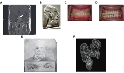
Acromegaly is a disorder of disproportionate skeletal, tissue, and organ growth and occurs with an annual incidence of approxi- mately five cases per one million individuals. Although the disor-der has been recognized since antiquity, the pathology of pituitary “prosopectasia” was first described by Andrea Verga in 1864 and the clinical features of acromegaly by Pierre Marie in 1886. Disease pathogenesis involves growth hormone (GH) hypersecretion by tumorous pituitary somatotroph cells, and the diagnosis is invari-ably preceded by about 10 years of active but unrecognized disease (1–3). Clinical presentation of acromegaly, in descending frequency as determined in a study of approximately 600 patients, includes acral and facial changes, hyperhidrosis (abnormally increased perspiration), headaches, paresthesia (“pins and needles” tingling sensation), sexual dysfunction, hypertension, goiter, and rarely, visual field defects (4) (see Sidebar 1). Subtle skeletal and acral overgrowth and soft tissue enlargement may occur inexorably over years (refs. 4, 5, S1; supplemental material available online with this article; doi:10.1172/JCI39375DS1), with frontal skull bossing (resulting in an unusually prominent forehead and heavy brow ridge), mandibular prognathism (protruding lower jaw), jaw mal-occlusion and overbite, skin thickening, and increased shoe and ring size (Figure 1). Chronic exposure to GH and IGF1 hypersecre-tion leads to soft tissue swelling of tongue, heart, kidney, colon, and vocal cords and periarticular and cartilaginous thickening, resulting ultimately in painful large-joint osteoarthritis. Up to 60% of patients exhibit spinal kyphoscoliosis (outward curvature of the spine) and diffuse skeletal hyperostosis (overgrowth of bone). Dis- ease duration, IGF1 levels, and concurrent hypogonadism deter-mine the prevalence of vertebral fractures (S2). Elevated levels of the hormone prolactin (PRL), observed in approximately 30% of patients, can be ascribed to mixed tumor GH and PRL cosecre-tion or to pituitary stalk impingement by the tumor mass. Rarely, plurihormonal tumors cosecrete the thyroid-stimulating hormone thyrotropin (TSH), leading to hyperthyroxinemia (elevated circu-lating thyroxine levels), or adrenocorticotropin (ACTH), leading to hypercortisolemia (elevated circulating cortisol levels).
This article reviews recent scientific discoveries that have had an impact on our understanding of acromegaly pathogenesis and clini-cal features. Novel approved and experimental therapies have evolved from these fundamental insights and are discussed in the context of providing added benefit to patient care and disease control.
Diagnosis
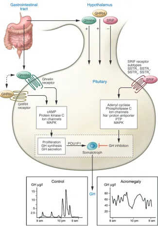
The diagnosis of acromegaly requires demonstration of dysregu-lated and enhanced GH secretion as well as elevated IGF1 levels, reflective of peripheral tissue exposure to tonically elevated GH concentrations (6). In acromegaly, basal GH secretion is tonically elevated with relatively blunted bursts (Figure 2). Accordingly, a random GH value of less than 0.04 μg/l effectively excludes the diagnosis of acromegaly. Importantly, an elevated randomly obtained GH measurement may not necessarily imply excessive integrated GH secretion. Net GH secretion is attenuated after age 60 (when 24-hour GH secretion is less than 50% of that in younger subjects) and by elevated BMI.
Older GH radioimmunoassays were relatively insensitive and poorly reproducible. Newer immunoradiometric assays and immu- noluminometric assays are based on the use of two-site monoclo-nal antibodies, and although they detect GH concentrations of less than 0.05 μ g/l, these assays are beset by challenges of reproducibil-ity. Lack of universal standards, nonuniform antibody recognition of GH isoforms, and the presence of circulating GH-binding pro- teins contribute to method-dependent and patient-specific vari-ability of GH measurements. In a multicenter study of GH nadir after an oral glucose tolerance test (OGTT), reference ranges varied by approximately 50%, and 30% of obtained results were inconsis-tent with the correct diagnosis (7). There is a compelling need for reliable GH assays based on robust reference standards.
A functional hallmark of a GH-secreting pituitary adenoma is the inability to respond appropriately to a glucose-induced neuro- endocrine suppressive signal. The inability to suppress GH secre-tion to less than 1 μg/l during 2 hours after an oral glucose load (75 grams) is the current consensus for diagnosing acromegaly (8). However, this cutoff may in fact be insensitive, and patients have been identified with clinical features of acromegaly, elevated IGF1 levels, and nadir postglucose GH levels of less than 1 μg/l (9). Using ultrasensitive GH assays, a nadir GH cutoff of less than
Acromegaly pathogenesis and treatment
Shlomo Melmed
Department of Medicine, Cedars-Sinai Medical Center, Los Angeles, California, USA
Dysregulated growth hormone (GH) hypersecretion is usually caused by a GH-secreting pituitary
adenoma and leads to acromegaly — a disorder of disproportionate skeletal, tissue, and organ
growth. High GH and IGF1 levels lead to comorbidities including arthritis, facial changes,
prog-nathism, and glucose intolerance. If the condition is untreated, enhanced mortality due to
car-diovascular, cerebrovascular, and pulmonary dysfunction is associated with a 30% decrease in life
span. This Review discusses acromegaly pathogenesis and management options. The latter include
surgery, radiation, and use of novel medications. Somatostatin receptor (SSTR) ligands inhibit GH
release, control tumor growth, and attenuate peripheral GH action, while GH receptor antagonists block GH action
and effectively lower IGF1 levels. Novel peptides, including SSTR ligands, exhibiting polyreceptor subtype affinities
and chimeric dopaminergic-somatostatinergic properties are currently in clinical trials. Effective control of GH and
IGF1 hypersecretion and ablation or stabilization of the pituitary tumor mass lead to improved comorbidities and
lowering of mortality rates for this hormonal disorder.
Conflict of interest: The author receives preclinical research grant support from Novartis and is a research consultant for Ipsen.
0.3 μg/l is effectively more discriminatory. Failure to suppress GH levels may also be encountered in patients with diabetes, renal or hepatic failure, and obesity or those receiving estrogen replace-ment or who are pregnant.
Screening of IGF1 levels is useful in obtaining a surrogate reflec- tion of integrated GH secretion. IGF1 levels are relatively stable, cor- relate with clinical features of acromegaly (10), and exhibit a log-lin-ear relationship with elevated GH levels (S3). Measured circulating IGF1 concentrations plateau when GH levels are greater than 20 μg/l, and subtle GH elevations do not uniformly induce IGF1. Accurate IGF1 evaluation requires age-matched control values, especially as levels decrease about 14% per decade with aging (11). Malnourished patients and those with liver and renal failure or those receiving estrogen exhibit lower IGF1 levels. The importance of appropriate age-adjusted normal IGF1 values was highlighted in a recent study using 4 different assays in 40 acromegaly patients; result variances were minimized with increased numbers of appropriately matched controls (12). Importantly, robust IGF1 assays may exhibit up to 30% within-subject variance in healthy subjects (13).
Morbidity impact of exposure to excess GH/IGF1
Mortality determinants
Large retrospective studies of acromegaly patients (14) indicate an average 10-year reduction in life expectancy, with at least a doubling of standardized mortality rates (SMRs) due to cardiovascular, cere- brovascular, metabolic, and respiratory comorbidities. Mean weight-ed SMRs in treated patients were 1.72 (1.62–1.83, 95% CI), with earlier studies showing higher ratios (15). Overall, achieving a post-treatment GH level of less than 2.5 μ g/l results in maintaining nor-mal life expectancy rates (2, 15). Independent survival determinants include the last recorded GH level (P < 0.001), hypertension (P < 0.02), heart disease (P < 0.03), and disease duration (P < 0.04) (14). In an outcomes study of 419 patients, cerebrovascular SMRs were elevated at 2.68 (1.73–4.15, P < 0.007), while malignancy-associated mortal-ity was not enhanced (16). These results likely reflect the positive impact of recent multimodal therapy on mortality outcomes.
Radiotherapy directed at the GH-secreting pituitary adenoma may also be associated with increased mortality (P < 0.005) (16), especially from cerebrovascular disease. Although overall cancer
incidence is not enhanced, there is a moderate risk of develop-ing colorectal cancer (S4). Uncontrolled GH levels likely provide a growth advantage to neoplasms, resulting in more aggressive disease and increased cancer-associated mortality. Colonoscopy shows increased colon length and mucosal hypertrophy; up to 25% of polyps are right-sided and recur within 3 years.
A potential determinant of acromegaly mortality could be iat-rogenic or endogenous hypopituitarism (failure of the pituitary gland to produce normal amounts of one or more of its hor-mones), resulting in deficiencies of pituitary-target hormone axes. Ideally, achievement of rigorously controlled GH and IGF1 levels would be expected to normalize SMRs. Accordingly, comorbidities associated with musculoskeletal degeneration and disfigurement, large organ hyperplasia, and cardiac and vascular dysfunction remain as therapeutic challenges.
Comorbidities
The constellation of hypertension, cardiac arrhythmias, glucose intolerance, and diastolic dysfunction leads to heart failure, which may be intractable, especially if GH levels remain uncontrolled. Biventricular cardiac hypertrophy manifests early in response to elevated GH levels and is present in 20% of young acromegaly patients and in up to 90% of patients with long-standing disease independent of the presence of hypertension. Postexercise ventric-ular ejection fraction is increased in approximately 70% of patients (17), and approximately 50% are at intermediate-to-high risk for coronary arteriosclerosis (S5). The pathogenesis of hypertension is associated with plasma volume expansion and increased cardiac output (18). Although hypertension has been ascribed to increased peripheral vascular resistance, vessel growth and intimal thickness are not uniformly dysregulated. GH exerts antinatriuretic effects, leading to increased extracellular volume, soft tissue swelling, and organomegaly. GH acts at the aldosterone-sensitive distal neph-ron, and transepithelial sodium transport is attenuated by a GH receptor (GHR) antagonist, while cortical collecting duct epithe-lial sodium channel subunit transcription is induced by GH (19). Insulin resistance caused by GH excess results in glucose intoler-ance and diabetes (5), further exacerbating renal dysfunction.
Airway obstruction consequent to macroglossia (tongue enlarge- ment) and hypertrophy of laryngeal and pharyngeal mucosal tis-sues lead to upper airway obstruction, hypoventilation, snoring, and sleep apnea in approximately 50% of patients (S6).
Sidebar 1
Impact of long-term GH and IGF1 exposure
Organ/tissue Clinical feature
Bone and joint Acral changes, gigantism, prognathism, arthritis, osteopenia, vertebral fractures, carpal tunnel syndrome Heart Cardiomyopathy, hypertension, arrhythmias, valvulopathy, heart failure
Skin Tags, excessive oily perspiration Pancreas Insulin resistance, diabetes Lung Obstructive sleep apnea
Kidney Antinatriuresis, fluid retention, increased aldosterone, renal failure
Gonads Hypogonadism
Thyroid Goiter
Muscle Proximal myopathy
Colon Polyps
Fat Lipolysis
GH secretion and action
Anterior pituitary development follows highly specialized precur- sor stem cell commitment, with restricted differentiation of hor-mone-secreting cell types. Somatotrophs account for more than 50% of pituitary hormone-secreting cells, and transcription fac-tors paired-like homeodomain factor 1 (PROP1) and POU class 1 homeobox 1 (POU1F1 ) determine cell differentiation and commit-ment to synthesizing and secreting GH (20, 21). A family of genes located on the long arm of chromosome 17 encodes the GH pep-tides, encompassing pituitary human GH, a placental variant of human GH (hGH) known as hGH-V, placental lactogen, and PRL (22). An alternatively spliced pituitary GH molecule is devoid of aa 32–46 and is designated as 20-kDa GH. Structural characteristics of the 191-aa GH molecule that are important for peptide func-tion include the third α-helix, comprising amphiphilic domain elements important for signaling, and integrity of the large helical loop is required for growth-promoting actions (23). GH mediates linear skeletal growth and also regulates carbohydrate, lipid, and mineral metabolism (24). Most of the growth-promoting actions of GH are enabled by IGF1.
Hypothalamic GH-releasing hormone (GHRH), ghrelin (mainly gut-derived), and somatostatin (SRIF) traverse the pituitary portal system to regulate GH production by anterior pituitary somato- troph cells (25) (Figure 2). GHRH, acting via the GHRH G pro- tein–coupled receptor, induces and maintains somatotroph tro-phic function and induces GH gene transcription and secretion (26). Ghrelin, a gut-derived GH secretagogue (27), acts mainly at
the hypothalamus and signals through the ghrelin secretagogue receptor type Ia (GHS-RIa) to induce GH secretion in synergy with GHRH (S7). GHRH also signals via the ghrelin receptor (28), act- ing as an allosteric coagonist for the GHS-RIa. GHRH and ghre-lin thus act coordinately to regulate pituitary function as well as energy homeostasis. SRIF, acting via pituitary SSTR2 (where SSTR
denotes SRIF receptor subtype) and SSTR5 subtypes, attenuates both
the timing and amplitude of GH secretory pulses. GH secretion is characterized by sporadic secretory pulses interspersed with most- ly minimal basal secretion determined by age, sex, specific nutri-ents, neurotransmitters, exercise, and stress. Random daytime GH measurements are usually very low for approximately 80% of the day and may range from undetectable to secretory peaks of up to 15 μg/l or higher in normal subjects, observed mainly at night. Increased BMI and obesity attenuate GH secretion, while malnu- trition and prolonged fasting result in elevated GH pulse frequen-cy and amplitude (29).
GH signaling
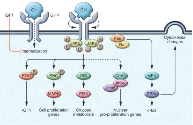
[image:4.585.80.506.79.332.2]The gene encoding the GHR, a class I pleiotropic cytokine receptor (30), is ubiquitously expressed, especially in liver, fat, and muscle. The GH molecule interacts with a preformed dimer of identical GHR pairs, which undergoes rotation and triggers ligand-recep-tor complex signaling (31) (Figure 3). As a consequence, two JAK2 molecules undergo autophosphorylation and also phosphorylate the GHR cytoplasmic domain (S8). Subsequent JAK2-dependent and -independent intracellular signal transduction pathways Figure 1
evoke pleiotropic cell responses including IGF1 synthesis, glucose metabolism, cell proliferation, and cytoskeletal changes.
STAT5b is the key intracellular molecule required for GH media- tion of postnatal growth, adipose tissue function, and sexual dimor-phism of hepatic gene expression (24). Importantly, GH-activated STAT5b induces IGF1 gene transcription (S9), and several lines of evidence point to this pathway as being critical for initiating and maintaining skeletal growth. Male Stat5b–/– mice exhibit impaired growth, attenuated circulating IGF1 levels, and insensitivity to injected GH (S10). Hepatic IGF1 is induced by constitutively active STAT5b, while a dominant negative STAT5b construct prevents GH-induced IGF1 expression (S11). In humans, STAT mutations result in relative GH insensitivity and growth retardation (32). GH also induces early response genes that precede cell growth and dif-ferentiation signals (33) mediated by CCAAT enhancer–binding protein β and serum response element sites on the c-fos promoter.
The GHR may also translocate to the nucleus by the importin α/β pathway in conjunction with coactivator activator (CoAA). Gene targets for nuclear-mediated GHR action are predomi-nantly proproliferative. Forced GHR targeting to the cell nucleus also enhances cell proliferation and transformation responsive-ness to autocrine-derived GH. Thus, CoAA and activated STAT5 are both required for GH-dependent proproliferative actions of nuclear GHR (34).
STAT5b mediates sexually dimorphic GH signals. Females exhibit more frequent GH secretory pulses and shorter interpulse nadir intervals, leading to relative desensitization of female hepatic STAT5 induction by GH as compared with that of males. Targeted disruption of STAT5b leads to male-selective reduced growth rates and loss of gender-specific hepatic gene induction (35).
[image:5.585.46.371.78.543.2]GHR insensitivity may occur as a consequence of extracellular receptor domain cleavage as well as toxin-induced proteolysis,
Figure 2
Normal and disrupted GHRH–GH–IGF1 axis and molecular targets for therapy. Pituitary somatotroph cell development and gene expression are determined by the POU1F1 transcription factor. Net GH secretion is deter-mined by integration of hypothalamic, nutri-tional, hormonal, and intrapituitary signals. GH synthesis and secretion are induced by hypothalamic GHRH and gut-derived ghre-lin. GHRH may also act as a coagonist for the ghrelin receptor (28). Hypothalamic SRIF suppresses GH secretion mainly by high-affinity binding to SSTR2 and SSTR5 receptor
subtypes expressed on somatotrophs (90). SSTR ligands (SRLs) signal through SSTR2
and SSTR5 to control GH hypersecretion and
which abrogates signaling (S11). GHR cell-surface translocation is also directly inhibited by IGF1, likely contributing to a local feedback loop (36) (Figure 3).
IGF1
IGF1, the polypeptide target hormone for GH, is synthesized in the liver and extrahepatic tissues (principally bone, muscle, and kidney) and also in the pituitary gland itself. IGF1 mediates most of the growth-promoting actions of GH (37). Acting at both endocrine and paracrine levels, IGF1 exerts negative feedback regulation of GH synthesis and secretion (38). Approximately 80% of circulating IGF1 originates from the liver, and high-affin-ity binding proteins including IGF-binding protein 3 (IGFBP3) and acid-labile subunit (ALS) transport and also mediate IGF1 peptide activity by regulating IGF1 cell-surface receptor access (39). As IGF1 receptors are ubiquitously expressed, widespread enhanced cell proliferation as well as metabolic actions are trig-gered by elevated IGF1 concentrations. IGF1 acts in an endocrine fashion to mediate tissue growth, or locally synthesized IGF1 acts in an autocrine/paracrine manner to regulate local GH target tissue growth. Ultimately, organ growth responses to IGF1 are determined by the intrinsic replicative potential of local tissues. Observations that doubly mutant Ghr–/–Igf1–/– mice exhibit more severe growth retardation than animals with either single-gene deletion alone indicate that anabolic actions of GH, especially on muscle, may also be distinctively direct and not necessarily IGF1 dependent (40). GH acts directly to induce germinal epiphyseal cells, while IGF1 acts to induce chondrocyte proliferation (41), and based on results derived from transgenic mice with respec-tive deletions of GHR or IGF1 (42), both GH- and IGF1-mediated signaling appear additive in enabling growth, while IGFI may attenuate metabolic effects of GH (43).
GH and IGF1 signaling in acromegaly
In acromegaly, cellular responses elicited by high GH levels over-whelm intracellular mechanisms attenuating GH signaling, including those mediated by SOCS, Src kinases, and tyrosine phosphatase pathways (24).
An in-frame deletion in exon 3 results in a GHR isoform devoid of 22 aa (known as d3-GHR), which is associated with enhanced GH responsiveness, as evidenced by higher STAT5 activation and acceler- ated growth (44). d3-GHR is also associated with a more florid clini-cal and biochemical acromegaly phenotype and relative resistance of IGF1 levels to acromegaly treatment interventions (45, S12).
Although mice overexpressing transgenic GH or IGF1 exhibit enhanced somatic growth reminiscent of acromegaly, several dis-tinctive features point to unique independent target functions for GH and IGF1 (46, S13). For example, transgenic mice overexpress- ing GH, but not IGF1, exhibit liver, spleen, and kidney enlarge-ment with features of renal glomerulosclerosis. In contrast, mice overexpressing IGF1 are obese, unlike GH transgenics (S13). This phenotype recapitulates acromegaly with reduced fat mass and increased lean body mass. To what extent GH-induced hyperin- sulinemia, manifest in GH transgenic mice but not in IGF1 trans-genic animals, contributes to the hypersomatotrophic phenotype is unclear. The body of experimental evidence indicates that GH actions in bone and soft tissue require IGF1 to enable a maximally robust tissue response (47).
Somatotroph adenoma pathogenesis
Pituitary tumors are commonly encountered monoclonal adeno-mas that account for approximately 15% of all intracranial tumors. These invariably benign tumors arise from highly differentiated anterior pituitary cells expressing hormone gene products includ- ing GH, PRL, ACTH, TSH, and the gonadotropins follicle-stimulat-ing hormone (FSH) and luteinizing hormone (LH). These tumors may secrete hormones excessively, leading to characteristic clinical features including acromegaly, Cushing disease, and hyperprolac- tinemia. More commonly, they are nonfunctional and lead primar-ily to hypogonadism and compressive pituitary failure (48).
[image:6.585.44.373.81.295.2]Mechanistic studies of human pituitary tumors have been con- strained due to inaccessibility of the gland for biopsy, lack of func-tional cell lines, and unique differentiated tumor subtype behavior. In most cases of acromegaly, GH hypersecretion is derived from somatotroph cell tumors (see Sidebar 2). Autonomous GH secre-tion by distinct somatotroph adenomas derived from the POU1F1
Figure 3
transcription factor cell lineage characterizes a distinct balance of GH secretion versus somatotroph trophic activity. GH-secreting tumor formation ensues as a consequence of unrestrained somato-troph proliferation associated with intrinsic cell-cycle dysfunction as well as altered endocrine and or paracrine factors regulating GH synthesis, GH secretion, and somatotroph cell growth.
GH-secreting adenomas very rarely exhibit activating ras muta-tions in invasive or metastatic lesions (49, S14). Uniquely, pituitary mitotic activity is relatively low, even in invasive adenomas. Sev-eral growth factors, including dysregulated receptors for fibroblast growth factors, dopamine, estrogen, and nerve growth factor (50), have been implicated predominantly in prolactinoma pathogen-esis, but not uniformly in acromegaly (Table 1).
cAMP signaling
Several lines of evidence support the role of the GHRH-cAMP signaling pathway in mediating somatotroph tumorigenesis (Figure 4). Ectopic GHRH production by peripheral carcinoid tumors (51) leads to somatotroph hyperplasia and GH hyper-secretion, but rarely adenoma formation. GHRH signals via the GHRH receptor (GHRH-R), a G protein–coupled receptor, by inducing cAMP, which induces GH transcription mediated by cAMP response element–binding protein (CREB). A constitu-tively activated murine pituitary-directed G protein subunit (Gs) transgene resulted in high GH levels and gigantism (S15). G ua-nine nucleotide–binding protein, α stimulating (GNAS) encodes the stimulatory Gs (Gsα), and activating GNAS mutations lead to constitutively elevated cAMP levels, protein kinase A activity, and GH synthesis and secretion (52). Postzygotic GNAS muta-tions result in a mosaic pattern of organ specificity with clinical features of McCune-Albright syndrome (OMIM 174800), includ-ing pigmented skin lesions and polyostotic fibrous dysplasia, and endocrine dysfunction including precocious puberty, thy- rotoxicosis, and GH and ACTH hypersecretion (53). GH hyper-secretion likely occurs as a consequence of pituitary hyperplasia, and true pituitary adenomas have rarely been identified (53). About 40% of patients with sporadic acromegaly harbor GNAS mutations with aa substitutions at Arg201 or Gln227, leading to constitutively elevated cAMP (52) with no distinctive clinical phenotype. The gene is imprinted in pituitary tissue, and muta-
tions occur in the expressed maternal allele (S16). CREB is con-stitutively activated in GH-secreting adenomas independently of the GNAS mutation (54) and likely a final common pathway for cAMP signaling for a subset of adenomas.
Inactivating mutations of the protein kinase cAMP-dependent, regulatory type 1, α (PRKAR1A) (55) gene, which encodes the type 1A regulatory subunit of PKA, lead to a rare syndrome of spotty skin pigmentation, mucosal and cardiac myxomas (benign neoplasms), and acromegaly (OMIM 160908). Although most identified patients exhibit elevated GH, IGF1, and often PRL levels, clinical manifes-tations of acromegaly are usually subtle. Mechanisms underlying acromegaly in these patients likely involve enhanced PKA activity in pituitary somatotroph cells. Analogous to the clinical phenotype, pituitary-specific transgenic deletion of Prkarla resulted in forma-tion of murine tumors derived from the POU1F1 lineage (S17).
Cell-cycle disruption
Cyclin D1–dependent kinase 4 (CDK4) is required for postnatal somatotroph and lactotroph proliferation, and Cdk4-null mice are resistant to the trophic effects of GHRH (56). In contrast, retino-blastoma gene (Rb) inactivation leads to endocrine tumorigenesis, and Rb+/– mice develop spontaneous pituitary tumors with almost 100% penetrance (57). Rb acts as a G1
/S cell-cycle checkpoint con-trol: cyclin-dependent kinases (CDKs) phosphorylate Rb, triggering the release of members of the E2F family of transcription factors, enabling the progression of S phase and cell proliferation. Loss of E2F1 reduces the frequency of pituitary tumors in the Rb+/–E2f11–/– mouse, further indicating that site-selective tumorigenesis in Rb+/– mice results from dysregulated E2F transcriptional activity (57).
MEN1 . The multiple endocrine neoplasia type I (MEN1) syn- drome (OMIM 131100) is an autosomal dominant disorder asso-ciated with germ-line mutations in MEN1, a tumor suppressor gene located on chromosome 11q13. The syndrome comprises a predisposition to parathyroid hyperplasia, pancreatic endocrine tumors, and pituitary adenomas. Up to 40% of affected individu-als harbor pituitary tumors, and these comprise prolactinomas (60%), GH-secreting adenomas (20%), ACTH-secreting adeno-mas (<10%), and nonfunctional adenomas (<10%) (58, S18). The MEN1 nuclear protein controls genome stability by repression of telomerase activity via telomerase reverse transcriptase (S19). Mechanisms for pituitary tumor pathogenesis in patients with MEN1 syndrome and disrupted MEN1, apparent from animal Sidebar 2
Glossary of GH-expressing lesions
Densely granulated GH-cell adenoma Pure GH secreting; usually in older patients with minimally elevated GH levels
Sparsely granulated GH-cell adenoma Pure GH secreting; usually in younger patients with high GH levels and aggressive growth Mixed GH and PRL PRL and GH secretion by monomorphous mammosomatotrophs or mixed somatotrophs mammosomatotroph adenomas and lactotrophs. May occur with gigantism.
Acidophilic stem cell adenomas PRL and GH secretion by precursor cell tumor; usually aggressive and may occur in younger patients with gigantism
Plurihormonal GH-cell adenoma Secretes GH plus PRL, ACTH, or rarely, TSH Silent GH-secreting adenoma Expresses GH without hypersecretion or acromegaly
Somatotroph hyperplasia Usually caused by tumor secreting ectopic GHRH (e.g., carcinoid) Ectopic GH-cell adenoma Arises in remnant nasopharyngeal pituitary tissue
Empty sella tumor Tumor remnants secreting GH arising in rim of pituitary tissue GH-cell carcinoma Exceedingly rare with extracranial metastases
GH-secreting extrapituitary tumor Abdominal, pancreatic, or lymphoma; very rare
studies, include regulation of p27 and p18, both of which are implicated in pituitary tumor growth in transgenic mice mod-els. Mice devoid of p27 exhibit striking features of gigantism with multiorgan hyperplasia and intermediate lobe pituitary tumors (S20). Thus, MEN1 enables suppression of pituitary and related neuroendocrine tumor formation, and disrupted MEN1 gene could facilitate development of these tumors. Mutations of MEN1, p18, or p27 have not been encountered in patients harbor-ing sporadic pituitary adenomas.
When p18 homozygote mutant mice were crossed with heterozygous Men1 mutants, development of pituitary, parathy-roid, thyroid, and pancreatic tumors was markedly accelerated (59). A germ-line mutation in p27 (also known as CDK inhibitor 1B [CDKNIB]) has been reported in a family exhibiting features of a recessive MEN1-like phenotype (60). The index patient harbored a GH-secreting pituitary adenoma and a parathyroid adenoma. This mutation may thus account, at least in part, for the subset of apparent MEN1 subjects who do not exhibit MEN1 mutations.
HMGA2 . Several lines of evidence support the role of high-mobil-ity group AT-hook 2 (HMGA2), a nuclear architectural protein, in murine and human pituitary tumorigenesis. Transgenic mice overexpressing HMGA2 exhibit highly prevalent pituitary tumors induced by (a) displacing histone deacetylase from the pRB com- plex; (b) acetylation and liberation of E2F1; and (c) driving pitu-itary cells into S phase (61). HMGA2 also induces pituitary tumor cyclin B2 (CCNB2) and directly induces CCNB2 promoter tran-scriptional activity. GH-secreting tumors coexpress high levels of CCNB2, HMGA1, and HMGA2 (61, 62).
PTTG. Pituitary tumor–transforming protein (PTTG), isolated from pituitary tumor cells, facilitates the spindle checkpoint by acting as a securin to inhibit separase and enable faithful sister chromatid separation (63, 64). Pttg mediates in vitro transforma-tion and in vivo tumorigenesis in mice, and PTTG overexpression induces aneuploidy with dysregulated G2
/M checkpoint surveil-lance, resulting in abnormal mitosis and chromosomal instability (65). PTTG modulates p53, participating in DNA damage/repair
and apoptosis (66–68). PTTG is abundantly expressed in pituitary adenomas (69) and correlates with tumor invasiveness and recur- rence; it is induced early in estrogen-induced pituitary tumorigen-esis. PTTG elicits pituitary tumorigenesis in a transgenic model of pituitary-directed Pttg overexpression, resulting in focal pituitary hyperplasia and functional adenoma formation (70).
Pituitary senescence. Pituitary carcinomas are exceedingly rare, and only isolated cases of pituitary metastases derived from GH-secret-ing adenomas have been reported. GH-secreting adenomas thus represent an intriguing model for studying triggers of malignant transformation. Cellular senescence mediated by oncogenic path-ways is associated with the activation of inhibitors of cell-cycle progression (such as p53-mediated p21), which protect the cell from proproliferative signals and act as a buffer against malig-nant transformation (71). Premature senescence may account for the overwhelming predominance of benign versus malignant GH-secreting pituitary tumors, as more than 70% of GH-secreting tumors overexpress PTTG, leading to aneuploidy and induction of senescence markers including p21 and senescence-associated β-galactosidase (72). In contrast, p21 is weakly expressed in normal pituitary tissue and undetectable in pituitary carcinomas. Senes-cent features of GH-secreting pituitary adenomas likely constrain malignant transformation of these invariably benign adenomas. Slow replicative pituitary cell-cycle progression is distinct from the rapid cell cycle of skin or digestive tract regenerative tissues (56), consistent with observations that pituitary tumors rarely exhibit malignant phenotypes. Thus, accumulated pituitary DNA dam-age and senescence, hallmarks of GH-secreting adenomas, likely enable a benign phenotype.
Epigenetic mechanisms
[image:8.585.52.537.112.326.2]Loss of gene expression due to DNA hypermethylation of both alleles in GH-secreting adenomas exemplifies an epigenetic mechanism by which the loss of genes that inhibit cell prolif-eration results in pituitary cell proliferation (S21). CDKN2A encodes CDK inhibitor 2A (also known as p16), which blocks Table 1
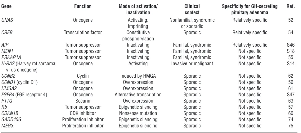
Genes that contribute to the molecular pathogenesis of GH-secreting adenomas
Gene Function Mode of activation/ Clinical Specificity for GH-secreting Ref. inactivation context pituitary adenoma
GNAS Oncogene Activating, Nonfamilial, syndromic Relatively specific 52 imprinting or sporadic
CREB Transcription factor Constitutive Sporadic Relatively specific 54 phosphorylation
AIP Tumor suppressor Inactivating Familial, syndromic Relatively specific S46 MEN1 Tumor suppressor Inactivating Familial, syndromic Not specific S18 PRKAR1A Tumor suppressor Inactivating Familial, syndromic Not specific 55 H-RAS (Harvey rat sarcoma Oncogene Activating Invasive or malignant Not specific S14 virus oncogene)
CCNB2 Cyclin Induced by HMGA Sporadic Not specific 62 CCND1 (cyclin D1) Oncogene Overexpression Sporadic Not specific 56 HMGA2 Oncogene Overexpression Sporadic Not specific 61 FGFR4 (FGF receptor 4) Oncogene Alternative transcription Sporadic Not specific S47 PTTG Securin Overexpression Sporadic Not specific 63
Rb Tumor suppressor Epigenetic silencing Sporadic Not specific 57
CDK4 from interacting with cyclin D1 and thereby preventing retinoblastoma protein (Rb) phosphorylation. Rb methylation in somatotroph adenomas is variable, with inconsistent effects on tumor proliferation (73). Epigenetic silencing of p16 and p27 expression or loss of heterozygosity on chromosome 13 is also associated with Rb inactivation in some human pituitary tumors (73). The growth arrest and DNA damage–inducible γ
(GADD45G) and maternal expressed 3 (MEG3 ) genes and mela-noma-associated antigen A3 are expressed in normal pituitary but not in pituitary adenomas (74–76).
Collectively, these observations suggest a model for pituitary adenoma growth whereby an initial proliferative phase occurs in response to growth stimuli and is then followed by irrevers-ible growth arrest of the benign tumor. Thus, the vital hormonal functioning of the somatotroph for maintaining homeostasis
control appears to be enabled by a senescent response to onco-genic stress restraining proliferation in an attempt to assure viable physiological functions.
[image:9.585.46.542.82.450.2]Familial isolated pituitary adenomas . Less than 5% of pituitary adeno- mas are inherited on a familial basis (77). In familial isolated pitu-itary adenoma (FIPA) families, prolactinomas account for about half of the adenomas, with GH-secreting and mixed GH- and PRL-secreting adenomas accounting for the remainder. Homogenous familial acromegaly (also known as isolated familial somatotropino-mas [IFS]) affects younger patients usually diagnosed as teenagers or in their 20s (78). About 25% of IFS patients present with gigan- tism and macroadenomas, with most not harboring a known germ- line mutation. Mutations in the tumor suppressor aryl hydrocar-bon receptor–interacting protein (AIP) predispose to somatotroph and lactotroph tumors in 15% of patients (79) (Table 1). Two of 21 Figure 4
Depiction of intracellular pathways associated with somatotroph transformation and proliferation. GH transcription and somatotroph prolifera-tion are induced by cAMP acting through CREB (26). SRIF inhibits cAMP and CREB activity (S43) to suppress GH secreprolifera-tion. Pituitary CDKs likely exhibit overlapping functions in G1 cell-cycle progression. Somatotroph mitogenic factors include POU1F1, GHRH, and GNAS as well as
families with heterogenous pituitary adenoma predisposition were shown to harbor relatively large intragenic AIP deletions (S22) and a 5818-bp deletion was detected in one of 7 families affected with acromegaly. Heterozygote germ-line AIP mutations were found in 47 subjects from 9 of 26 families with familial pituitary adenomas. Of these, 31 were diagnosed with gigantism or acromegaly (80).
Given the extremely low frequency of familial pituitary tumors, the small size of affected families, and the very low prevalence of mutations within these families, costly genetic screening is not yet convincingly warranted. Accurate relatively low-cost measurement of serum IGF1 levels is currently the most effective screening for affected patients with GH-secreting pituitary tumors.
Acromegaly treatment
Several treatment options are currently available for acromegaly (Table 2).
Surgery
Resection of GH-secreting pituitary adenomas is technically chal-lenging because of the anatomic inaccessibility of the pituitary and bony sellar confines and the proximity of vital brain and vas-cular structures. Functioning tumor microfoci often invade dural spaces, are not readily visible at surgery, and continue to secrete GH after tumor resection. GH-secreting tumors have a propen-sity to invade laterally into the cavernous sinus, precluding safe resection. Tumor-associated internal carotid artery tortuosity and microaneurysms also require surgical caution and alertness. Over 90% of resections are performed via an endonasal transsphenoidal approach, often with minimally invasive endoscopic techniques. Computerized image guidance and intraoperative MRI coupled with development of microinstrumentation and optics have resulted in safe, effective, and minimally traumatic procedures when performed by skilled and experienced neurosurgeons (S23). The goal of surgery is to balance maximal tumor mass resection with preservation of normal pituitary secretory function.
About 70% of patients harboring well-circumscribed GH-secret- ing microadenomas less than 10 mm in diameter achieve long-term biochemical control after surgery (81). Unfortunately, over
65% of GH-secreting adenomas are invasive macroadenomas at the time of diagnosis, and surgical outcomes for these patients are far less favorable, with an approximately 50% success rate reported from most experienced clinical centers (82, S24). Markers of surgi-cal remission include biochemical control, normal pituitary and parasellar MRI visualization, and recurrence-free postoperative duration. Determinants of surgical remission include the experi-ence of the surgeon in resecting these challenging adenomas (83), tumor size, and degree of invasiveness (82).
Transient side effects of surgery include local hemorrhage, CSF leak, diabetes insipidus, and rarely, local infection. Permanent side effects reported in less than 5% of patients include diabetes insipi- dus and pituitary hormone deficiency. Clearly, the major disad-vantage of surgery is persistent postoperative GH hypersecretion.
Radiotherapy
Conventional external-beam radiotherapy is administered up to a maximum of 4000–5000 cGy in 180-cGy weekly doses spread over six weeks. Overall, about 50% of patients achieve biochemical remis-sion (GH < 2 μg/l and normalized IGF1) after 10 years (84–86). In 77% of 884 irradiated patients, GH levels were attenuated to less than 2.5 μg/l by 20 years. The relatively long latency period required to achieve remission is a major disadvantage. Acquired residual pitu-itary damage is evident in approximately 50% of patients by 10 years. 27% exhibited TSH deficiency, 18% FSH/LH deficiency, and 15% ACTH deficiency (84). Rarely encountered local side effects include visual deficits, especially if the tumor abuts the optic chiasm, cere-bral radionecrosis, cerebrovascular damage, and cognitive deficits. One percent of patients develop secondary intracranial tumors, with a latency up to 24 years (86). The reported incidence of these side effects is imprecise, due to absence of well-controlled studies and heterogeneity of radiation methodology.
Stereotactic radiosurgery
Using a 60Cobalt source, relatively narrow beams of high-dose,
[image:10.585.55.534.109.270.2]focused γ radiation are delivered with stereotactic precision to a small tumor, and the approach is particularly effective in tumors less than 3 cm in diameter and distant from the optic tract. Five Table 2
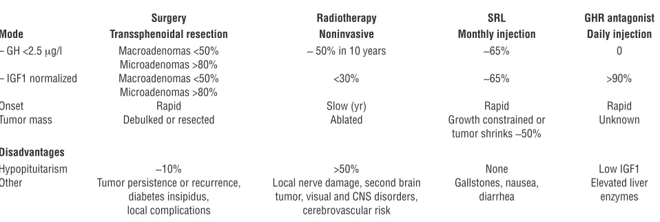
Acromegaly management
Surgery Radiotherapy SRL GHR antagonist
Mode Transsphenoidal resection Noninvasive Monthly injection Daily injection
– GH <2.5 μg/l Macroadenomas <50% ~ 50% in 10 years ~65% 0
Microadenomas >80%
– IGF1 normalized Macroadenomas <50% <30% ~65% >90%
Microadenomas >80%
Onset Rapid Slow (yr) Rapid Rapid
Tumor mass Debulked or resected Ablated Growth constrained or Unknown
tumor shrinks ~50% Disadvantages
Hypopituitarism ~10% >50% None Low IGF1
Other Tumor persistence or recurrence, Local nerve damage, second brain Gallstones, nausea, Elevated liver diabetes insipidus, tumor, visual and CNS disorders, diarrhea enzymes local complications cerebrovascular risk
years after treatment, post-OGTT serum GH levels are less than 1 μ g/l in approximately 50% of patients. Adenoma growth is arrest-ed, tumor shrinkage observed in most patients, and subsequent pituitary failure occurs in approximately 25% of patients. Employ-ing remission criteria of GH less than 2 μg/l and normalized IGF1, 17%–35% of patients remitted after 24–36 months (87). In 1567 patients undergoing radiosurgery, half of whom had prior conven-tional radiotherapy, 13 patients developed cerebral radionecrosis (S25). Factors determining the risk of radiation-induced pituitary failure include prior surgery, the precision of stereotactic tumor target resolution, and pituitary stalk exposure to radiation.
SSTR ligands
The two forms of endogenous SRIF, comprising 14 or 28 aa, respectively, elicit cellular responses by five ubiquitously expressed SSTR receptor subtypes (88). SSTRs act to inhibit both endocrine and exocrine hormone secretions and, less compellingly, attenuate neuroendocrine tumor cell proliferation. SSTR signaling is mainly mediated by Gα subunits to inhibit adenylyl cyclase and reduce cAMP generation. Other actions include regulating phosphotyro-sine phosphatase activity, K+ and Ca2+ channels, MAPK pathways,
and Na+/H+ exchange activities (S26). SSTR
2, SSTR3, and SSTR5
exhibit constitutive signaling to pituitary cells in a ligand-free environment (89). Thus, constitutive SSTR signaling may deter-mine ambient pituitary hormone secretion.
The availability of SSTR subtype–selective ligands has enabled elucidation of specific SST functions (88). Thus, SSTR2, and to a
lesser extent SSTR5
, determine secretion of GH, thyroid-stimu-lating hormone, and ACTH (90). GH-secreting adenomas exhibit heterogenous SSTR expression (SSTR2 >SSTR5 > SSTR1 > SSTR3),
while SSTR4 is notably undetectable in pituitary tumors (91, 92,
S27). Several lines of evidence point to a cooperative functional-ity of SSTR2 and SSTR5 in suppressing GH and ACTH secretion
(93–95). SSTR5 may also heterodimerize with SSTR2 to enhance
availability of cell membrane receptors (96). Thus, analogs that activate both SSTR2 and SSTR5 receptors are more efficacious
than respective monoselective SSTR analogs (91), and an SSTR2
antagonist reverses the GH-suppressive effects of biselective ago- nists or their respective combinations (95). Functional agonist-specific signaling may also determine cell responses of SSTR2,
SSTR4, and SSTR5 (S26).
Clinically available somatostatin receptor ligands. Octreotide, a cyclic octapeptide, is administered by s.c. or i.v. injection. Octreotide binds avidly to SSTR2 with a Kd of approximately
0.4 pM, and to a lesser extent to SSTR5. The starting dose is
100–250 μg every 8 hours, and up to 1.5 mg/24 hours can be safely administered in patients with acromegaly (97, S28). Peak drug concentrations are attained within 40 minutes of injection, and the ligand exhibits a circulating half-life of up to 2 hours, as compared with approximately 2 minutes for endogenous SRIF. The long-acting release (LAR) intramuscular formulation is encapsulated within biodegradable D, l-lactic, and glycolic
acid copolymer microspheres (88). The starting dose is usually 20 mg every 28 days, with safe maximal monthly doses up to approximately 60 mg or higher. Drug levels peak at 28 days, and plateau concentrations are sustained for approximately 14 days. When injected every 4 weeks, pharmacologically steady-state lev-els are achieved by the third injection. Lanreotide (BIM-23014) is incorporated into a biodegradable polymer for intramuscular injection (30 or 60 mg) every 7–14 days. With an approximately five-day half-life, the molecule exhibits high SSTR2 affinity and
also binds less avidly to SSTR5
. The long-acting lanreotide Auto- gel (Somatuline Depot in the USA) is available as a water-solu-ble, prefilled 60-, 90-, or 120-mg syringe for deep s.c. injection. Pharmacologically effective therapeutic levels of approximately 1 ng/ml are maintained for 28 days and, with a 23- to 29-day half-life, steady-state is achieved after four monthly injections. Both octreotide and lanreotide activate the SSTR2 receptor with
similar avidity, and head-to-head studies demonstrate nonsupe-riority for safety and efficacy of either formulation (98).
Ubiquitous tissue distribution of SSTR receptor targets under- lies the multitargeted therapeutic control elicited by somatosta-tin receptor ligands (SRLs) in acromegaly.
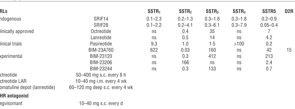
Hypothalamus. SRIF attenuates hypothalamic GHRH secretion and action by inhibiting GHRH induction of GH synthesis, secre- tion (99), and somatotroph cell replication (Figure 2) (S29). Ultra-Table 3
Acromegaly treatments
SRLs SSTR1 SSTR2 SSTR3 SSTR4 SSTR5 D2R
Endogenous SRIF14 0.1–2.3 0.2–1.3 0.3–1.6 0.3–1.8 0.2–0.9
SRIF28 0.1–2.2 0.2–4.1 0.3–6.1 0.3–7.9 0.05–0.4
Clinically approved Octreotide ns 0.4 35 ns 7
Lanreotide ns 0.5 14 ns 4.2
Clinical trials Pasireotide 9.3 1.0 1.5 >100 0.2
BIM-23A760 622 0.03 160 ns 42 15
Experimental BIM-23120 ns 0.3 412 ns 213
BIM-23206 ns 166 ns ns 2.4
BIM-23244 ns 0.3 133 ns 0.7
Octreotide 50–400 mg s.c. every 8 h Octreotide LAR 10–40 mg i.m. every 4 wk Somatuline depot (lanreotide) 60–120 mg deep s.c. every 4 wk GHR antagonist
Pegvisomant 10–40 mg s.c. every d
Experimental ligands depicted are either SSTR2 selective, SSTR5 selective, or biselective for both SSTR2 and SSTR5. D2R, dopamine receptor 2; ns,
[image:11.585.59.531.104.279.2]dian rat GH rhythm (where ultradian rhythms are recurrent periods or cycles repeated throughout a 24-hour circadian day) is mediated by tonic SRIF secretion, antagonizing GHRH action (100). SRLs also inhibit ectopic GHRH production by carcinoid tumors (S30).
Pituitary adenoma GH secretion is suppressed, leading to secondary suppression of circulating and perhaps tissue IGF1 levels . Clinical octreo-tide efficacy is predicted by tumor SSTR2 expression (101, S31).
Furthermore, SSTR2 gene transfer enhances octreotide respon-siveness in resistant GH adenoma cells (102). In patients treated for at least six months (103–106, S32, S33) and using random fasting GH levels of less than 2.5 μg/l and/or normalization of age-matched IGF1 levels as efficacy markers, approximately 65% of patients treated with octreotide LAR achieved control of GH secretion. If a more rigorous GH cutoff of less than 1 μg/l is used, approximately 33% of patients could be defined as controlled. In 36 patients followed for 3–18 years, GH of less than 2 μg/l and normal IGF1 were achieved in 70% of patients (107). Drug effi-cacy improves with prolonged treatment duration; tachyphylaxis has not been evident, and GH and IGF1 levels continue to decline, even after 9 years of sustained treatment (105). Headache, perspi-ration, fatigue, ring-finger thickness, and tissue swelling improve in up to 80% of patients. Efficacy may be moderately improved by adding the dopamine receptor agonist cabergoline to enhance GH suppression (108). SRLs are indicated in the following instances: (a) for first-line primary therapy; (b) for individuals too frail for anesthesia; (c) for those who decline surgery; (d) preoperatively to assure maximal surgical outcomes (109); (e) postoperatively for persistent and inevitable disease; (85) or (f) while awaiting radio-therapy impact to manifest. Biochemical control by primary SRL
treatment is not different from that achieved by surgery alone (110), and conversely, surgical adenoma debulking also enhances subsequent lanreotide efficacy (111).
Liver. Independently of pituitary action, SRLs act directly on the liver to regulate peripheral GH action by decreasing GHR binding and inhibiting hepatocyte IGF synthesis (112). In GH-deficient patients receiving a fixed GH replacement dose, octreotide also suppresses IGF1 levels, further supporting a pituitary-indepen-dent effect of the analog (113).
Pituitary adenoma growth . GH-secreting tumors, with rare excep-tions, do not continue growing during SRL administration, and tumor volumes may be reduced. About 75% of patients exhibit a greater than 20% reduction in tumor volume (106). Overall, approximately 70% of patients exhibit more than 20% tumor mass shrinkage, as evidenced by MRI measurement of greatest tumor diameter or calculated cuboidal mass (114). Biochemical responsiveness does not, however, invariably predict tumor shrink-age. The cellular basis for observed reversible tumor shrinkage is unclear, and several mechanisms have been proposed: Ki67 index is lower after octreotide, suggesting decreased cell cycling in GH adenoma cells (S34), and low adenoma levels of Raf kinase inhibi- tory protein are associated with attenuated octreotide responsive-ness (S35). SRLs also block rat adenalectomy-induced pituitary mitotic activity (S36).
Side effects . Up to 30% of patients develop asymptomatic gall-stones and transient gastrointestinal disturbances. Local injection site pain and sinus bradycardia may be encountered. A metaanaly-sis of 31 studies showed (115) that fasting plasma insulin levels were reduced, while glycosylated hemoglobin and fasting plasma glucose levels were unchanged.
New SRL molecules
Pasireotide binds with high affinity to SSTR1, SSTR2, SSTR3, and
SSTR5
(116). The molecule is currently being evaluated for treat-ment of octreotide-resistant GH-secreting adenomas (117). In addition to superior affinity for SSTR5
as compared with octreo-tide, pasireotide also acts to form unstable SSTR2 complexes with
β arrestin, resulting in rapid receptor recycling (S37).
Clinical trials are ongoing using chimeric molecules activating both SSTR2 and D2 receptors and potently suppressing both GH
and PRL. These hybrid molecules show comparable or superior GH suppression in hGH-secreting adenoma cells compared with cotreat-ment with monoselective D2 and SSTR2 analogs (118). Interestingly,
a D2 antagonist also blocks GH suppression by the hybrid molecule,
suggesting functional interaction between adenoma SSTR2 and the
D2R ligand. Although D2R and SSTR5 heterooligomerize in stably
transfected CHO-K1 cells (119), ligand-induced SSTR and D2
R het-erodimerization has not been shown in pituitary cells.
GHR antagonist
Pegvisomant is a 199-aa recombinant competitive GH antagonist mutated at Gly120Arg (Table 3). The drug abrogates GHR signal- ing and is pegylated to generate a stable molecule. PEG–hGH-G120K binds site 1 of the GHR and abrogates site 2 binding, preventing internal receptor conformational changes required for signaling. Eight additional mutations at site 1 enhance binding of the molecule to recombinant GH-binding protein (GHBP) (120). Covalent pegylation delays renal clearance, prolonging the half-life to approximately 100 hours (121). The drug thus blocks IGF1 gen-eration by specifically antagonizing peripheral GH action (122). Table 4
Treatment outcomes
Observed outcomes Treatment plan Biochemical and clinical control:
Nadir GH <1 μg/l after OGTT None or no change in current treatment Age-matched normal IGF1 level Evaluate pituitary axes
Tumor stable Annual MRI
No comorbidities Biochemical abnormality
Basal GH >0.4 μg/l Weigh treatment benefit vs. risks Nadir GH >1 μg/l after OGTT Consider new treatment
if being treated Elevated IGF1 level Evaluate pituitary axes
Tumor stable MRI as indicated
No comorbidities
Biochemically and clinically active:
Basal GH >0.4 μg/l Actively treat or change treatment Nadir GH >1 μg/l Evaluate pituitary function Elevated IGF1 level Assess cardiovascular, metabolic,
and tumoral comorbidity
Tumor growing MRI as indicated
Active comorbidities Treat