some don’t!
Bernice Lo, Michael J. Lenardo
J Clin Invest.
2011;
121(1)
:16-19.
https://doi.org/10.1172/JCI45664
.
Advances in DNA sequencing technologies have increased attention on genetic variation in
somatic tissues. Although long known to cause neoplastic diseases, somatic variation is
now being investigated as a pathogenetic mechanism for other diseases. Somatic changes
are genomic DNA variations that were not inherited but arise in tissues throughout life. In
this issue of the
JCI
, Magerus-Chatinet et al. explore somatic changes in patients with
autoimmune lymphoproliferative syndrome (ALPS), a congenital disease of defective
apoptosis and autoimmunity that is usually associated with germline heterozygous
mutations in the gene encoding the Fas death receptor. They explain why certain
individuals have severe disease manifestations by documenting somatic alterations in the
germline normal
FAS
allele in an unusual population of “double-negative” T cells found in
ALPS. Thus, the oncological concept of somatic loss of heterozygosity leading to selected
cell expansion also applies to autoimmune diseases.
Commentary
Find the latest version:
ings should be taken into consideration and evaluated thoroughly in better pre-clinical in vivo tumor models, if available. As rapid treatment resistance was shown with use of a Hh antagonist in a medullo-blastoma patient (12), further research into the mechanisms of ATO action on GLI pro-tein function may provide insights into the use of this agent either at the time of tumor relapse after Smo antagonist treatment or in combination with Smo antagonists upfront. In conclusion, as resistance to the current therapeutic modalities that are being used to treat cancer is a major hur-dle in the clinic, future preclinical studies similar to those described by Beauchamp et al. (13) may lead to additional treatment strategies for cancer patients who relapse despite aggressive interventions.
Acknowledgments
The author was a Fellow of the Pediatric Scientist Development Program (NICHD K12-HD00850) and is currently funded by a Clinical Scientist Award (NINDS K08NS066083).
Address correspondence to: G. Praveen Raju, Department of Pediatrics/Child Neu-rology, Weill Cornell Medical College, 525
East 68th Street, Box 91, New York, New York 10065, USA. Phone: 212.746.1279; Fax: 212.746.4001; E-mail: prr9001@med. cornell.edu.
1. Emadi A, Gore SD. Arsenic trioxide – An old drug rediscovered. Blood Reviews. 2010;24(4–5):191–199. 2. Sun HD, Ma L, Hu XC, Zhang TD. Ai-Lin I treated
32 cases of acute promyelocytic leukemia. Chin J Integrated Tradit West Med. 1992;12:170–172. 3. Soignet SL, et al. United States multicenter study
of arsenic trioxide in relapsed acute promyelocytic leukemia. J Clin Oncol. 2001;19(18):3852–3860. 4. Chen GQ, et al. In vitro studies on cellular and
molecular mechanisms of arsenic trioxide (As2O3)
in the treatment of acute promyelocytic leukemia.
Blood. 1996;88(3):1052–1061.
5. Porter AC, Fanger GR, Vaillancourt RR. Signal transduction pathways regulated by arsenate and arsenite. Oncogene. 1999;18(54):7794–7802. 6. Kapahi P, et al. Inhibition of NF-kappa B activation
by arsenite through reaction with a critical cyste-ine in the activation loop of Ikappa B kinase. J Biol Chem. 2000;275(46):36062–36066.
7. Jiang J, Hui CC. Hedgehog signaling in develop-ment and cancer. Dev Cell. 2008;15(6):801–812.
8. Teglund S, Toftgard R. Hedgehog beyond medullo-blastoma and basal cell carcinoma. Biochim Biophys Acta. 2010;1805(2):181–208.
9. Tremblay MR, McGovern K, Read MA, Castro AC. New developments in the discovery of small mol-ecule Hedgehog antagonists. Curr Opin Chem Biol. 2010;14(3):428–435.
10. Taylor MD, et al. Mutations in SUFU predispose to medulloblastoma. Nat Genet. 2002;31(3):306–310. 11. Zwerner JP, et al. The EWS/FLI1 oncogenic
transcription factor deregulates Gli1. Oncogene. 2008;27(23):3282–3291.
12. Rudin CM, et al. Treatment of medulloblastoma with hedgehog pathway inhibitor GDC-0449.
N Engl J Med. 2009;361(12):1173–1178.
13. Beauchamp EM, et al. Arsenic trioxide inhibits human cancer cell growth and tumor development in mice by blocking Hedgehog/GLI pathway. J Clin Invest. 2011;121(1):148–160.
14. Beauchamp E, et al. GLI1 is a direct transcrip-tional target of EWS-FLI1 oncoprotein. J Biol Chem. 2009;284(1):9074.
15. Gore SD, et al. Single cycle of arsenic trioxide-based consolidation chemotherapy spares anthracycline exposure in the primary management of acute promy-elocytic leukemia. J Clin Oncol. 2010;28(6):1047–1053. 16. Au WY, Tam S, Fong BM, Kwong YL. Determinants of
cerebrospinal fluid arsenic concentration in patients with acute promyelocytic leukemia on oral arsenic trioxide therapy. Blood. 2008;112(9):3587–3590. 17. Knipp S, Gattermann N, Schapira M, Kaferstein H,
Germing U. Arsenic in the cerebrospinal fluid of a patient receiving arsenic trioxide for relapsed acute promyelocytic leukemia with CNS involvement.
Leuk Res. 2007;31(11):1585–1587.
18. Kim J, Lee JJ, Kim J, Gardner D, Beachy PA. Arsenic antagonizes the Hedgehog pathway by preventing ciliary accumulation and reducing stability of the Gli2 transcriptional effector. Proc Natl Acad Sci U S A. 2010;107(30):13432–13437.
19. Bai LY, et al. Differential expression of Sonic hedgehog and Gli1 in hematological malignancies. Leukemia. 2008;22(1):226–228.
20. Lauth M, Bergstrom A, Shimokawa T, Toftgard R. Inhibition of GLI-mediated transcription and tumor cell growth by small molecule antagonists.
Proc Natl Acad Sci U S A. 2007;104(20):8455–8460. 21. Kimura H, Ng JM, Curran T. Transient inhibition
of the hedgehog pathway in young mice causes permanent defects in bone structure. Cancer Cell. 2008;13(3):249–260.
Gene defects in the soma:
some get it and some don’t!
Bernice Lo and Michael J. Lenardo Laboratory of Immunology, NIAID, NIH, Bethesda, Maryland, USA.
Advances in DNA sequencing technologies have increased attention on
genetic variation in somatic tissues. Although long known to cause
neoplas-tic diseases, somaneoplas-tic variation is now being investigated as a pathogeneneoplas-tic
mechanism for other diseases. Somatic changes are genomic DNA variations
that were not inherited but arise in tissues throughout life. In this issue of
the
JCI
, Magerus-Chatinet et al. explore somatic changes in patients with
autoimmune lymphoproliferative syndrome (ALPS), a congenital disease of
defective apoptosis and autoimmunity that is usually associated with
germ-line heterozygous mutations in the gene encoding the Fas death receptor.
They explain why certain individuals have severe disease manifestations by
documenting somatic alterations in the germline normal
FAS
allele in an
unusual population of “double-negative” T cells found in ALPS. Thus, the
oncological concept of somatic loss of heterozygosity leading to selected cell
expansion also applies to autoimmune diseases.
The pathological role of somatic genetic changes in cancer has long been recog-nized, but the contribution of somatic mutations in other diseases is only now
being uncovered. Somatic mutations are genetic alterations that are not inherited but arise in cells in the body over time. Recent reports have revealed an impor- tant role for somatic mutations in a vari-ety of diseases, including ichthyosis with confetti, Wiskott-Aldrich syndrome, and severe combined immunodeficiency syn-drome (1–3). Somatic mutations in highly proliferative tissues such as blood and epithelia may be more common than pre-viously appreciated. These somatic events may only be evident when they provide the altered cells with a selective survival or proliferative advantage and cause clinical manifestations in the context of disease. In this issue of the JCI, Magerus-Chatinet and colleagues report on their discovery of how somatic mutations underlie the variable Conflict of interest: The authors have declared that no
conflict of interest exists.
penetrance of clinical disease in certain cases of autoimmune lymphoproliferative syndrome (ALPS) (4).
ALPS
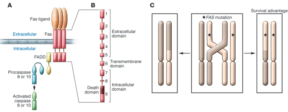
ALPS is an inherited disorder of lympho-cyte apoptosis. Defective apoptosis results in chronic lymphoproliferation, character- ized by lymphadenopathy and splenomeg-aly, autoimmunity, and the emergence of a normally rare population of TCRab+ T cells lacking CD4 or CD8 expression — so-called double-negative T cells (DNTs). The most common cause of ALPS is germline muta-tions in FAS. The second most common known cause of ALPS is somatic FAS muta-tions affecting primarily DNTs (5, 6). A smaller percentage of patients have germline mutations in genes encoding the Fas apop-tosis signaling pathway components caspase 10 and Fas ligand (Figure 1). Approximately 20% of patients diagnosed with clinical ALPS have no known mutation.
ALPS is usually dominantly inherited; thus, most individuals inheriting a FAS mutation display manifestations of dis-ease. The FAS mutations found in patients with ALPS often disrupt the signaling function of Fas by a dominant interfer-ing mechanism. Since Fas trimerizes and forms higher-order oligomers required for signal transduction, FAS mutations typi-cally encode defective proteins that enter complexes with the wild-type proteins and
disrupt function (Figure 1A). Since prop-er trimer function requires complexes of exclusively wild-type Fas molecules, only one in eight trimers will be fully functional in heterozygote individuals. Defective Fas function makes lymphocytes from patients bearing ALPS-causing FAS mutations resis-tant to apoptosis.
Although ALPS is typically a dominant-ly inherited disease, patients carrying a homozygous FAS mutation develop early-onset severe disease, whereas those with heterozygous FAS mutations can develop dis- ease of variable intensity. The variation in dis-ease severity is called variable “penetrance,” and even individuals from the same family with the same heterozygous FAS mutations can range from having severe autoimmunity and lymphoproliferation to having no dis-ease at all. Interestingly, T cells from ALPS patients and undiseased relatives harbor-ing the same FAS mutation exhibit similar degrees of resistance to apoptosis. Hence, the apoptosis defect is completely penetrant, but the clinical phenotype is incompletely pen-etrant. Disease manifestations can also vary depending on the location of the mutation within the FAS gene. Heterozygous muta-tions that affect the extracellular domain of the Fas protein result in less clinical pen-etrance and milder disease than mutations affecting the intracellular portion of the pro-tein (Figure 1B).
The cause of the variable penetrance of ALPS has been a long-standing ques-
tion in the field. Is it the result of genet-ics or environment? If genetic, then how precisely does it work? Is there a modifier gene or additional genetic factor that is necessary for disease manifestation? In this issue of the JCI, Magerus-Chatinet and colleagues demonstrate that the “two-hit” or “loss of heterozygosity” concept, often used to describe the somatic genetic basis of cancer, applies to disease penetrance in patients with heterozygous germline FAS mutations (4). The two-hit hypothesis pos-its that in individuals with a heterozygous germline mutation, usually in reference to a tumor suppressor gene, a somatic second hit (or loss) affecting the wild-type allele is necessary to produce disease.
Genetic mechanism for the variable penetrance of ALPS
In their study (4), Magerus-Chatinet and colleagues examined seven ALPS patients and their asymptomatic relatives, all of whom bore heterozygous germline muta-tions affecting the extracellular domain of the Fas protein. They discovered that the seven affected individuals had a somatic “hit” in the DNTs that impaired or elimi-nated the wild-type FAS allele. Three of the individuals had somatic mutations in their second FAS allele, and four had telomeric uniparental disomy resulting in loss of the wild-type FAS allele and duplication of the mutant allele. Telomeric uniparental diso- my is a rare event involving genetic recom-
bination resulting in a cell having two cop-Figure 1
[image:3.585.59.527.80.263.2]ies of a maternal or paternal chromosomal segment extending to the telomere and no copies of that segment from the other par-ent. These findings provide a striking new insight into a somatic genetic mechanism that can explain the onset of disease in ALPS patients carrying FAS mutations of incomplete penetrance. By exhibiting either compound heterozygous or homozygous
FAS mutations, these patients effectively manifested homozygous recessive loss-of-function mutations, at least in their DNTs. Thus, more severe disease evidently emerg-es from a more severe loss of Fas function.
The survival advantage of FAS mutations
Mutations impairing apoptosis pathways give cells a survival advantage. Defective apoptosis has been found to be a key fac-tor in the progression and development of cancers, especially those of hematopoi-etic origin (7). FAS mutations, in particular, result in the chronic premalignant lympho-proliferation characteristic of ALPS and an increased risk of lymphoma. Somatic muta-tions in FAS have also been found in various cases of leukemia, multiple myeloma, and lymphoma (8–10). Thus, it is no wonder that the same mechanisms often involved in malignant transformation also appear to be responsible for some cases of ALPS. The telomeric uniparental disomy described by Magerus-Chatinet et al. (4) is probably the result of mitotic recombination, a rare process of genetic alteration often linked to cancer in which maternal and paternal chromosomes in somatically dividing cells exchange segments of DNA. In the study by Magerus-Chatinet and colleagues (4), the healthy relatives carried only one copy of the
FAS mutation, which apparently was insuf-ficient to cause clinical disease on its own. Loss of heterozygosity in the affected ALPS patients due to telomeric uniparental diso-my led to homozygosity of the FAS muta-tion (Figure 1C). These data suggest that two copies of the mutant allele, with loss of the wild-type allele, lead to clinical ALPS.
Three of the patients described by Mag-erus-Chatinet et al. (4) had a second hit in their other originally wild-type FAS allele. One of the somatic mutations was predict- ed to result in a severely truncated nonfunc-tional protein, and the other two mutations were in exon 9, which encodes the death domain that is critical for downstream sig- naling. Mutations in exon 9 often substan- tially impair Fas function (11, 12). Conse-quently, the second hit effectively removed
any wild-type Fas protein. This unexpected genetic event presumably gave the mutant cells a greater survival advantage and pro-moted the onset of clinical disease.
The role of DNTs as “drivers” of ALPS
DNTs are a hallmark of ALPS. DNTs make up less than 2% of T cells in the blood of healthy individuals but can account for up to 40% in ALPS patients (13). Evidence suggests that the DNTs of ALPS patients may be derived from polyclonal chronical-ly activated cytotoxic T cells that have lost CD8 expression (14, 15). One unresolved question in the study of ALPS is whether DNTs are the “drivers” of disease or merely a manifestation of immune dysregulation. Previous findings that somatic FAS muta- tions in DNTs cause ALPS that is indistin-guishable from germline FAS mutations suggest that the DNTs trigger the disease (5, 6). This report by Magerus-Chatinet and colleagues provides further evidence for the centrality of their role in the disease, since the secondary event correlating with clinical disease occurred mainly or perhaps wholly within the DNTs (4).
However, studies have shown that DNTs are hyporesponsive and do not proliferate, which would imply that they may not be the source of autoaggression (14, 16). According to this view, the DNTs represent a terminal state and perhaps the remaining footprint of effector T cells that may have been the actu- al drivers of disease. Although ALPS-caus-ing somatic mutations are predominantly detected in the DNTs, the somatic muta-tions may exist in single-positive T cells but go undetected due to either the localization of these cells in lymphoid organs or to their small numbers compared with that of the unaffected T cells. In this scenario, DNTs are merely a sign or warning of the chronic lymphoproliferation that is occurring in vivo. Conceivably, there may not be a single cell subset that is the sole “driver” of disease. This is illustrated in a study by Stranges et al. (17), which demonstrated that a condi-tional knockout of FAS in T cells, B cells, or dendritic cells could result in systemic auto- immune disease. Further research is there-fore necessary to clarify the contribution of DNTs and other cell subsets to the immu-nological derangements in ALPS.
Concluding remarks
The report by Magerus-Chatinet et al. (4) is one of the first to provide a molecular explanation for the variable penetrance
seen in ALPS. The finding that a somatic mutation or loss of heterozygosity is a “modifier” of disease penetrance provides a new perspective on how autoimmune dis-eases may develop. Modifier alterations in other genes have been suspected for some time to be the basis of the variable pen-etrance seen in ALPS. The fact that the lpr mouse, which is a model of ALPS, shows variable degrees of autoimmunity depend-ing on the background mouse strain had long implied a role for modifier genes in disease (18, 19). The authors’ discovery that a second hit in FAS itself predisposes to clinical ALPS (4) supports the concept that a modifier gene or genetic factor is responsible for disease expression but with a twist in that the “modifier” mutation is somatic and in the same gene. Future stud-ies examining other ALPS families with incomplete penetrance may lead to the dis-covery of new modifier genes or a second hit in other genes involved in apoptosis or proliferation as well as other tolerance checkpoints. This two-hit model of disease expression may also help explain the onset of disease in other genetic disorders with selected effects on certain cell populations and incomplete penetrance.
Acknowledgments
This work was supported by the Division of Intramural Research, National Institute of Allergy and Infectious Diseases, NIH.
Address correspondence to: Michael Lena-rdo, Laboratory of Immunology, NIAID, NIH, Bldg. 10, Rm. 11N311, 10 Center Dr., MSC 1892, Bethesda, Maryland 20892-1892, USA. Phone: 301.496.6753; Fax: 301.402.8530; E-mail: [email protected].
1. Choate KA, et al. Mitotic recombination in patients with ichthyosis causes reversion of dominant muta-tions in KRT10. Science. 2010;330(6000):94–97.
2. Davis BR, Candotti F. Revertant somatic mosa-icism in the Wiskott-Aldrich syndrome. Immunol Res. 2009;44(1–3):127–131.
3. Rieux-Laucat F, et al. Inherited and somatic CD3zeta mutations in a patient with T-cell defi-ciency. N Engl J Med. 2006;354(18):1913–1921. 4. Magerus-Chatinet A, et al. Onset of autoimmune
lymphoproliferative syndrome (ALPS) in humans as a consequence of genetic defect accumulation.
J Clin Invest. 2011;121(1):106–112.
5. Dowdell KC, et al. Somatic FAS mutations are common in patients with genetically undefined autoimmune lymphoproliferative syndrome. Blood. 2010;115(25):5164–5169.
6. Holzelova E, et al. Autoimmune lymphoprolifera-tive syndrome with somatic Fas mutations. N Engl J Med. 2004;351(14):1409–1418.
7. Thompson CB. Apoptosis in the pathogen-esis and treatment of disease. Science. 1995; 267(5203):1456–1462.
WS. Mutations in the Fas antigen in patients with multiple myeloma. Blood. 1997;90(11):4266–4270. 9. Tamiya S, Etoh K, Suzushima H, Takatsuki K,
Matsuoka M. Mutation of CD95 (Fas/Apo-1) gene in adult T-cell leukemia cells. Blood. 1998; 91(10):3935–3942.
10. Beltinger C, Kurz E, Bohler T, Schrappe M, Ludwig WD, Debatin KM. CD95 (APO-1/Fas) mutations in childhood T-lineage acute lymphoblastic leukemia.
Blood. 1998;91(10):3943–3951.
11. Fisher GH, et al. Dominant interfering Fas gene mutations impair apoptosis in a human autoim-mune lymphoproliferative syndrome. Cell. 1995; 81(6):935–946.
12. Jackson CE, et al. Autoimmune lymphoprolif-erative syndrome with defective Fas: genotype
influences penetrance. Am J Hum Genet. 1999; 64(4):1002–1014.
13. Caminha I, et al. Using biomarkers to predict the presence of FAS mutations in patients with features of the autoimmune lymphoprolif-erative syndrome. J Allergy Clin Immunol. 2010; 125(4):946–949.
14. Bleesing JJ, et al. A composite picture of TcR alpha/ beta(+) CD4(–)CD8(–) T Cells (alpha/beta-DNTCs) in humans with autoimmune lymphoproliferative syndrome. Clin Immunol. 2002;104(1):21–30. 15. Landolfi MM, Van Houten N, Russell JQ, Scollay R,
Parnes JR, Budd RC. CD2-CD4-CD8- lymph node T lymphocytes in MRL lpr/lpr mice are derived from a CD2+CD4+CD8+ thymic precursor. J Immunol. 1993;151(2):1086–1096.
16. Sobel ES, Kakkanaiah VN, Rapoport RG, Eisenberg RA, Cohen PL. The abnormal lpr double-negative T cell fails to proliferate in vivo. Clin Immunol Immu-nopathol. 1995;74(2):177–184.
17. Stranges PB, et al. Elimination of antigen-present- ing cells and autoreactive T cells by Fas contrib-utes to prevention of autoimmunity. Immunity. 2007;26(5):629–641.
18. Izui S, Kelley VE, Masuda K, Yoshida H, Roths JB, Murphy ED. Induction of various autoantibod-ies by mutant gene lpr in several strains of mice.
J Immunol. 1984;133(1):227–233.
19. Wang Y, Nose M, Kamoto T, Nishimura M, Hiai H. Host modifier genes affect mouse autoimmu-nity induced by the lpr gene. Am J Pathol. 1997; 151(6):1791–1798.
A stimulating way to improve T cell responses
to poxvirus-vectored vaccines
Stuart N. Isaacs
University of Pennsylvania School of Medicine and Philadelphia VA Medical Center, Philadelphia, Pennsylvania, USA.
Vaccines remain one of the most cost-effective public health measures.
Despite ongoing efforts, protective vaccines against cancer and many
infec-tious diseases, including malaria, tuberculosis, and HIV/AIDS, are still not
in hand. Most investigators believe that to succeed against these difficult
targets, vaccines that generate potent T cell responses are needed. In this
issue of the
JCI
, Salek-Ardakani et al. show how the relative virulence of a
virus/vaccine vector affects the memory CD8
+T cells generated and how the
response may be enhanced. The work has important implications for the
development of future vaccines that aim to trigger CD8
+T cell responses.
Disease prevention is the key to public health. One of the most cost-effective ways to prevent disease is through vaccination, an approach that has enabled successful con-trol of many infectious diseases that were once common, including measles, diph-theria, and pertussis (whooping cough). In addition, a protective vaccine was key to the global eradication of smallpox, and it is believed that vaccinations may soon rel-egate polio to a historical memory. However, many infectious diseases, including malaria, tuberculosis, and HIV/AIDS, have proven to be formidable foes; despite intensive research efforts, we still do not have effica-cious protective vaccines. Most believe that to be successful, such vaccines — as well as vaccines that protect against cancer — will need to generate potent T cell responses.
Poxviruses are members of a large fam-
ily of DNA viruses that share a long his-tory with vaccination. Cowpox virus was the active agent in Jenner’s pioneering approaches to preventing smallpox in the 18th century and is closely related to vac-cinia virus, which was used as the vaccine that helped eradicate smallpox in the lat-ter half of the 20th century. Remarkably, vaccinia virus remains one of the leading vector candidates to be used in the devel-opment of vaccines against challenging infectious diseases and cancers, largely as a result of the promise seen in the earli-est studies that showed its utility as an expression vector (1, 2) and its ability to prime T cell responses to expressed for-eign antigens (3). However, the laboratory strains of vaccinia virus, and even the old conventional human smallpox vaccines, are likely not ideal vaccine vectors from a safety perspective, especially in current times, when greater numbers of immu-nocompromised people are likely to be vaccinated or inadvertently exposed; even in normal individuals, what is considered an acceptable risk/benefit ratio has shift-ed. This has led to the pursuit of more
attenuated and replication-incompetent poxvirus-based vaccine vectors that have significantly improved safety profiles. A recent achievement that highlights the promise of poxvirus-based vaccines is the somewhat unexpected results of the Thai HIV vaccine trial (4). In that study, vac-cination with a recombinant Canarypox virus vector expressing three HIV proteins along with boosts of HIV envelope pro-teins showed modest protection from HIV acquisition. While some worry about the reproducibility of the effect found, others are clearly encouraged by the results and are seeking to understand and improve upon the immune responses generated by the vaccination strategy. Since vaccine-induced sterilizing immunity (defined as antibody and innate immune responses that prevent actual infection) is a high bar to reach against the problematic organ-isms that cause malaria, tuberculosis, and HIV/AIDS, future vaccines will need to generate potent antibody and cytotoxic T cell responses.
In this issue of the JCI, Salek-Ardakani et al. show how the relative virulence of a virus/vaccine affects the memory CD8+ T cells that are generated and how one might manipulate costimulatory mole-cules to enhance the CD8+ T cell response (5). The work has important implications for development of future vaccines that aim to target the CD8+ T cell response and the stimulatory molecules that might be candidates to generate such responses. Conflict of interest: The author has declared that no
conflict of interest exists.