Alloantigen-specific regulatory T cells
generated with a chimeric antigen receptor
Katherine G. MacDonald, … , Raewyn Broady, Megan K.
Levings
J Clin Invest. 2016;
126(4)
:1413-1424.
https://doi.org/10.1172/JCI82771
.
Adoptive immunotherapy with regulatory T cells (Tregs) is a promising treatment for allograft
rejection and graft-versus-host disease (GVHD). Emerging data indicate that, compared
with polyclonal Tregs, disease-relevant antigen-specific Tregs may have numerous
advantages, such as a need for fewer cells and reduced risk of nonspecific immune
suppression. Current methods to generate alloantigen-specific Tregs rely on expansion with
allogeneic antigen-presenting cells, which requires access to donor and recipient cells and
multiple MHC mismatches. The successful use of chimeric antigen receptors (CARs) for the
generation of antigen-specific effector T cells suggests that a similar approach could be
used to generate alloantigen-specific Tregs. Here, we have described the creation of an
HLA-A2–specific CAR (A2-CAR) and its application in the generation of alloantigen-specific
human Tregs. In vitro, A2-CAR–expressing Tregs maintained their expected phenotype and
suppressive function before, during, and after A2-CAR–mediated stimulation. In mouse
models, human A2-CAR–expressing Tregs were superior to Tregs expressing an irrelevant
CAR at preventing xenogeneic GVHD caused by HLA-A2
+T cells. Together, our results
demonstrate that use of CAR technology to generate potent, functional, and stable
alloantigen-specific human Tregs markedly enhances their therapeutic potential in
transplantation and sets the stage for using this approach for making antigen-specific Tregs
for therapy of multiple diseases.
Technical Advance
Immunology
Transplantation
Find the latest version:
Introduction
The essential role of regulatory T cells (Tregs) in preventing autoimmunity and controlling responses to alloantigens is well established. Multiple Treg-based cell therapy approaches are now being tested in the clinic, with early promising results reported in prevention of graft-versus-host disease (GVHD) after allogeneic hematopoietic stem cell transplantation (HSCT) (1–3) and main-tenance of C-peptide levels in type 1 diabetes (4, 5). The results of these phase I trials indicate that Treg therapy seems to be well tol-erated and possibly efficacious, but that there may be a transient risk of generalized immunosuppression (6).
Data from animal studies indicate that the potency and speci-ficity of Treg therapy can be markedly enhanced by the use of antigen-specific cells. For example, in models of autoimmunity, antigen-specific Tregs are superior to polyclonal Tregs in reducing disease: Tregs isolated from pancreatic lymph nodes or pulsed with islet antigen are significantly better at preventing or curing type 1 diabetes than are polyclonal Tregs (7–11), and Tregs expressing an autoantigen-specific transgenic T cell receptor (TCR) are superior to polyclonal Tregs at suppressing central nervous system inflam-mation in a model of experimental autoimmune encephalomyelitis (EAE) (12). Similarly, alloantigen-specific Tregs, enriched by alloan-tigen-stimulated expansion in vitro or engineered to express a TCR
transgene, are more effective than polyclonal Tregs at preventing rejection of organ and tissue grafts (13–17). Although limited, there is some evidence that Tregs expanded with alloantigens also effec-tively prevent GVHD (18) and that in vivo induction of antigen-spe-cific Tregs promotes acceptance of hematopoietic allografts without GVHD (19). Humanized mouse models have shown similar results: alloantigen-expanded human Tregs are more potent suppressors of skin graft rejection than are polyclonal Tregs (20, 21).
An alternate approach to overexpressing transgenic TCRs or antigen-stimulated expansion to enrich for antigen-specific T cells is the use of chimeric antigen receptors (CARs), in which T cells are genetically engineered to express extracellular single-chain Ab (scFv) antigen–binding domains fused to intracellular signaling domains (22, 23). Tregs expressing CARs specific for model anti-gens have been tested (24–28), leading us to hypothesize that this approach could be used in the context of transplantation. Here, we describe what we believe is a new approach to generating potent alloantigen-specific Tregs, using a CAR targeting HLA-A2.
Results
Construction and validation of an A2-CAR. We aimed to generate a
new CAR specific for HLA-A2, as this is a commonly mismatched antigen in transplantation, with a prevalence of approximately 50% in those of mixed European descent (29), and HLA-A mis-matching is associated with poor outcomes after transplantation (30). As detailed in Methods, we generated lentiviral vectors encoding an HLA-A2–specific CAR by cloning and sequencing the heavy- and light-chain variable regions of the BB7.2 mAb and
fus-ing the resultfus-ing scFv to portions of CD8, CD28, and CD3ζ in a
second-generation CAR structure (ref. 31 and Figure 1, A and B). A
Adoptive immunotherapy with regulatory T cells (Tregs) is a promising treatment for allograft rejection and graft-versus-host disease (GVHD). Emerging data indicate that, compared with polyclonal Tregs, disease-relevant antigen-specific Tregs may have numerous advantages, such as a need for fewer cells and reduced risk of nonspecific immune suppression. Current methods to generate alloantigen-specific Tregs rely on expansion with allogeneic antigen-presenting cells, which requires access to donor and recipient cells and multiple MHC mismatches. The successful use of chimeric antigen receptors (CARs) for the generation of antigen-specific effector T cells suggests that a similar approach could be used to generate alloantigen-specific Tregs. Here, we have described the creation of an HLA-A2–alloantigen-specific CAR (A2-CAR) and its application in the generation of alloantigen-specific human Tregs. In vitro, A2-CAR–expressing Tregs maintained their expected phenotype and suppressive function before, during, and after A2-CAR–mediated stimulation. In mouse models, human A2-CAR–expressing Tregs were superior to Tregs expressing an irrelevant CAR at preventing xenogeneic GVHD caused by HLA-A2+ T cells. Together,
our results demonstrate that use of CAR technology to generate potent, functional, and stable alloantigen-specific human Tregs markedly enhances their therapeutic potential in transplantation and sets the stage for using this approach for making antigen-specific Tregs for therapy of multiple diseases.
Alloantigen-specific regulatory T cells generated
with a chimeric antigen receptor
Katherine G. MacDonald,1 Romy E. Hoeppli,1 Qing Huang,2 Jana Gillies,1 Dan S. Luciani,1 Paul C. Orban,1 Raewyn Broady,2 and Megan K. Levings1
1Department of Surgery and 2Department of Medicine, University of British Columbia, and Child and Family Research Institute, Vancouver, British Columbia, Canada.
Related Commentary: p. 1248
Authorship note: P.C. Orban and R. Broady contributed equally to this work.
Conflict of interest: The authors have declared that no conflict of interest exists.
Submitted: May 13, 2015; Accepted: February 4, 2016.
it was important to exclude the possibility that high expres-sion of a CAR in Tregs might result in loss of the expected Treg phenotype. A2-CAR– expressing Tregs maintained significantly higher expres-sion of FOXP3 than Tconvs and preserved a high degree of demethylation of the Treg-specific demethyled region (TSDR) of the FOXP3 locus (Figure 2, D and E), consistent with other reports of demeth-ylation observed in naive Tregs sorted by flow cytom-etry (38). High expression of other canonical Treg markers, including CD25, Helios, and CTLA-4, was also preserved in A2-CAR–expressing Tregs (Supplemental Figure 1A; supplemental material avail-able online with this article; doi:10.1172/JCI82771DS1). In comparison with CAR-expressing Tconvs, A2-CAR Tregs also did not contain significant proportions of IL-2– or IFN-γ–producing cells (Supplemental Figure 1B).
To determine whether A2-CAR Tregs preserved their in vitro suppressive function when activated via their endogenous TCR
(i.e., not via the A2-CAR), αCD3/CD28 mAbs were used to
stimu-late HLA-A2– peripheral blood mononuclear cells (PBMCs) alone
or in the presence of decreasing ratios of A2-CAR Tregs. As shown in Supplemental Figure 1C, A2-CAR Tregs suppressed the prolif-eration of CD8+ T cells in a dose-dependent manner. Finally,
cul-tures of Tregs expressing either CAR had high viability, ranging from 86% to 96% (Supplemental Figure 1D). Thus, expression of
a second-generation CAR comprising domains from scFv, CD8α,
CD28, and CD3ζ does not alter the phenotype, stability, or in vitro function of human Tregs.
A2-CAR–mediated stimulation activates Tregs. We next
inves-tigated how A2-CAR stimulation compares with traditional TCR activation of Tregs. We first tested the ability of TCR versus CAR activation to stimulate intracellular signaling pathways. A2-CAR Tregs or Tconvs were TCR stimulated with anti-CD3 mAbs or CAR stimulated by crosslinking the extracellular Myc-tag, and relative levels of phospho-ZAP70 were determined by flow cytometry. TCR and CAR stimulation activated ZAP70 equally, with no statistically significant differences between Tregs and Tconvs (Figure 3A).
A2-CAR–expressing Tregs or Tconvs were next left unstimu-lated or stimuunstimu-lated via the CAR, with K562 cells expressing
HLA-A2, or via the TCR, with K562 cells loaded with αCD3/CD28
mAbs, for 24 hours. CAR stimulation resulted in upregulation of the canonical activation marker CD69 in both Tregs and Tconvs to levels that tended to be higher than those of TCR-stimulated cells (Figure 3B). In contrast, in comparison with Tconvs, Tregs had significantly lower expression of CD154 (CD40L) upon activation second-generation CAR containing CD28 was chosen because of
the well-characterized importance of this costimulatory molecule in Treg development and function (32). A lentivector encoding a well-characterized HER2-specific CAR (HER2-CAR) (33) served as a negative control. Surface expression of the HLA-A2–specific CAR (A2-CAR) was confirmed by transient transfection of 293T cells and flow cytometric staining for the extracellular Myc epitope (Figure 1C). mAbs reformatted to scFv may have reduced antigen binding or specificity depending on their components (34, 35). We therefore used tetramers made from HLA-A2 or HLA-A24 to confirm that the specificity of binding to HLA-A2 was retained. As shown in Figure 1D, cells expressing the A2-CAR bound to HLA-A2 tetramers, but not to control HLA-HLA-A24 tetramers.
Generation of A2-CAR Tregs. To test the function of the A2-CAR
in Tregs, we sorted CD25hiCD45RA+ cells from peripheral blood,
as this population is known to have homogeneous expression of FOXP3 and to have the highest expansion potential (Figure 2A and ref. 36). CD25loCD45RA+ cells were sorted in parallel as the
con-ventional T cell (Tconv) control. As outlined in Figure 2B, Tregs (or control Tconvs) were stimulated, transduced, and purified as
ΔNGFR+ cells, then expanded for an additional 6 days. At the end
of 13 to 14 days of culture, cell-surface expression of A2-CAR or HER2-CAR on both Tregs and Tconvs was confirmed with flow cytometry (Figure 2C). Interestingly, while similar proportions of Treg and Tconv populations were positive for CAR expression, on a per-cell basis, A2-CAR expression appeared to be higher in both cell types than expression of the HER2-CAR. Even when CAR
expression intensity was normalized to the expression of ΔNGFR,
A2-CAR–expressing cells showed significantly higher levels of CAR expression than cells expressing HER2-CAR.
[image:3.585.41.416.56.240.2]The effect of CAR expression on the phenotype and function of human Tregs is unknown, and since high expression of CARs has been linked to undesired antigen-independent CAR activation (37),
Both TCR- and CAR-stimulated Tregs maintained the expected expression pattern of Treg lineage markers, with high expression of FOXP3 (both percentage and MFI) and CD25 and low expres-sion of CD127 (Figure 4A).
After 2 weeks, CAR stimulation resulted in a significantly higher fold expansion and increased viability of Tregs compared with TCR-expanded cells (Figure 4B) and retained the Tregs’ suppressive activ-ity (Figure 4C). TCR- and CAR-stimulated Tregs remained more than 85% FOXP3+ and had similar levels of FOXP3 on a per-cell basis,
with percentage of both FOXP3+ and MFI significantly higher than in
Tconvs (Figure 4D). However, there was a slight downregulation of the A2-CAR MFI in CAR-expanded cells, possibly due to receptor internalization, since the overall proportion of CAR+ cells was similar
after TCR or CAR expansion. Tregs expanded through their CAR also maintained high expression of CD25 and CTLA-4 (Figure 4E).
A2-CAR Tregs mediate HLA-A2–specific suppression. We then
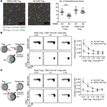
asked whether A2-CAR Tregs preferentially interacted with HLA-A2–expressing PBMCs. A2-CAR and HER2-CAR Tregs were
cul-tured with a mixture of HLA-A2+ and HLA-A2– PBMCs, which
were labeled with a red or green dye, respectively, and the number of interactions between the Tregs and the red versus green PBMCs was observed over time by fluorescence microscopy (Figure 5A). The total number of cells per field was similar, but we found sig-nificantly more interactions between A2-CAR Tregs and
HLA-A2+ PBMCs than between A2-CAR Tregs and HLA-A2– PBMCs or
with either TCR or CAR, consistent with the known low CD154 expression in human Tregs (39).
In comparison with TCR-mediated activation, A2-CAR–medi-ated stimulation of Tregs caused markedly greater upregula-tion of proteins associated with Treg funcupregula-tion. The proporupregula-tion of CTLA-4–expressing Tregs was significantly higher in CAR- versus TCR-stimulated cultures as was the mean fluorescence intensity (MFI) of expression (Figure 3C). CAR-stimulated Tregs also had significantly higher surface expression of latency-associated pep-tide (LAP) and glycoprotein A repetitions predominant (GARP), the inactive form of TGF-β and one of its receptors, respectively, compared with TCR-stimulated cells (Figure 3D). Importantly, A2-CAR–mediated Treg activation did not alter the cytokine phenotype of Tregs. Whereas A2-CAR–stimulated Tconvs pro-duced significant amounts of IFN-γ, TNF-α, and IL-2 (Figure 3E), A2-CAR–stimulated Tregs did not make significant amounts of any of these cytokines. Thus, short-term (24–48 hours) CAR stimula-tion does not alter the expected Treg phenotype.
A2-CAR–mediated stimulation and expansion do not alter the Treg phenotype. We next asked how long-term CAR-mediated
stim-ulation of Tregs affects their phenotype and function. To address this question, we analyzed the phenotype of A2-CAR–expressing Tregs or Tconvs (generated as in Figure 2B) stimulated through
their endogenous TCR (with K562.64 cells loaded with αCD3/28
[image:4.585.47.548.54.339.2]mAbs) or with K562 cells expressing HLA-A2 over multiple days.
To further confirm the antigen specificity of the A2-CAR Tregs, we used an autologous system consisting of an EBV-trans-formed B cell line (with and without ectopic HLA-A2 expres-sion), a tetanus toxoid–specific (TT-specific) T cell clone, and
A2-CAR Tregs from an HLA-A2– individual (Supplemental
Fig-ure 2A). This system enabled the Tregs and responding T cell clone to be activated individually, without polyclonal anti-CD3 stimulation (i.e., TCR), allowing precise testing of antigen speci-ficity. In the absence of HLA-A2, A2-CAR Tregs did not suppress TT-stimulated proliferation. In the presence of HLA-A2, only the A2-CAR, and not the HER2-CAR, Tregs suppressed prolif-eration. Finally, the presence of stimulated A2-CAR–expressing Tconvs enhanced proliferation.
HER2-CAR Tregs and HLA-A2+ PBMCs. These data suggest that
expression of A2-CAR enables Tregs to interact more specifically with HLA-A2–expressing cells.
To test the relative ability of A2-CAR Tregs to suppress T cell proliferation in response to allogeneic antigens, we set up mixed lymphocyte reactions (MLRs) in which either the responding T cells or the stimulating antigen-presenting cells (APCs) expressed HLA-A2. As shown in Figure 5, B and C, compared with HER2-CAR Tregs, A2-HER2-CAR Tregs were significantly better able to sup-press alloantigen-stimulated proliferation of CD8+ T cells
[image:5.585.59.520.57.483.2]regard-less of whether HLA-A2 was expressed on the responder or the stimulator cells. These data show that expression of A2-CAR on Tregs results in antigen-specific suppression.
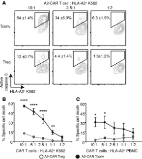
numbers of A2-CAR Tregs or Tconvs, and after 24 hours, the pro-portion of K562 cells expressing active caspase 3 was measured as an indicator of cell death (Figure 6A). A2-CAR Tconvs robustly induced cell death in HLA-A2+ K562 cells (but not HLA-A2– K562
cells, data not shown). Although a small proportion (<15%) of HLA-A2+ K562 cells were positive for active caspase 3 when cocultured
with high ratios of A2-CAR Tregs, A2-CAR Tregs mediated substan-tially less killing than Tconvs at all ratios tested (Figure 6B).
Possible Treg-mediated cytolytic activity was further tested using PBMCs as targets. Similar to data with K562 cells, coculture with A2-CAR Tconvs resulted in the presence of active caspase 3
in HLA-A2+ PBMCs. In contrast, in the presence of A2-CAR Tregs,
there was negligible active caspase 3 (Figure 6C).
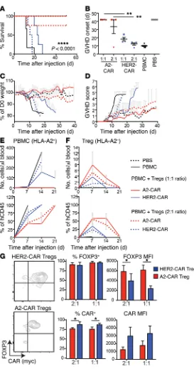
A2-CAR Tregs are superior to polyclonal Tregs at preventing xeno-geneic GVHD mediated by HLA-A2+ T cells. In order to test the
func-tional capacity of A2-CAR Tregs in vivo, we used a mouse model in which human PBMCs engrafted into immunodeficient NOD/ SCID IL-2Rγnull (NSG) mice cause xenogeneic GVHD (41). In these
experiments, 1 × 10⁷ PBMCs from an HLA-A2+ donor were
inject-In vivo, A2-CAR Tregs would have the potential to receive simultaneous signals through both their endogenous TCRs and the ectopically expressed CAR. To mimic this possibility,
HLA-A2+ PBMCs were stimulated with αCD3/28-coated beads in the
absence or presence of A2-CAR– or HER2-CAR–expressing Tregs. A2-CAR Tregs, which received stimulation from both the CAR
and the TCR (via αCD3/28 beads), were as suppressive as
HER2-CAR Tregs (which only received TCR stimulation), demonstrat-ing that combined CAR and TCR stimulation does not negatively affect suppressive function (Supplemental Figure 2B). A2-CAR– mediated suppressive activity was also found to require contact, as A2-CAR–stimulated Tregs were not suppressive in a Transwell suppression assay (Supplemental Figure 2C).
CAR-stimulated Tregs have minimal cytotoxic activity. CARs were
originally developed in the context of cytolytic T cells, and there are some reports that human Tregs may also employ cytotoxicity as one of their mechanisms of suppression (40), so we next determined whether CAR stimulation might induce Tregs to kill their targets.
[image:6.585.69.531.56.409.2]HLA-A2+ or HLA-A2– K562 cells were cocultured with increasing
Figure 4. A2-CAR–mediated stimulation and expansion maintains a Treg phenotype. A2-CAR Tregs or Tconvs were stimulated with K562.64 cells loaded with αCD3/28 mAbs or expressing HLA-A2. (A) Expression of FOXP3, CTLA-4, CD25, and CD127 was assessed over 5 days on Tregs and Tconvs (n = 4). (B) Fold expansion based on cell counts at day 0 of restimulation and day 14, and viability of CAR- and TCR-expanded Tregs (n = 4). (C) Suppressive capac-ity of CAR-expanded A2-CAR Tregs when stimulated through endogenous TCR was assayed by titrated Treg ratios with HLA-A2– PBMCs stimulated with
ed into irradiated NSG mice with or without the indicated type of Treg at a 1:1 or 2:1 ratio (i.e., 1 × 10⁷ Tregs or 5 × 10⁶ Tregs). Mice
were monitored for up to 7 weeks by clinical score as described in Methods. Consistent with previous reports, control Tregs express-ing the HER2-CAR, which would be stimulated via their endog-enous TCR by xenogeneic antigens (but not through the CAR), significantly improved survival of mice when infused at a 1:1 ratio (Figure 7A). Importantly, mice receiving A2-CAR Tregs at either a 1:1 or 1:2 ratio were significantly better protected. A2-CAR Tregs improved survival (Figure 7A) and delayed onset of xenogeneic
GVHD (Figure 7B) in comparison with results in mice receiving HER2-CAR–expressing Tregs. Even when xenogeneic GVHD developed in mice that received A2-CAR Tregs, weight loss was less (Figure 7C) and disease progression was slower (Figure 7D).
Engraftment of human T cells and survival of the infused Tregs was monitored by weekly blood draws. As shown in Fig-ure 7E, in comparison with mice receiving HER2-CAR Tregs, mice injected with A2-CAR Tregs had a lower absolute number
of human HLA-A2+CD45+ cells per μl of blood. Lower absolute
[image:7.585.68.513.57.501.2]numbers of circulating HLA-A2+CD45+ cells likely reflect the
Figure 5. A2-CAR Tregs preferentially interact with HLA-A2+ PBMCs and are superior to polyclonal Tregs at suppressing alloantigen-stimulated
failure of these cells to expand (as observed for mice coinjected
with HER-2 Tregs), since the proportion of HLA-A2+CD45+ cells
was the same at days 7 and 14 and corresponded to expansion and persistence of A2-CAR Tregs in mouse blood (Figure 7, E and F).
Notably, A2-CAR Tregs remained FOXP3+ and were detectable in
circulation for twice as long as HER2-CAR Tregs. Similar to find-ings from in vitro data (Figure 4C), cells that received CAR stimu-lation in vivo had a lower MFI of CAR expression, but also had a significantly higher FOXP3 MFI (Figure 7G), likely reflecting in vivo activation.
In the xenogeneic GVHD model discussed above, HLA-A2 is expressed on the responding T cells, but in human HLA-A2+
recipi-ents of hematopoietic stem cells or recipirecipi-ents of HLA-A2+ organs,
HLA-A2 would be expressed ubiquitously on tissues. It was there-fore important to exclude the possibility that the A2-CAR Tregs might cause tissue destruction. To test this possibility, HLA-A2
transgenic NSG mice were engrafted with HLA-A2– PBMCs in the
absence or presence of A2-CAR Tregs or A2-CAR Tconvs. Mice were sacrificed at 2 different time points (2 and 4 weeks after cell infusion), and histological analysis was performed to measure immune cell infiltration and tissue integrity (Supplemental Fig-ure 3). We found that mice injected with PBMCs, with or without
A2-CAR Tconvs, had substantial immune cell infiltra-tion and tissue destrucinfiltra-tion. In contrast, mice receiv-ing PBMCs and A2-CAR Tregs had less infiltration and most closely resembled the PBS control mice. Thus, injection of A2-CAR Tregs into mice that sys-temically express HLA-A2 does not result in tissue cytotoxicity, but rather activates their suppressive effect and limits PBMC-mediated tissue toxicity.
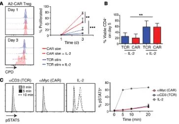
A2-CAR–stimulated Tregs require exogenous IL-2 for survival. A defining characteristic of human and
mouse Tregs is in vitro anergy, i.e., their inability to proliferate in the absence of exogenous IL-2 (39, 42). We therefore asked whether CAR stimulation affect-ed this phenotype, possibly overcoming a require-ment for exogenous IL-2. We found that, in contrast with TCR, CAR could indeed stimulate IL-2–inde-pendent proliferation in short-term (3 day) assays, but addition of exogenous IL-2 significantly enhanced CAR- and TCR-stimulated cell division (Figure 8A). Beyond 3 days, however, in the absence of exogenous IL-2, CAR- and TCR-stimulated Tregs had a signifi-cant decrease in viability (Figure 8B). The inability of CAR stimulation to maintain long-term cell viability is consistent with the inability of the CAR to stimulate STAT5 phosphorylation (Figure 8C). Thus, although CAR stimulation results in potent Treg activation, such as that in unmodified Tregs, CAR-Tregs require exogenous IL-2 for long-term survival.
Discussion
In this study, we show that the specificity of human Tregs can be redirected toward a transplant-relevant antigen using a CAR. Expression of an A2-CAR in Tregs enables antigen-specific activation and pro-liferation that is stronger than that stimulated by the endogenous TCR. Despite this relatively strong CAR-medi-ated activation and/or expansion, A2-CAR Tregs retained high expression of FOXP3 and other Treg markers and demethylation of the TSDR, had preserved suppression function in vitro, and did not have significant cytolytic activity. Adoptive transfer experi-ments revealed that A2-CAR Tregs that received in vivo CAR stimulation were markedly better than Tregs that only received stimulation through the endogenous TCR at preventing xeno-geneic GVHD. Collectively, these data show that human Tregs redirected to recognize a specific alloantigen are more effective at preventing GVHD than Tregs not thus directed, supporting the rationale for CAR modification of Tregs as a means of generating therapeutic antigen-specific cells.
Clinically applicable approaches to generating alloantigen-specific Tregs are currently limited to in vitro enrichment of allo-reactive T cells following stimulation with allogeneic APCs. For example, alloantigen-specific Tregs can be sorted on the basis of allogeneic APC–stimulated expression of CD69 and CD71 (20) or expanded with CD40-stimulated allogeneic B cells (21). Cells generated by these methods are more potently suppressive in vitro than polyclonal Tregs and protect skin allografts better than polyclonal Tregs. Both these approaches, however, may
[image:8.585.45.334.55.379.2]fer from limitations of cell numbers, as the frequency of alloantigen-specific cells decreases with increasing MHC matching. Indeed, such approaches may not be feasible at all in the context of HSCT, since most or all of the HLA alleles are matched and in vitro expansion of alloantigen-specific cells decreases with increasing HLA matching (21, 43). Moreover, this method requires sufficient APCs from the donor, or recipient in the case of HSCT, which are not always available. An additional consideration is that expansion of alloantigen-specific Tregs by APC stimulation requires extensive in vitro expansion, which could lead to loss of FOXP3 (44) and,
based on data from CD8+ T cells, decreased telomere
length and, hence, in vivo survival (45). The use of CAR technology overcomes all of these limitations.
A2-CAR–expressing Tregs can be robustly activated by a single HLA mismatch. Targeting of Tregs with a CAR toward MHC class I, and specifically HLA-A2, provides several advantages: it bypasses the need for direct or indi-rect antigen presentation by MHC class II; MHC class I is broadly expressed on all tissues, so stimulation would not be dependent on the presence of professional APCs; and HLA-A2 is expressed by a substantial proportion of the population, leading to many HLA-A2–mismatched transplants. In the context of solid organ transplantation, expression of HLA-A2 on the organ would likely stimulate localization of A2-CAR Tregs to the transplant, a possi-bility supported by our observed preferential colocaliza-tion of A2-CAR Tregs with HLA-A2+ cells. In the context
of GVHD, our experiments in HLA-A2–expressing NSG mice suggest that systemic expression of HLA-A2 may result in systemic activation of Treg suppression without consequent tissue damage.
Proof-of-concept work in mouse models has dem-onstrated the feasibility of redirecting the specificity of Tregs using CARs specific for model antigens. Expres-sion of a trinitrophenol–specific CAR with a CD28
trans-membrane domain and FCγR signaling domains enabled
mouse Tregs to home to the site of disease and protect from trinitrobenzene sulfonic acid–induced colitis (24, 25). Antigen-driven homing and protection from EAE was also observed in mouse T cells that were transduced with FOXP3 and a myelin oligodendrocyte
glycoprotein–spe-cific CAR containing domains from CD3γ and CD28 (26).
There is also a previous report of expression of a CAR spe-cific for carcinoembryonic antigen in human Tregs (27), but the low expression of FOXP3 and coproduction of IFN-γ and IL-10 in these cells makes it difficult to draw any conclusions about the impact of CAR expression on Treg biology. Our study represents what we believe is a marked advance over these previous findings, as we report the cre-ation of a human disease–relevant CAR and detailed data on the functionality of this protein in human Tregs.
[image:9.585.40.313.49.568.2]An important consideration is how the presence of the CAR might affect the function of the endogenous TCR. We found that CAR expression does not abrogate the normal suppressive function of Tregs whether they
In conclusion, here we have demonstrated the feasibility of redirecting Tregs to a transplant-relevant antigen with a CAR and shown that CAR Tregs remain functionally and phenotypically stable in vitro and in vivo. This work provides what we believe is the first proof-of-concept that CAR Tregs have the potential to be used therapeutically. In the setting of transplantation, a bank of HLA-specific CARs could be used to overcome all of the limita-tions of current strategies to generate alloantigen-specific Tregs. These findings also set the stage for using CARs to redirect the antigen specificity of Tregs toward auto- and/or tissue-specific antigens for therapeutic use in autoimmunity.
Methods
A2-CAR generation. Variable regions of the Ig heavy and light chains were cloned from the anti–HLA-A2 BB7.2 hybridoma (ATCC) using published methods (56) and converted into a single-chain Ab (scFv). The scFv was fused to a Myc epitope tag in the extracellular region to enable cell-surface detection by flow cytometry, a stalk region from human CD8α, the transmembrane and intracellular domains of human CD28, and human CD3ζ, as described (33). The HER2-CAR2 and the A2-CAR were cloned into a lentiviral vector that encodes cell-surface expression of a truncated nerve-growth-factor (TrkA) receptor (ΔNGFR) as a marker. Surface expression was determined by flow cytometry with transiently transfected HEK 293T cells (Lipo-fectamine 2000, Life Technologies). Viral particles were produced as described (57).
Treg sorting, transduction, and expansion. CD4+ T cells were
isolated from HLA-A2– donors via RosetteSep (STEMCELL
Tech-nologies) and enriched for CD25+ cells (Miltenyi Biotec) prior
to sorting into live CD4+CD45ROloCD45RAhiCD25hi Tregs and
CD4+CD45ROloCD45RAhiCD25lo Tconv), using a FACSAria II (BD
Biosciences). Sorted T cells were stimulated with artificial APCs (aAPCs) loaded with αCD3 mAbs as described (58) in 1,000 U/ml or 100 U/ml of IL-2, for Tregs or Tconvs, respectively. One day later, cells are stimulated via the TCR or the CAR. This finding provides
assurance that the “off-target” effects of CAR-expressing Tregs are not likely to be different from those of polyclonal Tregs and indeed should be minimized because the “on-target” effects can be obtained with smaller cell numbers.
Tregs are known to have intracellular signaling pathways dis-tinct from those of Tconvs, with a specific requirement for low levels of PI3K activity to retain high FOXP3 expression (46). The second-generation format A2-CAR used here includes domains from CD28
and CD3ζ, both of which are necessary for Treg activation (47),
suppressive capacity (48, 49), and homeostasis (50, 51). Although stimulation through the CAR did not cause abnormal signaling via ZAP70 or stimulate phosphorylation of STAT5, in short-term (2 to 3 days) assays, it did partially break the canonical anergy of Tregs in the absence of exogenous IL-2. Reversal of anergy, however, did not compromise lineage stability, as evidenced by stable expression of the expected Treg markers over time, including CD25, LAP, GARP, CTLA-4, and FOXP3, as well as suppressive function. In addition, exogenous IL-2 was necessary for long-term viability in vitro. Inter-estingly, activation of Tconv through the A2-CAR resulted in a Treg-like phenotype, with high expression of CD25, CTLA-4, LAP, and GARP (see Figure 4), potentially related to the ability of tonic sig-naling from CD28 to lead to exhausted and less-potent Tconvs (52). Since depletion of Tregs results in a return of inflammation in tolerized mice (53), long-term persistence may be essential for Treg therapy. In humans, infused Tregs have been observed in circulation for up to 14 days (3, 4), and in CAR T cell therapies, transferred cells are detectable in the blood for several weeks (54, 55). Although we observed A2-CAR Tregs in vivo for twice as long as polyclonal Tregs, they were not detectable in circulation after
2 weeks. Given that the HLA-A2+ PBMCs failed to expand in the
[image:10.585.43.407.54.307.2]presence of A2-CAR Tregs, it is possible that limited antigen, lim-ited IL-2, or both were insufficient to sustain the expansion and survival of A2-CAR Tregs.
Figure 8. A2-CAR–stimulated Tregs require exogenous IL-2 for long-term survival. (A) A2-CAR Tregs or Tconvs were labeled with CPD, then stimu-lated with K562.64 cells loaded with
αCD3/28 mAbs or expressing HLA-A2 in the absence or presence of 100 U/ml IL-2 for 5 days. Representative plots of FVD–CD4+ cells (left) and percentage of proliferated compared with unstimu-lated controls are summarized (right,
Proliferation, activation, cytokine production, and antigen-nonspe-cific suppression. To assess proliferation and activation, T cell lines were labeled with CPD (65-0840-85 or 65-0842-85, eBiosciences) and stimulated with K562.64 cells loaded by 1 hour of preincubation with αCD3 and α28 mAbs (1 μg/ml each), K562.64.HLA-A2 cells, or at a 1:2 (K562:T cell) ratio (K562.64 cells, a gift from James Riley, University of Pennsylvania, Philadelphia, Pennsylvania, USA, which were then transduced to express HLA-A2). Staining of CD154 was performed in culture for 6 hours prior to analysis. Suppression was assessed with allogeneic HLA-A2– or HLA-A2+ PBMCs labeled with
CPD450 and stimulated via αCD3/28-coated beads (Invitrogen) at a 1:8 or 1:16 bead to PBMC ratio for 96 hours. Percentage of suppres-sion of CD8+ cells was calculated using division index (DI) as follows:
(100 – [(DI PBMCs + test)/(DI PBMCs)] × 100). For Transwell assays, CAR T cells were cocultured with HLA-A2+ K562 cells at a 1:1 ratio in
an insert with a 4-μm pore membrane (Millipore).
To measure cytokine production, T cell lines were stimulated with the indicated K562 cells (1 K562:2 T cells) for 48 hours. Supernatants were collected and cytokine concentration was determined by the Human Th1/Th2/Th17 Cytokine Kit (BD Biosciences) and analyzed by FCAP Array Software v1.01 (Soft Flow).
Cytotoxicity assays. HLA-A2+ K562 cells or PBMCs were labeled with
PKH26 (Sigma-Aldrich, PKH26GL-1KT) and cocultured with A2-CAR– expressing cells at the indicated ratios for 24 hours. Activation of caspase 3 in PKH26+ cells was determined by flow cytometry and
percentage-spe-cific cell death was calculated by subtracting the percentage of cells with active caspase 3 in cultures with no CAR Tregs/Tconvs as described (60).
Suppression of MLRs and TT-specific T cells. Adherent cells from HLA-A2+ or HLA-A2– PBMCs from healthy donors were differentiated
into monocyte-derived DCs as described (61). For MLRs, HLA-A2+ or
HLA-A2– CD3+ responder T cells were labeled with CPD eF450
(eBio-sciences, 65-0842-85); then 1 × 10⁵ CD3+ responder T cells were
stim-ulated with 5 × 10⁴ HLA-A2–mismatched DCs, with increasing ratios
of A2-CAR or HER2-CAR Tregs. After 4 days, division of CD8+ T cells
was measured by flow cytometry.
For suppression of TT-specific proliferation, TT-specific CD4+ T
clones were isolated from an HLA-A2– individual as described (62).
An EBV cell line from the same donor was transduced with HLA-A2 using lentivirus. EBV cell lines were pulsed overnight with 50 μg/ml of TT (5 μg/ml, Enzo Life Science, ALX-630-108), irradiated at 150 Gy, and cocultured with CPD-labeled TT-specific CD4+ T clones in
the absence of presence of CAR-expressing Tregs of Tconvs. Prolif-eration was determined after 4 days,and percentage of suppression of TT-specific clones was calculated using percentage of prolifera-tion as follows: (100 – [(% proliferated TT + test)/(% proliferated TT alone)] × 100).
In vivo experiments. The 8- to 12-week-old female NSG mice (The Jackson Laboratory, bred in house) received whole-body irradiation (150 cGy, RS-2000 Pro Biological System) 1 day before injection of 1 × 10⁷ HLA-A2+ PBMCs with or without 1 × 10⁷ or 0.5 × 10⁷ of the
indicat-ed type of Tregs. Saline-injectindicat-ed mice servindicat-ed as controls. GVHD was scored based on weight, fur texture, posture, activity level, and skin integrity, with 0 to 2 points per category as described (63, 64). GVHD scoring was performed by 2 blinded investigators. Peripheral blood from the saphenous vein was centrifuged; then erythrocytes were lysed and leukocytes were measured by flow cytometry, and plasma was aspirated and frozen at –80°C until use.
were transduced with lentivirus at an MOI of 10 virus particles/cell. At day 7, ΔNGFR+ cells were purified with magnetic selection (Miltenyi
Biotec), then restimulated with aAPCs as above and expanded for 6 to 7 days. To test effects of A2-mediated stimulation, Tregs were restim-ulated with irradiated (10 Gy) K562.64 cells (59), K562.64.HLA-A2 cells, or K562.64.HER2 cells (derived from K562.64 cells transduced with a lentivirus encoding HER2 and GFP) at a 1:2 (K562/T cell) ratio for 2 weeks in the presence of 1,000 U/ml IL-2.
Flow cytometry. For phenotypic analysis, cells were stained with fixable viability dye (FVD) (65-0865-14 and 65-0866-14, eBioscience) and for surface markers before fix/perm with FOXP3/Transcription Factor Staining Buffer Set (eBioscience), followed by staining for intracellular proteins. For analysis of cytokine production, cells were stimulated with 10 ng/ml PMA and 500 ng/ml ionomycin, in the pres-ence of brefeldin A (10 μg/ml) (all Sigma-Aldrich) for 4 hours. Samples were read on an LSRII or Fortessa (BD Biosciences) and results ana-lyzed using FlowJo Software versions 8.7 and 10.0.6 (Tree Star).
Surface staining was performed for ΔNGFR (130-091-917, Milt-enyi Biotec), HLA-A2 (551285, BD Biosciences), Myc (2233S, Cell Signalling), CD4 (46-0047-42, eBioscience), CD8 (48-0087-41, eBioscience or 555635, BD Biosciences), CD45RA (11-0458-73, eBio-science), CD45RO (48-0457-42, eBioeBio-science), hCD45 (560777, BD Biosciences), mCD45 (25-0451-82, eBioscience), CD25 (120-001-311, Miltenyi Biotec and 25-0259-42, eBioscience), LAP (25-9829-42, eBioscience), GARP (563958, BD Bioscience), CD69 (LMHCD6901, Caltag), CD154 (555702 BD Biosciences), and CD127 (48-1278-42, eBiosciences). HLA-A2 and HLA-A24 tetramers were a gift from Ru Tan (University of British Columbia).
Intracellular staining was performed for FOXP3 (12-4777-42, eBio-science), Helios (137223, Biolegend), CTLA-4 (555855 and 562743, BD Biosciences), IL-2 (559334, BD Biosciences), IFN-γ (557844, BD Biosciences), and active caspase 3 (584098, BD Biosciences).
Microscopy. PBMCs were labeled with PKH26 or PKH67 (Sigma- Aldrich, PKH26GL-1KT and PKH67GL-1KT), and Tregs were labeled with cell proliferation dye (CPD) eFluor450 (eBiosciences, 65-0842-85) and then suspended in a 3D gel of 1.5% rat tail collagen type I (Ibidi) composed of 1× DMEM and 10% FCS per the manufactur-er’s general 3D gel protocol. The cell suspension was pipetted into a
μ-Slide Chemotaxis3D and allowed to polymerize for 30 minutes in a
humidified incubator at 35°C and 5% CO2 (Tokai Hit) on a Leica TCS SP8 confocal microscope. The outer chambers were then filled with 1× DMEM and images recorded using a ×10/0.30 objective every 2 minutes for 3 hours. eFluor450, PKH67, and PKH26 were excited at 405 nm, 488 nm, and 561 nm, and the fluorescence emission was col-lected at 415–470 nm, 495–525 nm, and 570–650 nm, respectively. The number of interactions between CAR-Tregs and either HLA-A2+
or HLA-A2– cells was quantified every 20 minutes. Cells that did not
move were excluded from the analysis. The total numbers of each labeled cell type per field of view were counted using the analyze par-ticles function in ImageJ (http://imagej.nih.gov/ij/).
and also contributed to experimental design and data interpreta-tion. KGM and MKL wrote the manuscript, which was critically reviewed by PCO and RB. RB and MKL secured funding. MKL conceived of and directed the research, analyzed data, and had overall oversight over the manuscript.
Acknowledgments
This work was supported by the Canadian Cancer Society Research Institute (to R. Broady and M.K. Levings) and the BC Transplant Research Foundation (to M.K. Levings). M.K. Levings, K.G. Mac-Donald, and R.E. Hoeppli receive salary awards from the Child &
Family Research Institute. In addition, K.G. MacDonald and R.E. Hoeppli were supported by a graduate studentship from the Cana-dian Institute for Health Research Training Program in Transplan-tation. We thank Jonathan Bramson for ongoing support and dis-cussions and John Schrader and Welson Wang for technical support in cloning the scFv. We also thank Lisa Xu for excellent flow cytom-etry support and Scott Patterson for assisting with in vivo experi-ments. We are grateful to Canadian Blood Services and donors for providing research samples for completion of this project.
Address correspondence to: Megan K. Levings, A4-186, 950 West 28th Ave., Vancouver, British Columbia, Canada, V5Z 4H4. Phone: 604.875.2000, ext. 4686; E-mail: [email protected]. For experiments with A2-NSG mice, 8- to 12-week-old female
NOD.Cg-Prkdcscid Il2rgtm1Wjl Tg(HLA-A/H2-D/B2M)1Dvs/SzJ mice
(The Jackson Laboratory) were engrafted with HLA-A2– PBMCs and
T cell populations as indicated. Mice were sacrificed at indicated time points, and tissue samples for histology were fixed in 10% formalin and embedded in paraffin. Sections were stained with H&E, and pic-tures were acquired on an Olympus-BX61 using Image-Pro 6.2.
Statistics. Analysis was performed using Prism 6 software (Graph-Pad). P < 0.05 was considered significant. Significance of fold expan-sion was determined by a 2-tailed, paired t test. Significance of survival was determined by log-rank (Mantel-Cox) test. Significance of GVHD onset was determined by ordinary 1-way ANOVA with Turkey’s mul-tiple comparisons test. All other significance was determined by 2-way ANOVA with Sidak’s multiple comparisons test or multiple t tests with Holm-Šídák comparison.
Study approval. For human cells, healthy volunteers gave written informed consent according to protocols approved by the University of British Columbia Clinical Research Ethics Board and Canadian Blood Services. Animal protocols were approved by the University of British Columbia Animal Care Committee.
Author contributions
KGM, REH, QH, JG, and DSL designed and performed experi-ments and analyzed data. PCO contributed essential reagents
1. Trzonkowski P, et al. First-in-man clinical results of the treatment of patients with graft versus host disease with human ex vivo expanded CD4+CD25+CD127– T regulatory cells. Clin Immunol. 2009;133(1):22–26.
2. Di Ianni M, et al. Tregs prevent GVHD and pro-mote immune reconstitution in HLA-haploidenti-cal transplantation. Blood. 2011;117(14):3921–3928. 3. Brunstein CG, et al. Infusion of ex vivo expanded
T regulatory cells in adults transplanted with umbilical cord blood: safety profile and detection kinetics. Blood. 2011;117(3):1061–1070. 4. Marek-Trzonkowska N, et al. Administration of
CD4+CD25highCD127– regulatory T cells preserves
beta-cell function in type 1 diabetes in children.
Diabetes Care. 2012;35(9):1817–1820.
5. Bluestone JA, et al. Type 1 diabetes immunother-apy using polyclonal regulatory T cells. Sci Transl
Med. 2015;7(315):315ra189.
6. Brunstein CG, et al. Adoptive transfer of umbili-cal cord blood-derived regulatory T cells and early viral reactivation. Biol Blood Marrow
Trans-plant. 2013;19(8):1271–1273.
7. Green EA, Choi YW, Flavell RA. Pancreatic lymph node-derived CD4(+)CD25(+) Treg cells: Highly potent regulators of diabetes that require TRANCE-RANK signals. Immunity. 2002;16(2):183–191.
8. Tang Q, et al. In vitro-expanded antigen-specific regulatory T cells suppress autoimmune diabe-tes. J Exp Med. 2004;199(11):1455–1465. 9. Tarbell KV, Yamazaki S, Olson K, Toy P,
Stein-man RM. CD25+ CD4+ T cells, expanded with
dendritic cells presenting a single autoantigenic peptide, suppress autoimmune diabetes. J Exp
Med. 2004;199(11):1467–1477.
10. Masteller EL, Warner MR, Tang Q, Tarbell KV,
McDevitt H, Bluestone JA. Expansion of func-tional endogenous antigen-specific CD4+CD25+
regulatory T cells from nonobese diabetic mice.
J Immunol. 2005;175(5):3053–3059.
11. Tarbell KV, et al. Dendritic cell-expanded, islet-specific CD4+ CD25+ CD62L+ regulatory T cells
restore normoglycemia in diabetic NOD mice.
J Exp Med. 2007;204(1):191–201.
12. Stephens LA, Malpass KH, Anderton SM. Curing CNS autoimmune disease with myelin-reactive Foxp3+ Treg. Eur J Immunol. 2009;39(4):1108–1117.
13. Tsang JY, et al. Conferring indirect allospecific-ity on CD4+CD25+ Tregs by TCR gene transfer
favors transplantation tolerance in mice. J Clin
Invest. 2008;118(11):3619–3628.
14. Sanchez-Fueyo A, et al. Specificity of CD4+CD25+
regulatory T cell function in alloimmunity.
J Immunol. 2006;176(1):329–334.
15. Nishimura E, Sakihama T, Setoguchi R, Tanaka K, Sakaguchi S. Induction of antigen-specific immunologic tolerance by in vivo and in vitro antigen-specific expansion of naturally arising Foxp3+CD25+CD4+ regulatory T cells. Int Immu-nol. 2004;16(8):1189–1201.
16. Joffre O, et al. Prevention of acute and chronic allograft rejection with CD4+CD25+Foxp3+
regula-tory T lymphocytes. Nat Med. 2008;14(1):88–92. 17. Golshayan D, Jiang S, Tsang J, Garin MI, Mottet C, Lechler RI. In vitro-expanded donor alloan-tigen-specific CD4+CD25+ regulatory T cells
promote experimental transplantation tolerance.
Blood. 2007;109(2):827–835.
18. Trenado A, et al. Ex vivo-expanded CD4+CD25+
immunoregulatory T cells prevent graft-versus-host-disease by inhibiting activation/ differentiation of pathogenic T cells. J Immunol. 2006;176(2):1266–1273.
19. Verginis P, McLaughlin KA, Wucherpfennig KW, von Boehmer H, Apostolou I. Induction of anti-gen-specific regulatory T cells in wild-type mice: visualization and targets of suppression. Proc
Natl Acad Sci U S A. 2008;105(9):3479–3484.
20. Sagoo P, Ali N, Garg G, Nestle FO, Lechler RI, Lombardi G. Human regulatory T cells with alloantigen specificity are more potent inhibitors of alloimmune skin graft damage than polyclonal regulatory T cells. Sci Transl Med. 2011;3(83):83ra42.
21. Putnam AL, et al. Clinical grade manufactur-ing of human alloantigen-reactive regulatory T cells for use in transplantation. Am J Transplant. 2013;13(11):3010–3020.
22. June CH, Riddell SR, Schumacher TN. Adop-tive cellular therapy: a race to the finish line. Sci
Transl Med. 2015;7(280):280ps7.
23. Gill S, June CH. Going viral: chimeric antigen receptor T-cell therapy for hematological malig-nancies. Immunol Rev. 2015;263(1):68–89. 24. Elinav E, Adam N, Waks T, Eshhar Z.
Ame-lioration of colitis by genetically engineered murine regulatory T cells redirected by antigen-specific chimeric receptor. Gastroenterology. 2009;136(5):1721–1731.
25. Elinav E, Waks T, Eshhar Z. Redirection of regu-latory T cells with predetermined specificity for the treatment of experimental colitis in mice.
Gastroenterology. 2008;134(7):2014–2024.
26. Fransson M, et al. CAR/FoxP3-engineered T regulatory cells target the CNS and suppress EAE upon intranasal delivery. J Neuroinflammation. 2012;9:112.
27. Hombach AA, Kofler D, Rappl G, Abken H. Redi-recting human CD4+CD25+ regulatory T cells
specificity. Gene Ther. 2009;16(9):1088–1096. 28. Blat D, Zigmond E, Alteber Z, Waks T, Eshhar Z.
Suppression of murine colitis and its associated cancer by carcinoembryonic antigen-specific reg-ulatory T cells. Mol Ther. 2014;22(5):1018–1028. 29. Collins MM, et al. The relative frequencies of
HLA-DRB1*01 alleles in the major US popula-tions. Tissue Antigens. 2000;55(1):48–52. 30. Park M, Seo JJ. Role of HLA in hematopoietic
stem cell transplantation. Bone Marrow Res. 2012;2012:680841.
31. Sadelain M, Brentjens R, Riviere I. The promise and potential pitfalls of chimeric antigen recep-tors. Curr Opin Immunol. 2009;21(2):215–223. 32. Bour-Jordan H, Bluestone JA. Regulating the
regulators: costimulatory signals control the homeostasis and function of regulatory T cells.
Immunol Rev. 2009;229(1):41–66.
33. Sadelain M, Brentjens R, Riviere I. The basic principles of chimeric antigen receptor design.
Cancer Discov. 2013;3(4):388–398.
34. Albrecht H, Denardo GL, Denardo SJ. Monospe-cific bivalent scFv-SH: effects of linker length and location of an engineered cysteine on production, antigen binding activity and free SH accessibility.
J Immunol Methods. 2006;310(1–2):100–116.
35. Gu X, et al. Molecular modeling and affinity determination of scFv antibody: proper linker peptide enhances its activity. Ann Biomed Eng. 2010;38(2):537–549.
36. Hoffmann P, et al. Only the CD45RA+
subpopula-tion of CD4+CD25high T cells gives rise to
homo-geneous regulatory T-cell lines upon in vitro expansion. Blood. 2006;108(13):4260–4267. 37. Frigault MJ, et al. Identification of chimeric
anti-gen receptors that mediate constitutive or induc-ible proliferation of T cells. Cancer Immunol Res. 2015;3(4):356–367.
38. Rainbow DB, et al. Epigenetic analysis of regula-tory T cells using multiplex bisulfite sequencing.
Eur J Immunol. 2015;45(11):3200–3203.
39. Levings MK, Sangregorio R, Roncarolo MG. Human cd25(+)cd4(+) t regulatory cells suppress naive and memory T cell proliferation and can be expanded in vitro without loss of function. J Exp
Med. 2001;193(11):1295–1302.
40. Grossman WJ, Verbsky JW, Barchet W, Colonna M, Atkinson JP, Ley TJ. Human T regulatory cells
can use the perforin pathway to cause autologous target cell death. Immunity. 2004;21(4):589–601. 41. Hippen KL, et al. Massive ex vivo expansion of
human natural regulatory T cells (T(regs)) with minimal loss of in vivo functional activity. Sci
Transl Med. 2011;3(83):83ra41.
42. Thornton AM, Shevach EM. CD4+CD25+
immu-noregulatory T cells suppress polyclonal T cell activation in vitro by inhibiting interleukin 2 pro-duction. J Exp Med. 1998;188(2):287–296. 43. Mickelson EM, Guthrie LA, Etzioni R, Anasetti
C, Martin PJ, Hansen JA. Role of the mixed lymphocyte culture (MLC) reaction in marrow donor selection: matching for transplants from related haploidentical donors. Tissue Antigens. 1994;44(2):83–92.
44. Hoffmann P, et al. Loss of FOXP3 expression in natural human CD4+CD25+ regulatory T cells
upon repetitive in vitro stimulation. Eur J
Immu-nol. 2009;39(4):1088–1097.
45. Rosenberg SA, Dudley ME. Adoptive cell therapy for the treatment of patients with metastatic mel-anoma. Curr Opin Immunol. 2009;21(2):233–240. 46. Han JM, Patterson SJ, Levings MK. The role of the PI3K signaling pathway in CD4(+) T cell differen-tiation and function. Front Immunol. 2012;3:245. 47. Huynh A, Zhang R, Turka LA. Signals and
path-ways controlling regulatory T cells. Immunol Rev. 2014;258(1):117–131.
48. Schmidt AM, et al. Regulatory T cells require TCR signaling for their suppressive function.
J Immunol. 2015;194(9):4362–4370.
49. Zhang R, Huynh A, Whitcher G, Chang J, Maltzman JS, Turka LA. An obligate cell-intrinsic function for CD28 in Tregs. J Clin Invest. 2013;123(2):580–593.
50. Vahl JC, et al. Continuous T cell receptor signals maintain a functional regulatory T cell pool.
Immunity. 2014;41(5):722–736.
51. Levine AG, Arvey A, Jin W, Rudensky AY. Contin-uous requirement for the TCR in regulatory T cell function. Nat Immunol. 2014;15(11):1070–1078. 52. Long AH, et al. 4-1BB costimulation ameliorates
T cell exhaustion induced by tonic signal-ing of chimeric antigen receptors. Nat Med. 2015;21(6):581–590.
53. Kendal AR, et al. Sustained suppression by Foxp3+ regulatory T cells is vital for
infec-tious transplantation tolerance. J Exp Med. 2011;208(10):2043–2053.
54. Maus MV, Grupp SA, Porter DL, June CH. Antibody-modified T cells: CARs take the front seat for hematologic malignancies. Blood. 2014;123(17):2625–2635.
55. Maude SL, et al. Chimeric antigen receptor T cells for sustained remissions in leukemia. N Engl
J Med. 2014;371(16):1507–1517.
56. Babcook JS, Leslie KB, Olsen OA, Salmon RA, Schrader JW. A novel strategy for gen-erating monoclonal antibodies from single, isolated lymphocytes producing antibodies of defined specificities. Proc Natl Acad Sci U S A. 1996;93(15):7843–7848.
57. Allan SE, et al. Generation of potent and stable human CD4+ T regulatory cells by
activation-independent expression of FOXP3. Mol Ther. 2008;16(1):194–202.
58. Himmel ME, MacDonald KG, Garcia RV, Steiner TS, Levings MK. Helios+ and Helios– cells coexist
within the natural FOXP3+ T regulatory cell subset
in humans. J Immunol. 2013;190(5):2001–2008. 59. Ye Q, et al. Engineered artificial antigen
present-ing cells facilitate direct and efficient expansion of tumor infiltrating lymphocytes. J Transl Med. 2011;9:131.
60. Jerome KR, Sloan DD, Aubert M. Measuring T-cell-mediated cytotoxicity using antibody to activated caspase 3. Nat Med. 2003;9(1):4–5. 61. Dijke IE, et al. Discarded human thymus is a
novel source of stable long-lived therapeutic reg-ulatory T cells. Am J Transplant. 2016;16(1):58–71. 62. Bacchetta R, Sartirana C, Levings MK,
Bor-dignon C, Narula S, Roncarolo MG. Growth and expansion of human T regulatory type 1 cells are independent from TCR activation but require exogenous cytokines. Eur J Immunol. 2002;32(8):2237–2245.