Postmortem human brain tissues are critical for the advance-ment of neuroscience. In basic science dealing with neuro-chemistry and signaling pathways as well as translational re-search pursuing therapeutic goals, the final step is often ver-ification through human brain tissues. Obtainment and storage of human brain tissues with reliable medical records and
clini-cal data are invaluable for neuroscience. Antemortem workup using relevant biomarkers and imaging techniques can result in clinical diagnosis, even to the level of specific disorder sub-types. Indeed, brain autopsy remains the gold standard for con-firmative diagnosis of neurodegenerative disorders.1 Adequate categorization of postmortem brain tissue according to the fi-nal pathologic diagnosis expedites proper use of limited hu-man materials.
In 2016, we established the national neuropathology refer-ence and diagnostic laboratory for Alzheimer’s disease (AD). This project aimed to standardize a neuropathology-based di-agnosis of dementia by establishing a country-wide brain tis-sue bank network. Herein, we reviewed brain autopsy proce-dures performed in brain banks within and outside the co-untry, and also designed an optimal brain autopsy procedure to be performed for brain bank in South Korea. This proposed guideline covers the overall processes of brain autopsy, diag-nostic processes, and quality control of stored tissues. The ethical, legal, and procedural issues associated with disburse-ment of postmortem human brain tissue are not considered,
Proposal Guidelines for Standardized Operating
Procedures of Brain Autopsy: Brain Bank in South Korea
Kyung-Hwa Lee
1*, Sang Won Seo
2*, Tae Sung Lim
3, Eun-Joo Kim
4, Byeong-Chae Kim
5, Yeshin Kim
2,
Ho-Won Lee
6, Jae Pil Jeon
7, Sung-Mi Shim
7, Duk L. Na
2, Gi Yeong Huh
8, Min-Cheol Lee
1, and Yeon-Lim Suh
9 Departments of 1Pathology and 5Neurology, Chonnam National University Medical School, Gwangju;Departments of 2Neurology and 9Pathology, Samsung Medical Center, Sungkyunkwan University School of Medicine, Seoul; 3Department of Neurology, School of Medicine, Ajou University, Suwon;
4Department of Neurology, Pusan National University Hospital, Busan;
6Department of Neurology, School of Medicine, Brain Science and Engineering Institute, Kyungpook National University, Daegu; 7Division of Brain Diseases, Center for Biomedical Sciences, Korea National Institute of Health, Osong;
8Department of Forensic Medicine, Pusan National University School of Medicine, Yangsan, Korea.
To obtain an in-depth understanding of brain diseases, including neurodegenerative diseases, psychiatric illnesses, and neoplasms, scientific approach and verification using postmortem human brain tissue with or without disease are essential. Compared to oth-er countries that have run brain banks for decades, South Korea has limited expoth-erience with brain banking; nationwide brain banks started only recently. The goal of this study is to provide provisional guidelines for brain autopsy for hospitals and institutes that have not accumulated sufficient expertise. We hope that these provisional guidelines will serve as a useful reference for pa-thologists and clinicians who are involved and interested in the brain bank system. Also, we anticipate updating the provisional guidelines in the future based on collected data and further experience with the practice of brain autopsy in South Korea.
Key Words: Dementia, neuropathology, guideline, standardization
pISSN: 0513-5796 · eISSN: 1976-2437
Received: April 7, 2017 Revised: April 24, 2017
Accepted: April 24, 2017
Co-corresponding authors: Dr. Min-Cheol Lee, Department of Pathology, Chon-nam National University Medical School, 160 Baekseo-ro, Dong-gu, Gwangju 61469, Korea.
Tel: 82-62-220-4303, Fax: 82-62-227-3429, E-mail: [email protected] and Dr. Yeon-Lim Suh, Department of Pathology, Samsung Medical Center, Sungkyunk-wan University School of Medicine, 81 Irwon-ro, Gangnam-gu, Seoul 06351, Korea. Tel: 82-2-3410-2800, Fax: 82-2-3410-0025, E-mail: [email protected]
*Kyung-Hwa Lee and Sang Won Seo contributed equally to this work.
•The authors have no financial conflicts of interest.
© Copyright: Yonsei University College of Medicine 2017
This is an Open Access article distributed under the terms of the Creative Com-mons Attribution Non-Commercial License (http://creativecomCom-mons.org/licenses/ by-nc/4.0) which permits unrestricted non-commercial use, distribution, and repro-duction in any medium, provided the original work is properly cited.
because these issues are beyond the scope of this article. We rather focused on the methodology for management and pa-thologic workup of autopsied human brains, and the section below provides recommendations about the acquisition of human brain tissue, dissection and processing of the tissue, his-tological evaluation to reach a pathologic diagnosis, and quality control of the tissue.
Dissection and preparation of frozen tissue
A brain donated to the brain bank should be processed as soon as possible after death in order to minimize autolytic effects dur-ing the postmortem interval. Degenerative processes in the brain are believed to begin at death, but a large body of evidence in-dicates that the effects of autolysis within 24 hours following death are smaller than antemortem hypoxic events.2-4 After re-moval of the brain, it should be examined, photographed, and weighed. Surface pH should be measured with a pH meter if available. Next, the brain should be separated into two spheres. In general, the dominant hemisphere (the left hemi-sphere for a right-handed person) should be fixed for thorough neuropathologic evaluation, while the contralateral hemi-sphere should be left unfixed, dissected into slabs, and imme-diately snap-frozen. However, if a unilateral lesion or more se-vere lesion with asymmetric changes is identified on gross examination, the side involved should be reserved for patho-logic workup and fixed in formalin.2
Before the two hemispheres are separated, the brainstem and cerebellum need to be removed from the cerebrum. To se-parate the brainstem, the mammillary body should be pressed softly with a cutting blade, and the midbrain is detached from the cerebral hemisphere transversely through the crus cerebri up to the dorsal aspect of the superior colliculus.2 When the red nuclei and substantia nigra are clearly visible, the cutting level is appropriate. The cerebellum should be separated from the brainstem by sequential sectioning through the superior, middle, and inferior cerebellar peduncles. The brain stem should then be cut sagittally into two parts 2-mm to the right of the midline. The bigger left side should be fixed in formalin and blocked for pathologic examination. The smaller right side should be left unfixed and immediately snap-frozen for stor-age at -80°C. The cerebellar hemispheres should be separated sagittally through the vermis. The right cerebellar hemisphere should be dissected parasagittally into three slabs and snap-frozen. The left hemisphere should be reserved and fixed in formalin. The left cerebral hemisphere, left cerebellar hemi-sphere, and left-side brainstem should be fixed in 10–20% neutral buffered formalin (pH 7.4) for 2–4 weeks before tissue blocks are prepared.
The unfixed cerebral hemisphere should be dissected im-mediately into slabs following the same steps as described for the fixed hemisphere. First, the hemisphere should be cut coro-nally into two parts at the level of the mammillary bodies. The sectioning should be performed gently with one stroke per
sagittal cut along an axis perpendicular to a ventrodorsal line that connects the temporal pole and occipital pole.2 The ante-rior part should then be dissected into 1-cm-thick slabs for-wardly and numbered “S, minus, and sequential number” from the mammillary bodies throughout the frontal pole; for exam-ple, S-1, S-2, S-3, and so on. The posterior portion should also be dissected into 1-cm-thick slabs backwardly and numbered “S, plus, and sequential number” from S0, S+1, S+2, and so on. Unfixed slabs should be placed on metal trays covered with powdered dry ice with their anterior surface facing down. They should then be wrapped in aluminum foil, individually put into labeled airtight storage bags, and stored in a -80°C freezer un-til investigational use. Frozen brain parenchyma aliquots when fresh or from stored frozen tissue on request for disbursement later are optional.2
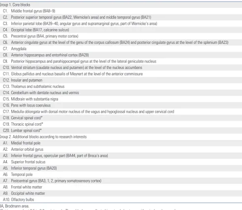
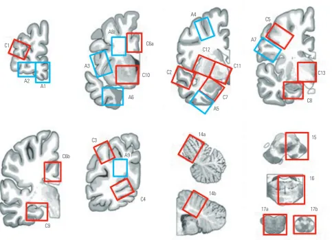
Block preparation following standard section list After sufficient fixation, the left hemisphere should be dissect-ed into 1-cm-thick slabs, and blocks are prepardissect-ed according to the proposed standard set of brain regions (Table 1, Fig. 1). By sharing the standard set of brain blocks, information on stored brains can easily be accessed by researchers through the data network hub. Two groups of block sites are listed in Table 1. Core blocks are crucial for pathologic confirmation of neurodegenerative diseases. Additional blocks are optional and should be based on the academic interests of pathologists or researchers. In these guidelines, we defined core blocks and additional blocks using previously published data. The recent National Institute on Aging-Alzheimer’s Association (NIA-AA) revision of criteria for the pathologic diagnosis of AD proposed a minimum set of 13 histologic sections for evaluation of ma-jor neurodegenerative diseases.5,6 In addition to the minimum set of 13 regions, brain section lists were obtained from the brain donation and neuropathology manual of the Alzheimer’s Disease Neuroimaging Initiative-Neuropathology Core (AD-NI-NPC) at Washington University (kindly provided by Dr. Cairns), the neuropathology section list from the University of California San Francisco (kindly provided by Dr. Seeley), and the neuropathology core manual from Northwestern Memo-rial Hospital (kindly provided by Dr. Bigio).
Consistently identifying the central sulcus from the dissect-ed slabs is important when blocking the motor cortex and sensory cortex. The precentral gyrus should be inked as need-ed. The blocks should be post-fixed for an additional 2–4 days and then processed in an automatic tissue processor. The tis-sue processing program should be adjusted to increase the time in ethanol tanks in order to allow sufficient dehydration of brain tissues.
Gross examination
semi-quantitatively. Regional atrophy should be assessed using a semi-quantitative method involving a four-tier system of none, mild, moderate, and severe. Atrophy of the hippocampus, cau-date, subthalamic nucleus, brainstem, cerebellum, and neo-cortices must be documented. Pallor of the substantia nigra and locus ceoruleus should be recorded. Unbalanced pallor in the substantia nigra and locus ceoruleus can help a conjectural diagnosis; AD is favored with greater pallor in the locus ceoru-leus, and the opposite is seen in frontotemporal lobar degen-eration (FTLD).1 Furthermore, the degree of degeneration in the cerebellar dentate nucleus and color changes of basal ganglia should be recorded.
Microscopic workup using immunohistochemical and special stains
The three most frequently encountered neurodegenerative dis-orders are AD, dementia with Lewy bodies (DLB) or Lewy body
disease (LBD), and FTLD.1 Elements of pathologic diagnosis of neurodegenerative diseases in our guidelines are based on recent guidelines released by the NIA-AA.5 The guidelines sum-marize remarkable advances made since publication of the NIA/Reagan Institute of the AA Consensus Recommendations for the Postmortem Diagnosis of AD or NIA-Reagan Criteria in 1997.7
[image:3.595.59.552.83.509.2]Alzheimer’s disease neuropathologic change (ADNC) should be evaluated with an “ABC” score along with Aβ (β-amyloid) plaque score based on Thal phases, Braak neurofibrillary tan-gle (NFT) stage, and Consortium to Establish a Registry for Al-zheimer’s Disease (CERAD) neuritic plaque score.5,6 Immu-nohistochemistry (IHC)-based analysis of Aβ should be used to assess Thal phases based on progressive Aβ deposition.8 IHC staining for phospho-tau can be used to assess NFTs.8-10 The scope of tau pathology observed in AD includes pretan-gles, neurophil threads in neuronal processes, and dystrophic Table 1. Block Sampling Sites
Group 1. Core blocks
C1. Middle frontal gyrus (BA8–9)
C2. Posterior superior temporal gyrus (BA22, Wernicke’s area) and middle temporal gyrus (BA21) C3. Inferior parietal lobe (BA39–40, angular gyrus and supramarginal gyrus, part of Wernicke‘s area) C4. Occipital lobe (BA17, calcarine sulcus)
C5. Precentral gyrus (BA4, primary motor cortex)
C6. Anterior cingulate gyrus at the level of the genu of the corpus callosum (BA24) and posterior cingulate gyrus at the level of the splenium (BA23) C7. Amygdala
C8. Anterior hippocampus and entorhinal cortex (BA28)
C9. Posterior hippocampus and parahippocampal gyrus at the level of the lateral geniculate nucleus C10. Ventral striatum (caudate nucleus and putamen) at the level of the nucleus accumbens C11. Globus pallidus and nucleus basalis of Meynert at the level of the anterior commissure C12. Insular and putamen
C13. Thalamus and subthalamic nucleus C14. Cerebellum with dentate nucleus and vermis C15. Midbrain with substantia nigra
C16. Pons with locus coeruleus
C17. Medulla oblongata with dorsal motor nucleus of the vagus and hypoglossal nucleus and upper cervical cord C18. Cervical spinal cord*
C19. Thoracic spinal cord* C20. Lumbar spinal cord*
Group 2. Additional blocks according to research interests A1. Medial frontal pole
A2. Anterior orbital gyrus
A3. Inferior frontal gyrus, opercular part (BA44, part of Broca‘s area) A4. Superior frontal sulcus
A5. Inferior temporal gyrus (BA20) A6. Temporal pole
A7. Postcentral gyrus (BA3, 1, 2, primary somatosensory cortex) A8. Frontal white matter
A9. Occipital white matter A10. Olfactory bulbs BA, Brodmann area.
neuritis in neuritic plaques, as well as NFTs in cell bodies.10 Bielschowsky or Gallyas silver or thioflavin-S fluorescent spe-cial staining can be performed for differential detection of neu-ritic plaques from Aβ deposits.11,12 By combining ABC scores, ADNC can be transformed into one of four levels: not, low, in-termediate, or high.6
LBD is a neuropathological term that encompasses the two clinical entities of Parkinson disease and DLB.13 Currently, LBD is classified as follows: brainstem-predominant, limbic (transi-tional), neocortical (diffuse), or amygdala-predominant.5,14 As-sessment of LBD pathology includes identification of Lewy bod-ies on H&E staining, mainly in neurons of the brainstem sections. IHC staining for α-synuclein is the preferred method because of its high sensitivity for revealing Lewy body pathology, includ-ing Lewy neurites and variable neuronal perikaryal inclusions that comprise the continuum of immunoreactive pathology leading to Lewy body formation.13α-synuclein IHC staining should be performed on amygdala and anterior cingulate sec-tions. If α-synuclein pathology is identified in the anterior cin-gulate, additional stains can be performed on neocortical sec-tions to determine the stage of LBD pathology.1,15
To explore FTLD pathology, tau, ubiquitin/p62, and transac-tive response DNA-binding protein 43 (TDP-43) staining of hip-pocampal and neocortex sections should be performed. To
spe-cifically examine FTLD-tau, such as corticobasal degeneration, progressive supranuclear palsy, and Pick disease, tau immunos-taining should be performed on the neocortex, basal ganglia, brainstem, and dentate gyrus to determine the pathognomi-nic inclusions of each entity.1 IHC for 3-repeat and 4-repeat tau can be beneficial in some cases. To diagnose TDP, FTLD-fused in sarcoma (FUS), or FTLD-ubiquitin-proteasome system (UPS), a set of immunostains of corresponding antibodies should be performed on the hippocampus, cerebellum, neocor-tex, and deep nuclei. For diagnostic details of a wider patho-logic spectrum, please refer to previously published articles.16,17 To evaluate amyotrophic lateral sclerosis, additional TDP-43 staining can be performed on spinal cord sections and medulla with hypoglossal and vagus nuclei as well as the motor cortex. Commonly used antibodies and manufacturer information are listed in Table 2. Basic stain sets for screening and an extended set for specific diagnosis are summarized in Table 3.
Neurodegenerative disorders are frequently accompanied by other forms that can contribute to cognitive impairment of the affected individuals.18 Nevertheless, comparative estima-tion for weighing of co-existing pathologic changes in terms of contribution to cognitive impairment is rarely proposed except for AD and LBD.14 It is demanding to gauge the extent to which each disease course might have contributed to cognitive
dys-C1
A2 A1
A3
A6 C10
C6a A8
C5
A7
C8 C13
C6b
C9
C3
A9
C4
14a
14b
15
16
17a 17b C2
A5 C7
C11 C12
[image:4.595.55.539.66.415.2]A4
function.5 Nonetheless, all observed pathologic changes should be described with regard to disorder type and severity.
As a progression of the molecular genetic study in the neu-rodegenerative diseses, DNA and RNA analysis from the au-topsy brain sections should be recommended.19-21
Cerebrovascular diseases that cause vascular brain injury are mainly atherosclerosis, arteriolosclerosis, and cerebral amy-loid angiopathy. Vascular brain injury manifests as hemor-rhages or infarcts. Infarcts are classified according to dimen-sions as territorial infarcts, lacular infarcts, and microinfarcts.5 All infarcts and hemorrhages observed should be document-ed by description of location, size, and chronicity, as a comor-bidity of other degenerative illnesses, or as an isolated lesion.
In conclusion, a nationwide brain bank system is being es-tablished in South Korea. Coordination of brain donation, pro-cessing, storage, and research steps will result in a synergistic chain that might help alleviate the burden of devastating neu-rodegenerative diseases.2 For efficient utilization of resources and to establish a functional and efficient system, we herein proposed tentative standard operating protocols for data
sys-temization. We hope that our proposed guidelines will lead to constructive debate and commentary that will facilitate the de-velopment of an advanced brain bank system in Korea. We an-ticipate that updated protocols and procedures will be devel-oped and shared with the medical community in the future.
ACKNOWLEDGEMENTS
This research was supported by a fund (2016-ER6202-00) by Research of Korea Centers for Disease Control and Prevention.
REFERENCES
1. Bigio EH. Making the diagnosis of frontotemporal lobar degener-ation. Arch Pathol Lab Med 2013;137:314-25.
2. Vonsattel JP, Del Amaya MP, Keller CE. Twenty-first century brain banking. Processing brains for research: the Columbia University methods. Acta Neuropathol 2008;115:509-32.
[image:5.595.54.558.86.248.2]3. Ross BM, Knowler JT, McCulloch J. On the stability of messenger RNA and ribosomal RNA in the brains of control human subjects and patients with Alzheimer’s disease. J Neurochem 1992;58:
Table 2. Antibodies Commonly Used in the Diagnosis of Major Neurodegenerative Disorders
Antibody Clone Manufacturer Catalogue no.
Tau AT8 Thermo Fisher MN1020
Beta amyloid 4G8 Covance SIG-39220
Phospho TDP-43 pS409/410-2 CosmoBio TIP-PTD-P02
Alpha-synuclein phospho S129 Abcam ab59264
p62 (SQSTM1) 2C11 Abnova H00008878-M01
Beta amyloid (1−40) IBL 18580
Beta amyloid (1−42) IBL 18582
3-R Tau (RD3) 8E6/C11 Merck Millipore 05-803
4-R Tau CosmoBio TIP-4RT-P01
FUS polyclonal Sigma-Aldrich HPA008784
Ubiquitin DAKO Z 0458
Dr. Bigio at Northwestern.
Table 3. Special and Immunohistochemical Stain Sets for Screening and an Extended Set for Specific Diagnosis
Site Sliver Aβ Screen TauFTLD-tau Screenα-Syn DLB Ubiquitinp62/ ScreenTDP-43FTLD-TDP/ALS
MFG V V V V V V V
STG V V V
IP V V V
Hippo V V V V V V V
Amyg V V V
Cing V V
Motor V
BG V V
Cbll V V
Stem V V
SC V
Aβ, beta amyloid; FTLD, frontotemporal lobar degeneration; α-Syn, alpha-synuclein; DLB, dementia with Lewy bodies; ALS, amyotrophic lateral sclerosis; MFG, middle frontal gyrus; STG, superior temporal gyrus; IP, inferior parietal lobe; Hippo, hippocampus; Amyg, amygdala; Cing, cingulate gyrus; Motor, motor cortex; BG, basal ganglia; Cbll, cerebellum; Stem, brainstem; SC, spinal cord.
[image:5.595.56.554.273.439.2]1810-9.
4. Bahn S, Augood SJ, Ryan M, Standaert DG, Starkey M, Emson PC. Gene expression profiling in the post-mortem human brain--no cause for dismay. J Chem Neuroanat 2001;22:79-94.
5. Hyman BT, Phelps CH, Beach TG, Bigio EH, Cairns NJ, Carrillo MC, et al. National Institute on Aging-Alzheimer’s Association guidelines for the neuropathologic assessment of Alzheimer’s disease. Alzheimers Dement 2012;8:1-13.
6. Montine TJ, Phelps CH, Beach TG, Bigio EH, Cairns NJ, Dickson DW, et al. National Institute on Aging-Alzheimer’s Association guidelines for the neuropathologic assessment of Alzheimer’s dis-ease: a practical approach. Acta Neuropathol 2012;123:1-11. 7. Hyman BT, Trojanowski JQ. Consensus recommendations for the
postmortem diagnosis of Alzheimer disease from the National In-stitute on Aging and the Reagan InIn-stitute Working Group on diag-nostic criteria for the neuropathological assessment of Alzheimer disease. J Neuropathol Exp Neurol 1997;56:1095-7.
8. Thal DR, Rüb U, Orantes M, Braak H. Phases of A beta-deposition in the human brain and its relevance for the development of AD. Neurology 2002;58:1791-800.
9. Thal DR, Rüb U, Schultz C, Sassin I, Ghebremedhin E, Del Tredici K, et al. Sequence of Abeta-protein deposition in the human me-dial temporal lobe. J Neuropathol Exp Neurol 2000;59:733-48. 10. Braak H, Alafuzoff I, Arzberger T, Kretzschmar H, Del Tredici K.
Staging of Alzheimer disease-associated neurofibrillary patholo-gy using paraffin sections and immunocytochemistry. Acta Neu-ropathol 2006;112:389-404.
11. Braak H, Braak E. Neuropathological stageing of Alzheimer-relat-ed changes. Acta Neuropathol 1991;82:239-59.
12. Mirra SS, Heyman A, McKeel D, Sumi SM, Crain BJ, Brownlee LM, et al. The Consortium to Establish a Registry for Alzheimer’s Dis-ease (CERAD). Part II. Standardization of the neuropathologic as-sessment of Alzheimer’s disease. Neurology 1991;41:479-86. 13. Lowe J, Kalaria R. Dementia. In: Love S, Perry A, Ironside J, Budka
H, editors. Greenfield’s neuropathology. 9th ed. London: CRC Press; 2015. p.858-973.
14. McKeith IG, Dickson DW, Lowe J, Emre M, O’Brien JT, Feldman H, et al. Diagnosis and management of dementia with Lewy bodies: third report of the DLB Consortium. Neurology 2005;65:1863-72. 15. Uchikado H, Lin WL, DeLucia MW, Dickson DW. Alzheimer
dis-ease with amygdala Lewy bodies: a distinct form of alpha-synucle-inopathy. J Neuropathol Exp Neurol 2006;65:685-97.
16. Mackenzie IR, Neumann M, Bigio EH, Cairns NJ, Alafuzoff I, Kril J, et al. Nomenclature and nosology for neuropathologic subtypes of frontotemporal lobar degeneration: an update. Acta Neuropathol 2010;119:1-4.
17. Mackenzie IR, Neumann M, Baborie A, Sampathu DM, Du Plessis D, Jaros E, et al. A harmonized classification system for FTLD-TDP pathology. Acta Neuropathol 2011;122:111-3.
18. Montine TJ, Larson EB. Late-life dementias: does this unyielding global challenge require a broader view? JAMA 2009;302:2593-4. 19. Glasel JA. Validity of nucleic acid purities monitored by 260nm/
280nm absorbance ratios. Biotechniques 1995;18:62-3.
20. Barbas CF 3rd, Burton DR, Scott JK, Silverman GJ. Quantitation of DNA and RNA. CSH Protoc 2007;2007:pdb.ip47.