1
Title: “EP4 as a therapeutic target for aggressive human breast cancer"
Authors: Mousumi Majumder 1, Pinki Nandi2, Ahmed Omar1, Kingsley Chukwunonso Ugwuagbo1 and
Peeyush K. Lala2,3,*
1 Department of Biology, Brandon University, Brandon, Manitoba R7A 6A9, Canada
2 Departments of Anatomy and Cell Biology, and 3 Oncology, Schulich School of Medicine and
Dentistry, University of Western Ontario, London, Ontario N6A5C1, Canada
Author Emails: MM; [email protected]; PN: [email protected]; AO: [email protected]; KU: [email protected]; PKL: [email protected]
*Corresponding author; Tel 1-519-661-3015; email: [email protected]
Abstract
G protein-coupled receptors (GPCRs, also called seven-transmembrane or heptahelical receptors) are a superfamily of cell surface receptor proteins that bind to many extracellular ligands and transmit signals to an intracellular guanine nucleotide-binding protein (G protein). When a ligand binds, the receptor activates the attached G-protein by causing the exchange of Guanosine-5'-triphosphate (GTP) for guanosine diphosphate (GDP). They play a major role in many physiological functions as well as in the pathology of many diseases including cancer progression and metastasis. Only a few GPCR members have been exploited as targets for developing drugs with therapeutic benefit in cancer. Present review deals with the Prostaglandin E receptor EP4, a member of the EP family of GPCR, as a promising newer therapeutic target for treating breast cancer. We show that aberrant over-expression of cyclooxygenase
2 (COX)-2, an inflammation-associated enzyme, occurring in 40-50% of breast cancer patients leads to tumor progression and metastasis due to multiple cellular events resulting from an increased prostaglandin (PG) E2 production in the tumor milieu. They include inactivation of host anti-tumor immune (NK and T) cells, increased immuno-suppressor function of tumor-associated macrophages, promotion of tumor cell migration, invasiveness and tumor-associated angiogenesis (due to upregulation of VEGF-A), lymphangiogenesis (due to upregulation of VEGF-C/D) and a stimulation of stem-like cell (SLC) phenotype in cancer cells. All these events were primarily mediated by activation of the PGE receptor EP4 on tumor or host cells. We show that selective EP4 antagonists (EP4A) could mitigate all these events tested with cells in vitro as well as in vivo in syngeneic COX-2 expressing mammary cancer bearing mice or immune-deficient mice bearing COX-2 over-expressing human breast cancer xenografts. We suggest that EP4A can avoid thrombo-embolic side effects of long term use of COX-2 inhibitors by sparing cardio-protective roles of PGI2 via IP receptor activation or PGE2 via EP3 receptor activation. Furthermore, we identified two COX-2/EP4 induced oncogenic and SLC-stimulating microRNAs - miR526b and miR655, one of which (miR655) appears to be a potential blood biomarker in breast cancer patients, for monitoring SLC-ablative therapies such as with EP4A. We suggest that EP4A will likely produce the highest benefit in aggressive breast cancers such as COX-2 expressing triple-negative breast cancers, when combined with other newer agents such as PD-1 or PD-L1 inhibitors.
Key words: COX-2, Breast Cancer, PGE2, EP receptors, Stem-like cells, Metastasis,
Angiogenesis, Lymphangiogenesis, MicroRNAs, Triple negative breast cancer
3 G protein coupled receptors (GPCRs) are a superfamily of receptors that transduce signals by their coupling with guanine nucleotide-binding proteins (G proteins). They include about 900 members, some with known ligands, others identified as orphan receptors. A diverse set of ligands including peptide hormones, neurotransmitters, and odor molecules bind to GPCRs. They represent the most notable family of validated pharmacological targets in a variety of diseases, including cancer. Numerous GPCRs such as receptors for chemokines, thrombin, lysophosphatidic acid (LPA), gastrin-releasing peptide, angiotensin, the sphingosine 1-phosphate, endothelin and prostaglandins have been reported to play a key role in cancer progression and metastasis [reviewed in 1, 2]. Present article will focus on
prostaglandin E receptor EP4 as a therapeutic target in aggressive breast cancer including triple negative breast cancer.
2. Breast Cancer: Needs to identify novel therapeutic targets.
Breast cancer accounts for the most frequent cancer in the female globally. It represents the second highest cause of cancer-related mortality in the western hemisphere due to resistance of some 25-30% of the patients to currently practiced therapies such as surgery, radiotherapy, chemotherapy, hormone therapy and HER2-trageted drugs, necessitating the search for newer therapy targets. Recent advances in cancer genomics have formed the basis of “Personalized medicine” in identifying therapeutic target(s) appropriate for the individual patient [3]. Genomic profiling of breast cancer by gene micro-array has recently been used to predict therapeutic outcome, that forms the basis for numerous commercially developed assays for use in the clinic [reviewed in 4, 5]. The remarkable advent of current
4 “triple negative breast cancer (TNBC)” reveal an upregulation of the inflammation-associated enzyme cyclooxygenase (COX)-2 which drives tumor progression and metastasis, and that prostaglandin E receptor EP4, a GPCR family member, presents as a promising newer therapeutic target in these patients.
3. Cyclo-oxygenase pathway.
5 only in a small minority of cells such as macrophages and some cells in the reproductive organs.
Typically, it is an inflammation-associated enzyme induced by inflammatory cytokines, mitogens and certain carcinogens. PGE2 production via COX-1 pathway occurs steadily at low local concentrations. In contrast, COX-2-mediated PGE2 production during inflammation occurs at high local concentrations and stops after withdrawal of the inflammatory stimulus. However, aberrant COX-2 activity that occurs in many epithelial cancers including breast cancer leads to persistent PGE2 production [7, 8].
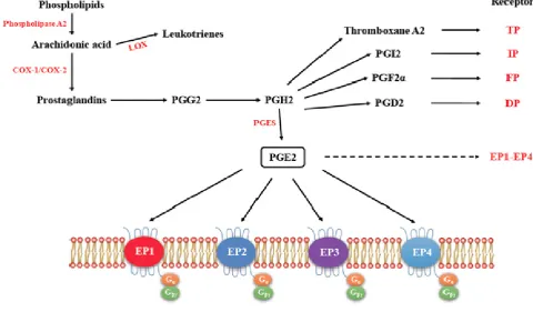
3.1. EP receptors: PGE2-mediated intracellular signaling depend on its binding of target cells to one or
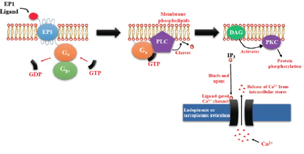
more of the specific prostaglandin E receptors (EP1-4) which are coupled to different G proteins. [9, 10, 11] (Figure 1). The activation or inactivation of G proteins occurs as follows (Figure 2). When a ligand binds, the receptor activates the attached G-protein by causing the exchange of GTP for GDP. The activated G-protein then dissociates into an alpha (G-α) and a beta-gamma (G-β/ϒ) complex. GTP bound G-α is active, and can diffuse along the membrane surface to activate (and sometimes inhibit) target proteins, typically enzymes that generate second messengers. Similarly, the G-β/ϒ complex is also able to diffuse along the inner membrane surface and affect protein activity. Intrinsic GTPase activity is responsible for inactivation of the G-Protein. After GTP hydrolysis, GDP bound G-α will re-associate with a β/ϒ complex to form an inactive G-protein that can again re-associate with a receptor. Signaling mediated by the EP family depends on the coupled G-protein. As shown in Figure 3, EP1 couples with Gq, activating phospholipase C (PLC) that cleaves PIP2, a membrane phospholipid, to generate secondary messengers, IP3 and diacylglycerol (DAG). IP3 is water soluble, diffusing through the cytosol to bind to and open a ligand-gated Ca++ channel in the endoplasmic reticulum (or
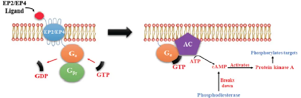
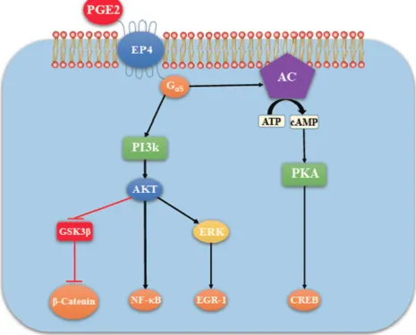
6 couple with GS, which activates the enzyme adenylyl cyclase (AC) and catalyzes the formation of the second messenger cyclic AMP (cAMP) (Figure 4). An activated AC can generate many molecules of cAMP within the cell to amplify the signal. The major effect of cAMP is to bind to and activate protein kinase A (PKA; also known as cAMP-dependent kinase). PKA then phosphorylates target proteins in the cell. cAMP is rapidly broken down by phosphodiesterases, limiting the length of the signal. Additionally, in contrast to EP2, EP4 also stimulates non-canonical pathways phosphatidylinositol 3 kinase (PI3K)/protein kinase B (PKB, also known as Akt) promoting cell survival, and extracellular regulated kinase (ERK), promoting migration and proliferation. The phosphorylation of EP4 receptor recruits β-arrestin-1, which in turn activates c-Src to initiate the transactivation of the epidermal growth factor receptor (EGFR) and subsequent downstream signaling through phosphatidyl inositol 3-kinase (PI3K) and Akt [12] (Figure 5). The activation of this signaling cascade has been proposed to regulate the migration and metastasis of colorectal carcinomas [13]. EP3 receptor mediated signaling (shown in Figure 6) depends on the coupling with several G-protein isoforms. Most are coupled with Gi inhibiting cAMP-PKA; those coupled with Gs stimulate cAMP-PKA; those coupled with G12/13 stimulate Rho family GTPases involved in cytoskeletal changes required for cellular migration. Exploitation of EP receptors as therapeutic targets by the development of selective agonists and antagonists has been elegantly reviewed [12, 13, 14]. Of them, the roles of EP4 receptor in health and disease have received much attention [15]. As documented below, COX-2 expressing breast cancers utilize the EP4 pathway for cancer cell survival and metastasis, making EP4 a targetable molecule for treating aggressive breast cancer patients.
7 Aberrant COX-2 expression promotes tumor initiation, progression and metastasis in most epithelial
cancers [16]. This has been shown by over-expression [17] and down regulation [18] of the COX-2 gene, and protective effects of the use of selective as well as non-selective COX-2 inhibitors from colorectal and mammary carcinogenesis [16, 19-23]. COX-2 overexpression is a phenotype shared by aggressive cancers of the colon [24], lungs [25], pharynx and larynx [26], pancreas [27], and the breast [28]. Elevated COX-2 expression noted in 40-50% of breast cancer, marks poor prognosis [29] resulting from the high levels of PGE2 in the tumor microenvironment.
4.1. Prostaglandin-inhibitors in cancer immunotherapy-- a historical perspective: We demonstrated
8 in activating both NK and T cells in situ and curing a high proportion of mice bearing a variety of
advanced syngeneic metastatic cancers, e.g. melanomas, mammary adenocarcinomas and fibro-sarcomas [34-36]. It was also effective in curing metastatic human melanomas grown in nude mice, which were NK cell competent [37]. Encouraged with these results, we tested this protocol in a single-center phase 2 human trial in advanced kidney cancer and melanoma patients with promising results [38-42]. However, high-dose systemic IL2 therapy soon became unpopular due to IL-2 mediated capillary leak syndrome (CLS), a major side effect leading to rapid fluid accumulation in tissues and serous cavities. We discovered that CLS resulted from IL2-mediated activation of inducible NO synthase (iNOS) leading to high Nitric Oxide (NO) production causing vascular leakage [43]. We showed that therapy with NOS inhibitors could ameliorate IL2 induced CLS in healthy mice [44] as well as in mammary tumor-bearing mice [45-47]. Interestingly, NOS inhibitors had anti-tumor effects on their own as well as in combination with IL2 [46-47]. This is because endogenous NO resulting from aberrant NOS activity in certain tumors including breast cancer, also promoted tumor progression and metastasis by stimulation tumor cell migration, invasiveness and tumor-associated angiogenesis
[48,49,50]. The pro-migratory and pro-invasive role of tumor-derived NO was mediated by an elevation of cyclic GMP followed by activation of the PI3K and ERK pathways. However, a combination of IL2 with NOS inhibitors was never translated to the human, because NO was shown to have both anti-tumor and pro-tumor roles depending on the tumor genotype and NO concentrations in the tumor milieu [51, 52].
4.2. COX-2/PGE2 mediated breast cancer progression: role of EP4 receptor. Elevated COX-2
9 invasiveness [53,55], associated angiogenesis [30] due to upregulation of VEGF-A and tumor-associated lymphangiogenesis [56-59] due to upregulation of VEGF-C and -D. These events were primarily due to activation of the PGE2 receptor EP4 on tumor and host cells, as listed below. EP4 activity on tumor cells promoted tumor cell migration, invasion, angiogenesis and lymphangiogenesis [53, 54, 56-59]. We further observed that in COX-2 expressing breast cancer cells, under inductive conditions, endogenous PGE2 upregulated iNOS by activation of EP4 to promote invasive functions [55]. Others reported that EP4 on host NK cells [60, 61] and T cells [62] blocked their killer functions. We found that EP4 on tumor-associated macrophages [58] promoted their lymphangiogenic function by upregulating VEGF-C or –D. Similarly EP4 activation on host lymphatic endothelial cells (LEC) promoted lymhangiogenesis, resulting from stimulated LEC proliferation, migration and tube formation triggered by upregulation of VEGF-C or –D and VEGFR3 [59]. EP4 activation on host macrophages also promoted their immunosuppressor functions [63]. EP2/EP4 activity on dendritic cells blocked their antigen-presenting function [64]. Finally we discovered that COX-2/EP4 activities also induced and sustained stem-like cell (SLC) phenotype in breast cancer cells in a syngeneic murine breast cancer model [58] and human breast cancer cells [65]. Similar findings have been reported in a different murine breast cancer model [66]. SLCs are a minor subpopulation of cells within tumors, which have an
10 4.3. Rationale for the choice of EP4 as a potential therapeutic target in breast cancer: While intake of COX-2 inhibitors can reduce breast cancer risk and morbidity [71-73], their reported cardiovascular side effects [74, 75] necessitated the search for alternative downstream target(s) that may spare
11 > 800 human arthritis patients was well tolerated in pharmacologically effective doses (300 mg orally twice daily) with no evidence of dose-limiting toxicity (Dr Yukinori Take, Ask/At, Japan, personal communication).
4.4. Functional roles of COX-2 in the absence or presence of HER-2 in breast cancer: Human
epidermal growth factor receptor (HER) 2 expressed by approximately 20 % breast cancer patients is another major driver of breast cancer progression. HER-2 is often co-expressed with COX-2 in human breast cancer [81], although the reverse in not true. Interestingly, most HER-2 actions e.g., upregulation of aromatase [82], angiogenesis [83], lymphangiogenesis [81] and anti-apoptotic action [84] were shown to be intermediated by COX-2. To define the functional roles of COX-2 in the absence or presence of HER-2, we stably transfected COX-2 gene into MCF-7 (COX-2-, 2-) and SKBR-3 (COX-2-, HER-2-high) human breast cancer cell lines [63]. Ectopic COX-2 over-expression in MCF-7 and SKBR-3 cell lines resulted in: increased migration/invasion/proliferation, epithelial-mesenchymal transition (EMT), elevated SLCs (spheroid formatting ability in vitro), increased ALDH activity- a recognized SLC marker [85] and co-localization of COX-2 with numerous SLC markers (ALDH1A, CD44, β-Catenin, NANOG, OCT3/4 and SOX-2) in spheroids. These changes were reversed with COX-2-inhibitor or
12 MCF-7-COX-2 cells (as compared to control cells) showed up-regulation of angiogenic
/lymphangiogenic factors VEGF-A/C/D, Vimentin (mesenchymal marker) and phospho-AKT (an EP4 signaling marker), down-regulation of epithelial marker E-Cadherin and an enrichment of SLC marker positive and spheroid forming cells. Findings in primary human breast cancer tissues were supportive of the findings in mice as noted above. Expression of COX-2, EP4 and ALDH1A mRNA in these tissues were highly correlated with one other, more marked in progressive stage of disease. In situ
immunostaining of the tissues revealed co-localization of SLC markers with COX-2, supporting SLC induction by COX-2. Finally, high COX-2/EP4 mRNA expression was linked with reduced survival. These preclinical and clinical data strongly suggest that EP4 represents a novel therapeutic target to inhibit tumor growth, metastasis and eradicate SLCs in human breast cancer [63]. This contention was fully validated by us in mouse breast cancer models [57, 58] with two EP4 antagonists (ONO-AE3-208, ONO pharma, Japan; and RQ-15986, currently renamed as AAT 007, Ask/At Pharma, Japan). Treating mice bearing syngeneic COX-2 expressing, highly metastatic C3L5 mammary tumors with EP4A at non- toxic doses inhibited tumor growth, spontaneous metastasis and eradicated SLCs in residual tumors [57, 58]. This finding has been duplicated in another murine breast cancer model [66] with AAT 007. Similarly therapeutic efficacy of ONO-AE3-208 was reported in a castration-resistant prostate cancer model [87].
4.5. SLC-linked microRNAs induced by COX-2/EP4 activity as breast cancer biomarkers: There are
very few reliable blood biomarkers for breast cancer that are useful to monitor the disease. Levels of specific miRNAs in blood plasma remain as a newer family of cancer biomarkers. miRNAs are single stranded non-coding RNAs (20-24 nucleotides) that down-regulate specific genes at the
13 and transit in the blood. Recently, levels of a panel of 7 candidate miRNAs were measured in tissue and blood specimens of 148 patients with minimally invasive breast cancer and 44 age-matched and disease free control individuals [92]. The authors found increased levels of blood miR-195 in breast cancer patients, which decreased to control levels following curative tumor resection. The circulating miRNAs correlated with certain clinic-pathological variables, namely nodal status and estrogen receptor status. We conducted differential gene and miRNA expression micro arrays using control
MCF-7-Mock-tranfected vs. MCF-7-COX-2 transfected cell lines, which identified downregulation of six miRNAs and an upregulation of two miRNAs (miR-655 and miR-526b)by COX-2. Both COX-2 upregulated
14 MiR-655 expression also led to TGFβ resistance for Smad3 phosphorylation [94]. Tail vein injection of ectopic miR-526b or miR-655 over-expressing MCF7 and SKBR3 cells into NOD/SCID/GUSB-null mice revealed increased lung colony growth and micro-metastases to other organs. Expression of both miRNAs was strongly correlated with each other in human breast cancer tissues, was higher than in non-tumor tissues, and associated with reduced patient survival [93, 94]. Thus they could serve as prognostic breast cancer biomarkers for monitoring SLC-reduction during therapies. In support, our preliminary data reveal that miR-655levels are significantly higher in the plasma of breast cancerbearing than in patients withbenign tumors (unpublished; manuscript in preparation). In summary, we found that aberrant COX-2 activity in human breast cancer leads to tumor progression and metastasis by utilizing multiple signaling pathways in which EP4 activation plays a pivotal role, and two COX-2/EP4
upregulated miRNAs are important partners (schema presented in Figure 8).
4.6. Triple negative breast cancers (TNBC) are mostly COX2 expressing: TNBC represents the most
deadly type of breast cancer, which resist cytotoxic therapies. In an earlier study [81], designed to identify the roles of COX-2 and HER2 in VEGF-C expression and lymphangiogenesis, we used 65 human breast cancer tissue samples and multiple human breast cancer cell line genetically manipulated for COX-2 and HER2 expression. We concluded that COX-2 was a primary driver of
15 patients. Our future goal is to use EP4A as an adjunct in metastatic TNBC patients. However it is
currently unknown whether EP4A as a single agent will provide any benefit, as observed in our syngeneic murine breast cancer models[57, 58]. We suggest that a combination therapy with other agents such as immune checkpoint inhibitors holds a greater promise.
4.7. Proposed combination of an EP4 antagonist with an immune checkpoint inhibitor for treating
TNBC. Programmed cell death (PD)-1 is a checkpoint protein on T cells that normally acts as an “off
16 inactivation of host antitumor immunity was shown to be due to EP4 binding on multiple immune cell classes: NK cells, T cells, macrophages, and dendritic cells. EP4 antagonists were shown to be highly effective in abrogating all these events in animal models leading to tumor cell killing [57, 58, 60]. As outlined earlier, immune check point inhibitors work via different non-overlapping mechanisms. Thus it is expected that a combination of the two should cast the net far wider to block multiple tumor and host cell mediated pathways, leading to a synergistic action. Indeed a synergistic action on tumor regression and animal survival was shown with an EP4 antagonist in combination of either of two checkpoint inhibitors, anti-CTLA4 and anti-PD-1 antibodies, in murine colon and breast cancer models [101]. 4.8. EP4 antagonist in the breast cancer clinic. Recently a phase 2 human trial with the EP4 antagonist AAT 007 (AskAt, Japan) was registered by Dr Martin Edleman at the University of Maryland (currently moved to the FOX Chase Cancer Centre) in advanced solid tumors including prostate, breast or non-small cell lung cancer (Clinical Trials.gov Identifier: NCT02538432, last update posted on June 6, 2017). The trial will test (a) whether the administration of the study drug AAT 007 can decrease circulating tumor cells or myeloid-derived suppressor cells; and (b) whether the drug may improve outcome on its own in these solid tumors or when combined with a cytotoxic drug gemcitabine in patients with prostate or lung cancer, if the disease worsened with AAT 007 alone. No patient registration or outcome has yet been reported.
Acknowledgment:
17 (NSERC) to PKL and NSERC and Brandon University Research Committee (BURC) new faculty grants to MM.
Author contributions:
MM and PN: Performed some of the cited studies and wrote parts of the Review; AO and KU :
Prepared Figures; PKL: Wrote and finalized the Review.
Conflict of interest statement:
The authors declare no conflict of interest.
References:
1. Lappano, R.; Maggiolini, M., G protein-coupled receptors: novel targets for drug discovery in cancer. Nat Rev Drug Discov 2011, 10 (1), 47-60.
2. Liu, Y.; An, S.; Ward, R.; Yang, Y.; Guo, X. X.; Li, W.; Xu, T. R., G protein-coupled receptors as promising cancer targets. Cancer Lett 2016, 376 (2), 226-39.
3. Cho, S.-H.; Jeon, J.; Kim, S. I., Personalized Medicine in Breast Cancer: A Systematic Review. J Breast Cancer. 2012, 15 (3), 265–272.
4. Fan, C.; Oh, D. S.; Wessels, L.; Weigelt, B.; Nuyten, D. S.; Nobel, A. B.; van't Veer, L. J.; Perou, C. M., Concordance among gene-expression-based predictors for breast cancer. N Engl J Med 2006, 355 (6), 560-9.
5. Dai, X.; Li, T.; Bai, Z.; Yang, Y.; Liu, X.; Zhan, J.; Shi, B., Breast cancer intrinsic subtype classification, clinical use and future trends. Am J Cancer Res. 2015, 5 (10), 2929–2943. 6. Simmons, D. L.; Botting, R. M.; Hla, T., Cyclooxygenase isozymes: the biology of
prostaglandin synthesis and inhibition. Pharmacol Rev 2004, 56 (3), 387-437.
18 8. Greenhough, A.; Smartt, H. J.; Moore, A. E.; Roberts, H. R.; Williams, A. C.; Paraskeva,
C.; Kaidi, A., The COX-2/PGE2 pathway: key roles in the hallmarks of cancer and adaptation to the tumour microenvironment. Carcinogenesis 2009, 30 (3), 377-86. 9. Breyer, R. M.; Bagdassarian, C. K.; Myers, S. A.; Breyer, M. D., Prostanoid receptors:
subtypes and signaling. Annu Rev Pharmacol Toxicol 2001, 41, 661-90.
10. Fujino, H.; Xu, W.; Regan, J. W., Prostaglandin E2 Induced Functional Expression of Early Growth Response Factor-1 by EP4, but Not EP2, Prostanoid Receptors via the Phosphatidylinositol 3-Kinase and Extracellular Signal-regulated Kinases. The Journal of Biological Chemistry 2003, 278, 12151-12156.
11. Sugimoto, Y.; Narumiya, S., Prostaglandin E Receptors. The Journal of Biological Chemistry 2007, 282, 11613-11617.
12. Prostaglandin E2 and the EP receptors in malignancy: possible therapeutic targets? Br J Pharmacol. 2015, 172 (22), 5239–5250.
13. Buchanan, F. G.; Gorden, D. L.; Matta, P.; Shi, Q.; Matrisian, L. M.; DuBois, R. N., Role of β-arrestin 1 in the metastatic progression of colorectal cancer. PNAS 2006,103 (5), 1492-1497.
14. Markovič, T.; Jakopin, Ž.; Dolenc, M. S.; Mlinarič-Raščan, I., Structural features of subtype-selective EP receptor modulators. Drug Discovery Today 2017, 22 (1), 57-71. 15. Konya, V.; Marsche, G.; Schuligoi, R.; Heinemann, A., E-type prostanoid receptor 4
(EP4) in disease and therapy. Pharmacol Ther. 2013, 138 (3), 485–502.
16. Harris, R., COX-2 Blockade in Cancer Prevention and Therapy. 1 ed.; Humana Press: New York, 2003; p X -371.
17. Liu, C. H.; Chang, S. H.; Narko, K.; Trifan, O. C.; Wu, M. T.; Smith, E.; Haudenschild, C.; Lane, T. F.; Hla, T., Overexpression of cyclooxygenase-2 is sufficient to induce tumorigenesis in transgenic mice. J Biol Chem 2001, 276 (21), 18563-9.
19 19. Gupta, A., Aberrant crypt foci: are they intermediate endpoints of colon carcinogenesis in
humans? Curr Opin Gastroenterol 2009, 25 (1), 59-65.
20. Howe, L. R.; Dannenberg, A. J., COX-2 inhibitors for the prevention of breast cancer. J
Mammary Gland Biol Neoplasia 2003,8 (1), 31-43.
21. Sharpe, C. R.; Collet, J. P.; McNutt, M.; Belzile, E.; Boivin, J. F.; Hanley, J. A., Nested case-control study of the effects of non-steroidal anti-inflammatory drugs on breast cancer risk and stage. Br J Cancer 2000,83 (1), 112-20.
22. Harris, R. E.; Chlebowski, R. T.; Jackson, R. D.; Frid, D. J.; Ascenseo, J. L.; Anderson, G.; Loar, A.; Rodabough, R. J.; White, E.; McTiernan, A.; Women's Health, I., Breast cancer and nonsteroidal anti-inflammatory drugs: prospective results from the Women's Health Initiative. Cancer Res 2003,63 (18), 6096-101.
23. Oshima, M.; Dinchuk, J. E.; Kargman, S. L.; Oshima, H.; Hancock, B.; Kwong, E.; Trzaskos, J. M.; Evans, J. F.; Taketo, M. M., Suppression of Intestinal Polyposis in ApcΔ716 Knockout Mice by Inhibition of Cyclooxygenase 2 (COX-2). Cell 1996,87 (5), 803-809.
24. Tsujii, M.; Kawano, S.; DuBois, R. N., Cyclooxygenase-2 expression in human colon cancer cells increases metastatic potential. Proc Natl Acad Sci U S A 1997,94 (7), 3336-40.
25. Hida, T.; Yatabe, Y.; Achiwa, H.; Muramatsu, H.; Kozaki, K.-i.; Nakamura, S.; Ogawa, M.; Mitsudomi, T.; Sugiura, T.; Takahashi, T., Increased Expression of Cyclooxygenase 2 Occurs Frequently in Human Lung Cancers, Specifically in Adenocarcinomas. Cancer Res 1998,58 (17), 3761-3764.
26. Chan, G.; Boyle, J. O.; Yang, E. K.; Zhang, F.; Sacks, P. G.; Shah, J. P.; Edelstein, D.; Soslow, R. A.; Koki, A. T.; Woerner, B. M.; Masferrer, J. L.; Dannenberg, A. J.,
Cyclooxygenase-2 Expression Is Up-Regulated in Squamous Cell Carcinoma of the Head and Neck. Cancer Res 1999,59 (5), 991-994.
20 28. Parrett, M.; Harris, R.; Joarder, F.; Ross, M.; Clausen, K.; Robertson, F.,
Cyclooxygenase-2 gene expression in human breast cancer. 1997,10 (3), 503-507.
29. Ristimäki, A.; Sivula, A.; Lundin, J.; Lundin, M.; Salminen, T.; Haglund, C.; Joensuu, H.; Isola, J., Prognostic Significance of Elevated Cyclooxygenase-2 Expression in Breast Cancer. Cancer Res 2002,62 (3), 632-635.
30. Parhar, R. S.; Lala, P. K., Changes in the host natural killer cell population in mice during tumor development: 2. The mechanism of suppression of NK activity. Cellular
Immunology 1985,93 (2), 265-279.
31. P.K.Lala; R.S.Parhar; P.Singh, Indomethacin therapy abrogates the prostaglandin-mediated suppression of natural killer activity in tumor-bearing mice and prevents tumor metastasis. Cellular Immunology 1986,99 (1), 108-118.
32. Lala, P. K.; Al-Mutter, N.; Orucevic, A., Effects of chronic indomethacin therapy on the development and progression of spontaneous mammary tumors in C3H/HEJ mice.
International Journal of Cancer 1997,73 (3), 371-380.
33. Rosenberg, S. A.; Lotze, M. T.; Yang, J. C.; Aebersold, P. M.; Linehan, W. M.; Seipp, C. A.; White, D. E., Experience with the use of high-dose interleukin-2 in the treatment of 652 cancer patients. Annal of Surgery 1989,210 (4), 474-484.
34. Parhar, R. S.; Lala, P. K., Amelioration of B16F10 melanoma lung metastasis in mice by a combined therapy with indomethacin and IL 2. J. Exp. Med. 1987,165, 14-28.
35. Lala, P. K.; Parhar, R. S., Cure of B16F10 melanoma lung metastasis in mice by chronic indomethacin therapy combined with repeated rounds of interleukin 2: characteristics of killer cells generated in situ. Cancer Res 1988,48 (5), 1072-9.
36. Lala, P. K., PGE2-mediated inactivation of potentially tumoricidal effector cells of the host during tumor development: relevance to metastasis and immunotherapy. In
Carcinogenesis and Dietary Fat Abraham, S., Ed. Kluwer Academic Publishers: Bodton /
Dordrecht / London, 1989; pp 219-232.
21 38. Mertens, W. C.; Bramwell, V. H. C.; Gwadry-Sridhar, F.; Romano, W.; Banerjee, D.;
Lala, P. K., Effect of indomethacin plus ranitidine in advanced melanoma patients on high-dose interleukin-2. The Lancet 1992,340 (8816), 397-398.
39. Mertens, W. C.; Bramwell, V. H. C.; Lala, P. K.; Banerjee, D.; Gwadry-Sridhar, F.; Romano, W., Continuous indomethacin and ranitidine with interleukin-2 in advanced renal carcinoma and melanoma. Can J Infect Dis 1992,3 (Suppl. B), 133B-137B. 40. Mertens, W. C.; Bramwell, V. H. C.; Banerjee, D.; Gwadry-Sridhar, F.; Al-Mutter, N.;
Parhar, R. S.; Lala., P. K., Sustained Oral Indomethacin and Ranitidine with Intermittent Continuous Infusion Interleukin-2 in Advanced Renal Cell Carcinoma. Cancer Biotherapy
2009,8 (3), 229-233.
41. W.C.Mertens; V.H.C.Bramwell; D.Banerjee; F.Gwadry-Sridhar; P.K.Lala, Sustained indomethacin and ranitidine with intermittent continuous infusion interleukin-2 in
advanced malignant melanoma: A phase II study. Clinical Oncology 1993,5 (2), 07-113. 42. Mertens, W. C.; Banerjee, D.; Al-Mutter, N.; Stitt, L.; Bramwell, V. H. C.; Lala, P. K.,
High-dose continuous venous infusion of interleukin-2: Influence of dose and infusion rate on tumoricidal function and lymphocyte subsets. Cancer Immunology, Immunotherapy
1995,41 (5), 271 - 279.
43. Orucevic, A.; Hearn, S.; Lala, P., The role of active inducible nitric oxide synthase expression in the pathogenesis of capillary leak syndrome resulting from interleukin-2 therapy in mice. Laboratory Investigation 1997,76 (1), 53-65.
44. Orucevic, A.; Lala, P., NG-nitro-L-arginine methyl ester, an inhibitor of nitric oxide synthesis, ameliorates interleukin-2-induced capillary leak syndrome in healthy mice. J
Immunother Emphasis Tumor Immunol 1995,18 (4), 210-220.
45. Orucevic, A.; Lala, P. K., NG-nitro-L-arginine methyl ester, an inhibitor of nitric oxide synthesis, ameliorates interleukin 2-induced capillary leakage and reduces tumour growth in adenocarcinoma-bearing mice. Br J Cancer. 1996,73 (2), 189–196.
22 47. Orucevic, A.; Lala, P. K., Effects of NG-methyl- L -arginine, an inhibitor of nitric oxide
synthesis, on interleukin-2-induced capillary leakage and antitumor responses in healthy and tumor-bearing mice. Cancer Immunology, Immunotherapy 1996,42 (1), 38-46. 48. Orucevic, A.; Bechberger, J.; Green, A. M.; Shapiro, R. A.; Billiar, T. R.; Lala, P. K.,
Nitric-oxide production by murine mammary adenocarcinoma cells promotes tumor-cell invasiveness. International Journal of Cancer 1999,81 (6), 889-896.
49. Lorraine C. Jadeski; Kathleen O. Hum; Chandan Chakraborty; Lala, P. K., Nitric oxide promotes murine mammary tumour growth and metastasis by stimulating tumour cell migration, invasiveness and angiogenesis. 2000,86 (1), 30–39.
50. Lorraine C. Jadeski; Chandan Chakraborty; Lala, P. K., Nitric oxide-mediated promotion of mammary tumour cell migration requires sequential activation of nitric oxide synthase, guanylate cyclase and mitogen-activated protein kinase. nt. J. Cancer 2003,106 (4), 496– 504.
51. Lala, P. K.; Chakraborty, C., Role of nitric oxide in carcinogenesis and tumour progression. Lancet Oncol 2001,2 (3), 149-56.
52. Vannini, F.; Kashfi, K.; NiharikaNath, The dual role of iNOS in cancer Redox Biology
2015,6, 334-343.
53. Jerry G. Rozic; Chandan Chakraborty; Lala, P. K., Cyclooxygenase inhibitors retard murine mammary tumor progression by reducing tumor cell migration, invasiveness and angiogenesis. Int. J. Cancer 2001,93 (4), 497–506.
54. Timoshenkoa, A. V.; Xu, G.; Chakrabarti, S.; Lala, P. K.; Chakraborty, C., Role of
prostaglandin E2 receptors in migration of murine and human breast cancer cells. Exp Cell
Res. 2003,289 (2), 265-74.
55. Timoshenko, A. V.; Lala, P. K.; Chakraborty, C., PGE2-mediated upregulation of iNOS in murine breast cancer cells through the activation of EP4 receptors. Int J Cancer 2004,108 (3), 384-9.
56. Timoshenko, A. V.; Chakraborty, C.; Wagner, G. F.; Lala, P. K., COX-2-mediated stimulation of the lymphangiogenic factor VEGF-C in human breast cancer. British
23 57. Xin, X.; Majumder, M.; Girish, G. V.; Mohindra, V.; Maruyama, T.; Lala, P. K.,
Targeting COX-2 and EP4 to control tumor growth, angiogenesis, lymphangiogenesis and metastasis to the lungs and lymph nodes in a breast cancer model. Lab Invest 2012,92 (8), 1115-28.
58. Majumder, M.; Xin, X.; Liu, L.; Girish, G. V.; Lala, P. K., Prostaglandin E2 receptor EP4 as the common target on cancer cells and macrophages to abolish angiogenesis,
lymphangiogenesis, metastasis, and stem-like cell functions. Cancer Sci 2014,105 (9), 1142-51.
59. Nandi, P.; Girish, G. V.; Majumder, M.; Xin, X.; Tutunea-Fatan, E.; Lala, P. K., PGE2 promotes breast cancer-associated lymphangiogenesis by activation of EP4 receptor on lymphatic endothelial cells. BMC Cancer. 2017,17 (11).
60. Holt, D.; Ma, X.; Kundu, N.; Fulton, A., Prostaglandin E2 (PGE2) suppresses Natural Killer cell function primarily through the PGE2 receptor EP4. Cancer Immunol
Immunother. 2011,60 (11), 1577–1586.
61. Ma, X.; Holt, D.; Kundu, N.; Reader, J.; Goloubeva, O.; Take, Y.; Fulton, A. M., A prostaglandin E (PGE) receptor EP4 antagonist protects natural killer cells from PGE2-mediated immunosuppression and inhibits breast cancer metastasis. Oncoimmunology.
2013,2 (1), e22647.
62. Okano, M.; Sugata, Y.; Fujiwara, T.; Matsumoto, R.; Nishibori, M.; Shimizu, K.; Maeda, M.; Kimura, Y.; Kariya, S.; Hattori, H.; Yokoyama, M.; Kino, K.; Nishizaki, K., E prostanoid 2 (EP2)/EP4-mediated suppression of antigen-specific human T-cell responses by prostaglandin E2. Immunology 2006,118 (3), 343-52.
63. Albu, D. I.; Wang, Z.; Wu, J.; Huang, K.-c.; Li, W.; Liu, D.; Kuznetsov, G.; Chen, Q.; Bao, X.; Woodall-Jappe, M., Abstract 275: ER-886046, an antagonist of PGE2 receptor type-4, induces an effective antitumor immune response in mice by attenuating
intratumoral MDSCs and TAMs. In AACR 106th Annual Meeting, Cantley, L. C., Ed. Pennsylvania Convention Center Philadelphia, Pennsylvania, 2015; Vol. 75.
24 65. Majumder, M.; Xin, X.; Liu, L.; Tutunea-Fatan, E.; Rodriguez-Torres, M.; Vincent, K.;
Postovit, L. M.; Hess, D.; Lala, P. K., COX-2 Induces Breast Cancer Stem Cells via EP4/PI3K/AKT/NOTCH/WNT Axis. Stem Cells 2016,34 (9), 2290-305.
66. Kundu, N.; Ma, X.; Kochel, T.; Goloubeva, O.; Staats, P.; Thompson, K.; Martin, S.; Reader, J.; Take, Y.; Collin, P.; Fulton, A., Prostaglandin E receptor EP4 is a therapeutic target in breast cancer cells with stem-like properties. Breast Cancer Res Treat 2014,143 (1), 19-31.
67. Wicha, M. S.; Liu, S.; Dontu, G., Cancer stem cells: an old idea--a paradigm shift. Cancer Res 2006,66 (4), 1883-90; discussion 1895-6.
68. Tysnes, B. B., Tumor-Initiating and -Propagating Cells: Cells That We Would Like to Identify and Contro. Neoplasia. 2010,12 (7), 506–515.
69. Li, X.; Lewis, M. T.; Huang, J.; Gutierrez, C.; Osborne, C. K.; Wu, M. F.; Hilsenbeck, S. G.; Pavlick, A.; Zhang, X.; Chamness, G. C.; Wong, H.; Rosen, J.; Chang, J. C., Intrinsic resistance of tumorigenic breast cancer cells to chemotherapy. J Natl Cancer Inst 2008,
100 (9), 672-9.
70. Visvader, J. E.; Lindeman, G. J., Cancer stem cells: current status and evolving complexities. Cell Stem Cell 2012,10 (6), 717-28.
71. Howe, L. R.; Dannenberg, A. J., COX-2 inhibitors for the prevention of breast cancer. J
Mammary Gland Biol Neoplasia. 2003,8 (1), 31-43.
72. Sharpe, C. R.; Collet, J.-P.; McNutt, M.; Belzile, E.; Boivin, J.-F.; Hanley, J. A., Nested case–control study of the effects of non-steroidal anti-inflammatory drugs on breast cancer risk and stage. Br J Cancer. 2000,83 (1), 112–120.
73. Harris, R. E.; Chlebowski, R. T.; Jackson, R. D.; Frid, D. J.; Ascenseo, J. L.; Anderson, G.; Loar, A.; Rodabough, R. J.; White, E.; McTiernan, A., Breast cancer and nonsteroidal anti-inflammatory drugs: prospective results from the Women's Health Initiative. Cancer
Res. 2003,63 (18), 6096-101.
74. Garret A. FitzGerald, Coxibs and Cardiovascular Disease. N Engl J Med. 2004,351 (17), 1709-11.
25 76. Cathcart, M. C.; Tamosiuniene, R.; Chen, G.; Neilan, T. G.; Bradford, A.; O'Byrne, K. J.;
Fitzgerald, D. J.; Pidgeon, G. P., Cyclooxygenase-2-linked attenuation of hypoxia-induced pulmonary hypertension and intravascular thrombosis. J Pharmacol Exp Ther 2008,326 (1), 51-8.
77. Xiao, C. Y.; Hara, A.; Yuhki, K. i.; Fujino, T.; Ma, H.; Okada, Y.; Takahata, O.; Yamada, T.; Murata, T.; Narumiya, S.; Ushikubi, F., Roles of Prostaglandin I2 and Thromboxane A2 in Cardiac Ischemia-Reperfusion Injury: A Study Using Mice Lacking Their
Respective Receptors. Circulation 2001,104 (18), 2210-2215.
78. Martin, M.; Meyer-Kirchrath, J.; Kaber, G.; Jacoby, C.; Flogel, U.; Schrader, J.; Ruther, U.; Schror, K.; Hohlfeld, T., Cardiospecific overexpression of the prostaglandin EP3 receptor attenuates ischemia-induced myocardial injury. Circulation 2005,112 (3), 400-6. 79. Thiemermann, C.; Zacharowski, K., Selective activation of E-type prostanoid3-receptors
reduces myocardial infarct size. Pharmacology & Therapeutics 2000,87 (1), 61-67. 80. Hishikari, K.; Suzuki, J.; Ogawa, M.; Isobe, K.; Takahashi, T.; Onishi, M.; Takayama, K.;
Isobe, M., Pharmacological activation of the prostaglandin E2 receptor EP4 improves cardiac function after myocardial ischaemia/reperfusion injury. Cardiovasc Res 2009,81 (1), 123-32.
81. Bhattacharjee, R. N.; Timoshenko, A. V.; Cai, J.; Lala, P. K., Relationship between cyclooxygenase-2 and human epidermal growth factor receptor 2 in vascular endothelial growth factor C up-regulation and lymphangiogenesis in human breast cancer. Cancer Sci
2010,101 (9), 2026-32.
82. Subbaramaiah, K.; Howe, L. R.; Port, E. R.; Brogi, E.; Fishman, J.; Liu, C. H.; Hla, T.; Hudis, C.; Dannenberg, A. J., HER-2/neu status is a determinant of mammary aromatase activity in vivo: evidence for a cyclooxygenase-2-dependent mechanism. Cancer Res
2006,66 (10), 5504-11.
83. Howe, L. R.; Chang, S. H.; Tolle, K. C.; Dillon, R.; Young, L. J.; Cardiff, R. D.; Newman, R. A.; Yang, P.; Thaler, H. T.; Muller, W. J.; Hudis, C.; Brown, A. M.; Hla, T.;
26 84. Simeone, A.-M.; Li, Y.-J.; Broemeling, L. D.; Johnson, M. M.; Tuna, M.; Tari, A. M.,
Cyclooxygenase-2 is essential for HER2/neu to suppress N- (4-hydroxyphenyl)retinamide apoptotic effects in breast cancer cells. Cancer Res. 2004,64 (4), 1224-8.
85. Croker, A. K.; Goodale, D.; Chu, J.; Postenka, C.; Hedley, B. D.; Hess, D. A.; Allan, A. L., High aldehyde dehydrogenase and expression of cancer stem cell markers selects for breast cancer cells with enhanced malignant and metastatic ability. Journal of Cellular
and Molecular Medicine 13 (8b), 2236–2252.
86. Al-Hajj, M.; Wicha, M. S.; Benito-Hernandez, A.; Morrison, S. J.; Clarke, M. F., Prospective identification of tumorigenic breast cancer cells. PNAS 2003,100 (7), 3983-3988.
87. Xu, S.; Zhang, Z.; Ogawa, O.; Yoshikawa, T.; Sakamoto, H.; Shibasaki, N.; Goto, T.; Wang, L.; Terada, N., An EP4 antagonist ONO-AE3-208 suppresses cell invasion, migration, and metastasis of prostate cancer. Cell Biochem Biophys 2014,70 (1), 521-7. 88. <http://www.mirbase.org/cgi-bin/mirna_summary.pl?org=hsa>.??
89. Calin, G. A.; Croce, C. M., MicroRNA signatures in human cancers. Nat Rev Cancer
2006,6 (11), 857-66.
90. Esquela-Kerscher, A.; Slack, F. J., Oncomirs - microRNAs with a role in cancer. Nat Rev
Cancer 2006,6 (4), 259-69.
91. Kosaka, N.; Iguchi, H.; Ochiya, T., Circulating microRNA in body fluid: a new potential biomarker for cancer diagnosis and prognosis. Cancer Sci 2010,101 (10), 2087-92. 92. Heneghan, H. M.; Miller, N.; Lowery, A. J.; Sweeney, K. J.; Newell, J.; Kerin, M. J.,
Circulating microRNAs as novel minimally invasive biomarkers for breast cancer. Ann
Surg 2010,251 (3), 499-505.
93. Majumder, M.; Landman, E.; Liu, L.; Hess, D.; Lala, P. K., COX-2 Elevates Oncogenic miR-526b in Breast Cancer by EP4 Activation. Mol Cancer Res 2015,13 (6), 1022-33. 94. Majumder, M.; Dunn, L.; Liu, L.; Hasan, A.; Vincent, K.; Brackstone, M.; Hess, D.; Lala,
P. K., COX-2 induces oncogenic micro RNA miR655 in human breast cancer. Sci Rep
27 95. Mosalpuria, K.; Hall, C.; Krishnamurthy, S.; Lodhi, A.; Hallman, D. M.; Baraniuk, M. S.;
Bhattacharyya, A.; Lucci, A., Cyclooxygenase-2 expression in non-metastatic triple-negative breast cancer patients. Mol Clin Oncol 2014,2 (5), 845-850.
96. Zou, W.; Wolchok, J. D.; Chen, L., PD-L1 (B7-H1) and PD-1 pathway blockade for cancer therapy: Mechanisms, response biomarkers, and combinations. Sci Transl Med
2016,8 (328), 328rv4.
97. He, J.; Hu, Y.; Hu, M.; Li, B., Development of PD-1/PD-L1 Pathway in Tumor Immune Microenvironment and Treatment for Non-Small Cell Lung Cancer. Sci Rep 2015,5, 13110.
98. Lipson, E. J.; Forde, P. M.; Hammers, H.-J.; Emens, L. A.; Taube, J. M.; Suzanne L. Topalian, Antagonists of PD-1 and PD-L1 in Cancer Treatment. Semin Oncol. 2015,42 (2), 587–600.
99. Gangadhar, T. C.; Salama, A. K., Clinical applications of PD-1-based therapy: a focus on pembrolizumab (MK-3475) in the management of melanoma and other tumor types. 2015,
8, 929—937.
100. Bertucci, F.; Gonçalves, A., Immunotherapy in Breast Cancer: the Emerging Role of PD-1 and PD-LPD-1. Curr Oncol Rep. 2017,19 (10), 64.
101. Bao, X.; Albu, D.; Huang, K.-C.; Wu, J.; Twine, N.; Nomoto, K.; Woodall-Jappe, M., Combination of EP4 antagonist and checkpoint inhibitors promotes anti-tumor effector T cells in preclinical tumor models. Journal for ImmunoTherapy of Cancer 2015,3 (Suppl 2), P350.
Figure legends:
Figure 1. The pathway for the synthesis of prostaglandins, their respective receptors and signaling. (Adapted from Markovicˇ T et al 2017; ref 14). Arachidonic acid acts as the substrate for COX-1 and COX-2 to produce Prostagandins PGE2, Thromboxane A2, PGI2, PGF2α and PGD2, all of which exert functions by binding to their respective receptors.
28 a β/ϒ complex. GTP bound Gα is active. Intrinsic GTPase activity leads to inactivation of the G-Protein. GDP bound Gα re-associates with a β/ϒ complex to form the inactive G-protein that can again associate with a receptor.
Figure 3. EP1-mediated signaling events. EP1 couples with Gq, activating PLC that cleaves PIP2, to generate second messengers, IP3 and diacylglycerol (DAG). IP3 binds to and opens a ligand-gated Ca++ channel in the endoplasmic reticulum leading to an increase in cytosolic Ca++. Ca++ in the cytosol exerts its effects by binding to Ca++-binding proteins.
Figure 4. Shared pathway of EP2/EP4 mediated Signaling. There is activation of adenylyl cyclase (AC) leading to a rise in the second messenger cAMP in the cytosol that activates Protein kinase A (PKA). PKA in turn activates a transcription factor CREB (cAMP response element-binding protein).
Figure 5. EP4 mediated signaling (in addition to PKA activation) not shared by EP2. (Adapted from Callaghan and Houston, 2015; reference 12) There is non-canonical activation of the PI3K-Akt and ERK pathways. Akt, also called protein kinase B (PKB) promotes cell survival by activating the transcription factor NF-κB. ERK is primarily a promoter of cell proliferation and migration. Cell proliferation depends on ERK mediated activation of the transcription factor EGR-1
Figure 6. EP3 mediated signaling (adapted from Callaghan and Houston, 2015; reference 12). EP3 has multiple isoforms, most of which are coupled with the inhibitory G protein Gi that acts by inhibiting AC-cAMP-PKA pathway. Those coupled with Gs stimulate AC-cAMP-PKA pathway. Those coupled with G12/13 are involved in Rho family GTPase signaling utilized in cell migration by cytoskeleton remodeling.
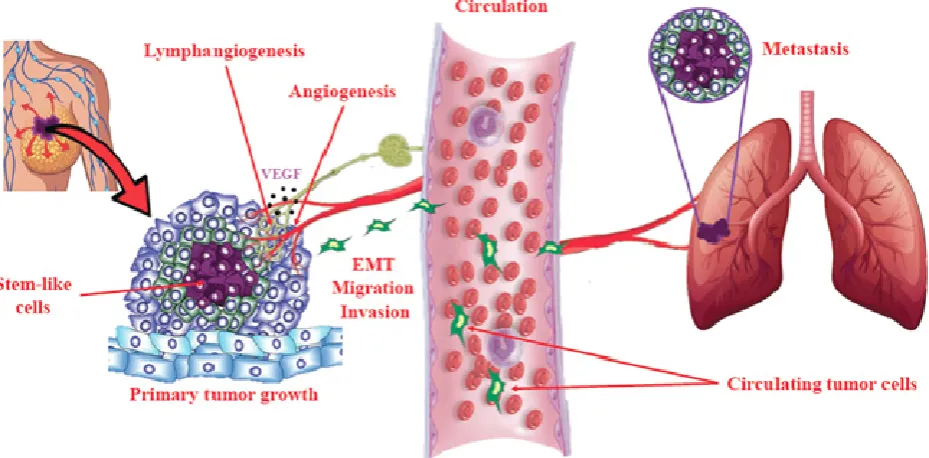
Figure 7. Schema of cellular events in tumor progression and metastasis. Primary tumor growth depends on proliferation of tumorigenic cells, some of which adopt a stem-like cell (SLC) phenotype under the influence of genetic and epigenetic (micro-environmental) mechanisms. Local tumor growth is
dependent on angiogenesis (formation of new blood vessels), which also facilitates tumor cell egress into the circulation. In addition, many epithelial tumors undergo intra-tumoral and/or peri-tumoral lymphangiogenesis (formation of new lymphatic vessels) that helps tumor cells to migrate to lymph nodes and then enter circulation. Epithelial-mesenchymal transition (EMT) is a phenotypic change in epithelial tumor cells utilized for invasion and migration out of the local confines. These cellular events are stimulated in COX-2 expressing breast tumors by activation of EP4 on tumor cells and tumor-associated host cells (immune cells, endothelial cells), so that EP4 presents as a therapeutic target to block multiple cellular events in tumor progression.
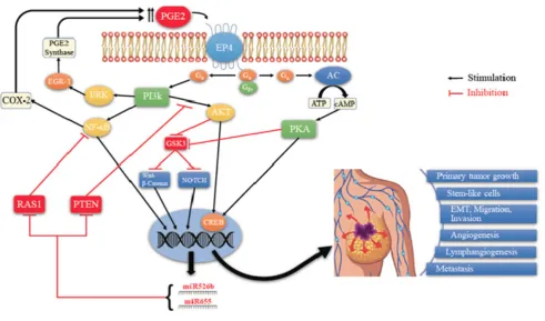
Figure 8. Schema of EP4 mediated signaling pathways in COX-2 expressing breast cancer. Aberrant COX- 2 activity leads to tumor progression and metastasis by utilizing multiple signaling pathways in which EP4 activation plays a pivotal role, and two COX-2/EP4 upregulated miRNAs (miR526b and miR655) are important partners. EP4 activation (like EP2) results in cAMP-dependent PKA activation leading to phosphorylation of the transcription factor CREB. PKA also upregulates WNT/β-catenin and NOTCH pathways by inhibiting GSK3. Furthermore, unlike EP2, EP4 also utilizes the non-canonical PI3K/AkT and ERK signaling pathways, respectively promoting cell survival and
29 PI3K/Akt activation and WNT/β-catenin / NOTCH pathways. While COX-2 induces these miRNAs, the miRNAs, in turn, upregulated COX-2. We suggest that these occurs via upregulation of NF-κB, a well-known upregulator of COX-2 under certain conditions. Predicted targets of these miRNAs include NF-κB repressor genes. Thus there appears to exist a positive feedback loop for
COX-2/EP4/NF-κB/miRNA/COX-2-mediated SLC perpetuation.