University of South Carolina
Scholar Commons
Theses and Dissertations
2017
The Role of Lipocalin-2 in the Hepatic
Microenvironment of Colorectal Cancer
Metastasis
Daniel Titus Hughes
University of South Carolina
Follow this and additional works at:https://scholarcommons.sc.edu/etd Part of theBiology Commons
Recommended Citation
T
HER
OLE OFL
IPOCALIN-2
IN THEH
EPATICM
ICROENVIRONMENT OFC
OLORECTALC
ANCERM
ETASTASISby
Daniel Titus Hughes
Bachelor of Science
North Greenville University, 2007
Submitted in Partial Fulfillment of the Requirements
For the Degree of Doctor of Philosophy in
Biological Sciences
College of Arts and Sciences
University of South Carolina
2017
Accepted by:
Maria Marjorette O. Peña, Major Professor
Hexin Chen, Chair, Examining Committee
DEDICATION
This work is dedicated to my beautiful and intelligent wife, Erin, who is the
best person I know and love. She has always been there for me through this
ACKNOWLEDGEMENTS
To my advisor, Dr. Peña, thank you for giving me the opportunity to carry
out research in your laboratory and for guiding me over the years. To my
committee members Hexin Chen, Frank Berger, David Reisman, and Michael
Wyatt, thank you for all your time, work, and guidance over the years. To those in
and around the Peña lab, Yu Zhang, Grishma Acharya, Nikeya Tisdale, John
Bonaparte, Karen Barbour, Sapana Shah, Vivek Vaish, Kristen Larsen, and
Maydelis Minaya, thank you for being a pleasure to work with and know. To Yu
Zhang and Vivek Vaish, thank you for all the hands-on teaching and
experimental analysis methods you taught me. To Tia Davis, thank you for all
your hard work over many years as our technician.
To Mom, Dad, Max, and Krysten, thank you for all that you have done
over the years, raising me and providing a lot of great memories. To Joel and
Cheryl Chandler and Pat and Jerry Neal, thank you all for proving support and
kindness over the years of this journey. To the rest of my friends and family,
thank you for all for your kindness over the years. I am privileged to have you all
in my life. “Remember, no man is a failure who has friends (Goodrich et al.).”
ABSTRACT
Colorectal cancer (CRC) is the second leading cause of cancer deaths in
the United States. The major cause of death is metastasis and the frequent
target organ is the liver. When diagnosed early at a localized stage, the five year
survival rate after resection is 90%. However, after metastasis has occurred, this
drops to less than 12%. Metastasis is often asymptomatic and diagnosed at the
final stage when therapeutic options are limited. Because of this, the genetic and
cellular mechanisms regulating metastasis are still poorly understood. Recent
studies have shown that prior to the arrival of cancer cells at the secondary
organ, molecular signals from the tumor direct the recruitment of bone marrow
derived cells (BMDCs) to create a pre-metastatic niche where cancer cells can
attach and develop into a metastatic lesion. Identifying and understanding these
signals can lead to the development of methods for early diagnosis or identifying
targets for intervention.
Using an orthotopic mouse model of CRC liver metastasis, we performed
with poor prognosis. The role of LCN2 in the tumor microenvironment has not
thoroughly been studied. Our studies show overexpression of Lcn2 in mouse
colon cancer cells had little impact on tumor growth or invasiveness; however,
invasion assays show that Lcn2 from some stromal cells increases the
invasiveness of colon tumor cells. These studies will allow us to better elucidate
the role of Lcn2 in tumor and stromal cells in the early stages of CRC metastasis
TABLE OF CONTENTS
DEDICATION ...iii
ACKNOWLEDGEMENTS ... iv
ABSTRACT ... v
LIST OF FIGURES... ix
LIST OF ABBREVIATIONS ... xi
CHAPTER 1:INTRODUCTION ... 1
1.1COLORECTAL CANCER ... 1
1.2METASTASIS ... 3
1.3TUMOR MICROENVIRONMENT AND THE IMMUNE CELL COMPARTMENT ... 4
1.4THE PRE-METASTATIC NICHE ... 6
1.5MOUSE MODELS OF COLORECTAL CANCER METASTASIS ... 9
1.6LIPOCALIN-2 IN COLORECTAL CANCER AND METASTASIS... 10
1.7GOALS OF THE PROJECT ... 13
CHAPTER 2:LIPOCALIN-2 IS OVEREXPRESSED IN THE LIVER OF COLORECTAL CANCER BEARING MICE ... 19
3.2RESULTS ... 27
3.3SUMMARY AND DISCUSSION... 31
CHAPTER 4:THE IN-VITROROLE OF TUMOR MICROENVIRONMENT DERIVED LCN2 IN STROMAL CELLS INVASION ... 39
4.1INTRODUCTION... 39
4.2RESULTS ... 42
4.3SUMMARY AND DISCUSSION... 46
CHAPTER 5:THE ROLE OF LIPOCALIN-2 IN SHAPING THE METASTATIC STROMA OF COLORECTAL CANCER IN-VIVO... 53
5.1INTRODUCTION... 53
5.2RESULTS ... 54
5.3SUMMARY AND FUTURE DIRECTIONS... 62
CHAPTER 6:MATERIALS AND METHODS ... 75
REFERENCES ... 90
LIST OF FIGURES
Figure 1.1 The metastatic cascade ... 14
Figure 1.2 The seed and soil organotropism of metastasis. ... 15
Figure 1.3 Working model of pre-metastatic niche formation. ... 16
Figure 1.4 Mouse models of liver metastasis. ... 17
Figure 1.5 The known mechanisms of Lipocalin-2 ... 18
Figure 2.1 Microarray analysis of genetic changes in the seed and soil of colorectal cancer bearing mice ... 24
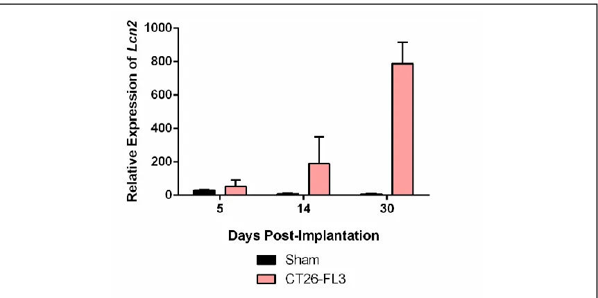
Figure 2.2 Liver “soil” mRNA expression of Lcn2 in tumor bearing mice ... 25
Figure 2.3 Circulating levels of Lcn2 in tumor progression. ... 25
Figure 3.1 Lcn2 overexpression in CT26 and CT26-FL3... 34
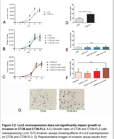
Figure 3.2 Lcn2 overexpression does not significantly impact growth or invasion in CT26 and CT26-FL3 ... 35
Figure 3.3 Lcn2 overexpression in CT26 and CT26-FL3 splenic injection ... 36
Figure 3.4 CT26-FL3 overexpressing Lcn2 in WT and Lcn2-/- BALB/C mice... 36
Figure 3.5 MC38-luc cells overexpressing Lcn2 ... 37
Figure 4.3 Lcn2 expression from TIB-73 hepatocytes directly affects
invasiveness of CT26 tumor cells ... 50
Figure 4.4 Lcn2 overexpression in neutrophil cell line appears to increase invasiveness of CT26 tumor cells ... 51
Figure 4.5 Raw264.7 co-culture increases CT26 and CT26-FL3 invasiveness but Lcn2 dampens MC38-luc invasiveness shown by a co-culture invasion assay with Lcn2 knockout BMDMs polarized to an M2 phenotype ... 52
Figure 5.1 Electroporation of pV1J-Lcn2 increases tumor burden in a breast cancer and melanoma cell lines... 64
Figure 5.2 IHC Staining of liver metastasis tissue for Lcn2 and Mpo ... 65
Figure 5.3 Liver metastasis tissue stained by H&E and in-situ RNA hybridization of Lcn2 mRNA... 66
Figure 5.4 In-situ RNA stain for Lcn2 compared to IHC stained for immune cells in liver metastasis tissue ... 67
Figure 5.5 Confocal microscopy adjacent sections from liver metastasis of MC38-luc bearing mice electroporated with pV1J or pV1J-Lcn2 showing liver (L) and tumor metastasis (M) regions and MPO+, F4/80+, MCT+, and CD45+ cell populations... 67
Figure 5.6 Quantification of confocal results at metastatic tumor-liver periphery from pV1J and pV1J-Lcn2 electroporated MC38-luc bearing mice ... 68
Figure 5.7 Quantification of flow cytometry results shows fold change of pV1J-Lcn2 over pV1J electroporation in cell populations of the spleen, liver, and liver metastasis compartments of tumor-bearing mice only... 68
Figure 5.8 Flow analysis of Cd45+ immune cells... 69
Figure 5.9 Flow analysis of F4/80+ macrophages ... 70
Figure 5.10 Flow analysis of Cd11b+/Ly-6G+ neutrophils and G-MDSCs ... 71
LIST OF ABBREVIATIONS
24p3... Lipocalin-2
24p3R ...Lipocalin-2 receptor
BMDM ... Bone Marrow Derived Macrophage
CRC ...Colorectal Cancer
DFO ... Deferoxamine
ELISA ... Enzyme-Linked Immunosorbent Assay
FeCl3...Ferric Chloride
IHC... Immunohistochemistry
Lcn2 ... Lipocalin-2
MCT ... Mast Cell Tryptase
M-CSF ...Macrophage Colony Stimulating Factor
MMP-9...Matrix Metalloproteinase 9
MPO... Myeloperoxidase
NGAL...Neutrophil Gelatinase Associated Lipocalin
RCF ... Relative Centrifugal Field
CHAPTER
1
INTRODUCTION
1.1
COLORECTAL
CANCER
Colorectal cancer (CRC), which consists of cancers originating in the
colon or rectum, is one of the deadliest cancers both in the United States and in
the world. The tumors are often slow growing, beginning as a benign polyp and
developing into a malignant tumor over multiple years or even decades. Three
types of polyps, adenomatous, hyperplastic, and dysplastic, may all form, but
adenomatous polyps most frequently develop into a harmful adenocarcinoma
(American Cancer Society, 2015).
In the United States, CRC ranks as the fourth most diagnosed cancer and
the second leading cause of cancer related deaths (Howlader et al., 2014).
According to the 2012 GLOBOCAN report by the World Health Organization
(WHO), CRC was diagnosed in 1,361,000 cases and responsible for 694,000
deaths worldwide, accounting for approximately 10% of all cancer cases (WHO).
As the global population continues to live longer due to better healthcare, the
incidence of colorectal cancer will, as a result, also rise significantly.
Colorectal cancer is problematic not only because of its slow early growth,
are usually well contained in the colon and the localized tumors can easily
be surgically resected, contributing to a five year survival rate of 90.1%
(Howlader et al., 2014). However, if the diagnosis occurs after the tumor has
spread to the lymph nodes, the tumor is classified as stage II or III and the
five-year survival drops to 60% and 40%, respectively. In Stage IV cancer where the
tumors have metastasized to distant organs, surgical resection of disseminated
metastatic lesions is difficult and tumor cell populations have increased
heterogeneity and drug resistance. The average five-year survival for all
colorectal cancer diagnoses is 64.9%, but for patients with metastatic disease,
the survival rate is a mere 13.1% (Howlader et al., 2014). Furthermore, nearly
70% of patients dying from CRC burden have metastasis present as revealed by
autopsy (Hugen et al., 2014; Welch and Donaldson, 1979). Colonoscopies
currently remain as the best screening method for early detection of colorectal
cancer development and the removal of polyps; however, less invasive methods
such as stool DNA testing (e.g. Cologuard) are quickly becoming reliably
accurate and significantly cheaper, providing a viable alternative to a
colonoscopy (American Cancer Society, 2014).The primary reason for mortality
in CRC and many other solid tumors, which includes all leukemic and
1.2
M
ETASTASISOne of the hallmark properties of cancer cells is the ability to activate
invasion and metastasis (Hanahan and Weinberg, 2011). This is the ability of
cancer cells to spread from the primary tumor to lymphatic organs and into
circulation and subsequently grow in distant organs. Treatment of a primary
tumor frequently only requires surgery and/or adjuvant chemotherapy; however,
metastatic tumors that have spread throughout the body are difficult to remove
surgically. They form heterogeneous populations that typically harbor additional
mutations as compared to the primary tumor cells and that confer variable
responses to chemotherapy. Patients with solid tumors that already present with
metastasis upon diagnosis are given a stage IV diagnosis that correlates with
low five-year survival rate as seen in the following cancers: ovarian (28.3%),
breast (25.2%), colon (13.1%), and lung (4.2%) (Howlader et al., 2014).
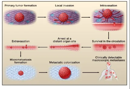
Metastasis is a complex, multistep process that involves growth and
vascularization of the primary tumor, invasion and entry into the submucosal
stromal compartment, intravasation into and survival in circulation, avoidance of
immune surveillance, arrest at a distant site, extravasation into the host tissue
bed, and development and proliferation into a clinically detectable metastatic
lesion (>2mm) (Figure 1, Valastyan and Weinberg, 2011).
The most detrimental aspect of metastasis is the general lack of
symptoms until the metastatic tumor is well established and becomes clinically
stage of the disease when intervention can have an impact on its progression.
This might also lead to the identification of critical biomarkers that might be used
for early diagnosis or as therapeutic targets to block progression or alleviate
morbidity and mortality from the disease.
1.3
T
UMORM
ICROENVIRONMENT AND THEI
MMUNEC
ELLC
OMPARTMENTIt is now fully appreciated that a tumor is not simply comprised of a mass
of rapidly proliferating malignant cancer cells (Hanahan and Weinberg, 2011).
Rather, a tumor is comprised of both neoplastic tumor cells and normal
non-neoplastic stromal cells that are recruited into and infiltrate the tumor bed. These
supporting stromal cells comprise the tumor microenvironment (TME) and play a
significant role in the growth of all solid tumors.
Host tissue is comprised of two primary cell types, parenchymal and
stromal. Parenchymal cells carry out the primary function of a specific organ; for
example, hepatocytes in the liver and splenocytes in the spleen. Stromal cells,
are located throughout the organ and provide support for the biological function
of the parenchymal cells. In the tumor, stromal cells include blood and lymphatic
endothelial cells, mesenchymal stem cells, cancer-associated fibroblasts,
to activate transcriptional programs that promote uncontrolled proliferation. They
create a permissive tumor microenvironment that is essential to the development
and advancement of many tumors. In a metastatic lesion, parenchymal cells are
infiltrated both by tumor and stromal cells, all of which are involved in signaling
crosstalk which can both promote and inhibit tumor growth. Additionally, many of
these stromal cells, such as the immune cells, can be polarized into either a
pro-tumor/immune-suppressive or anti-tumor phenotype and recent clinical evidence
indicates that polarizing normal cells towards their anti-tumor phenotype has
drastic effects on improving survival rates (Suzuki et al., 2016).
The immune cell component of the stroma is highly important to
understanding the tumor microenvironment. In a classic immune response to an
infection with a foreign antigen, T-cells, B-cells, and myeloid lineage cells are
mobilized to clear the infection and then return to an inactivated state (Chaplin,
2010). In the context of cancer, however, immune cells are recruited to a tumor
that they infiltrate where they may exert a pro-tumor phenotype as a
consequence of their plasticity. The primary immune cells found in the tumor
microenvironment include mast cells, myeloid-derived suppressor cells,
macrophages, neutrophils, and T-regulatory cells among other immune cell types
(Hanahan and Weinberg, 2011). Mast cells, the primary cell type responding to
an allergy stimulus, often infiltrate a tumor, correlate with poor prognosis, and
assist in chemotherapeutic drug resistance (Maciel et al., 2015; Oldford and
growing tumor (Arbab and Achyut, 2016; De Vlaeminck et al., 2016; Moses and
Brandau, 2016). Macrophages and neutrophils arise from the myeloid lineage
and are both highly plastic. Macrophages and neutrophils have a classically
activated M1/N1 anti-infection and anti-tumor phenotype, and an alternatively
activated M2/N2 wound healing and pro-tumor phenotype (Galdiero et al., 2013;
Kim and Bae, 2016). These activated states are extremes on a spectrum of
possible phenotypes, but must be differentiated to study their biological context
accurately. When infiltrating a tumor, macrophages and neutrophils are
designated as “tumor associated” macrophages or neutrophils, TAMs and TANs,
respectively. TAMs and TANs are frequently polarized towards a pro-tumor
phenotype and thus provide a potential target for therapy. In a healthy patient,
these wound healing phenotypes prevent the body from being overwhelmed by
too much inflammation; however, in the context of cancer, immune cell function
can be hijacked into a pro-tumor phenotype that supports the growth of the
tumor.
1.4
T
HEP
RE-
METASTATICN
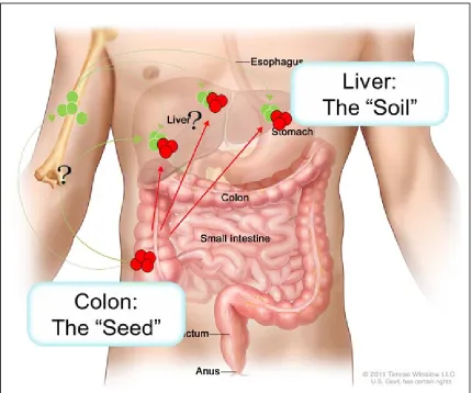
ICHEMetastasis is not a random process (Figure 1.2). As early as 1889 the
however, none of his autopsy patients presented with metastasis in the spleen.
He proposed the “seed and soil hypothesis,” wherein certain “seeds”, i.e., the
primary tumor cells have a preference and will only grow in certain “soil”, i.e., the
secondary organ that provides a permissive environment for the growth of
disseminated tumor cells.
In the 1930’s James Ewing challenged Paget’s hypothesis by suggesting
that metastatic dissemination can be explained solely by hematogenous
circulation (Ewing, 1928). The prevailing theory was that metastasis was
controlled by the pattern of blood and lymph circulation and that capillary beds,
such as those prevalent in the lung, liver, or spleen, were the only targets for
metastatic growth. Prior to this it was thought that tumors only metastasized
purely from the vasculature of organs although some physicians had speculated
such organotropism. Although it has been shown that vasculature has an effect,
the seed and soil hypothesis provides a viable explanation for discrepancies
between blood flow and metastatic growths in an organ (Fidler and Hart, 1982).
In the 1970’s, Isaiah Fidler’s work reignited interest in the seed-and-soil
hypothesis, and in 2005, David Lyden’s group was the first to show that the
formation of a pre-metastatic niche (PMN) in the target organ prior to the arrival
of metastatic cells promoted the establishment and progression of metastasis
(Kaplan et al., 2005). He showed that clusters of immune cells, specifically bone
marrow derived cells (BMDCs) expressing VEGFR1, c-kit, CD133, and CD34,
where they provided sites for attachment and a favorable environment for the
tumor cells to grow.
The current understanding of the pre-metastatic niche in liver metastasis
is well described in both Azizidoost, et al. and Obenauf and Massagué and is
schematically summarized in Figure 1.3 (Azizidoost et al., 2015; Obenauf and
Massagué, 2015). The liver is the most common site for metastasis of many solid
tumors and is primarily seen in the metastasis of colorectal, pancreatic, and
gallbladder cancers. Liver metastasis is also seen frequently in breast and lung
cancers. The hepatic portal system partially explains colorectal liver metastasis
because the veins of the gastrointestinal tract drain through the capillaries in the
liver, which provides ample endothelial surface area for the tumor cell to
extravasate, arrest, and grow.
Some of the mechanistic details of how the pre-metastatic niche is
developed have begun to unfold over the past decade. Exosomes have been
shown to induce TGF-β production, which activates hepatic stellate (ito) cells and
leads to BMDC recruitment to the liver. IL-6 and CCL2 have also been shown to
induce the recruitment of BMDCs to the liver (Obenauf and Massagué, 2015).
Neutrophils can produce MMP9, which degrades collagen in the basement
2012). By understanding the kinetics and cellular and molecular mechanisms
regulating the organotropism of metastasis and pre-metastatic niche formation, it
may be possible to develop methods to restrict the growth of metastasis in
patients whose tumors have not yet metastasized.
1.5
M
OUSEM
ODELS OFC
OLORECTALC
ANCERM
ETASTASISIn order to study the cellular and molecular factors effecting metastasis
during colon cancer progression, a clinically relevant and ethically appropriate
model, which can recapitulate the many traits of human metastasis from the
growth of the primary tumor to establishment of metastasis, is essential. Mice
provide an ideal model that has been used by many researchers for cancer
studies. In studies designed to test drugs directly on human tumors, a
patient-derived xenograph model (PDX) using immune-deficient mice have been very
useful; however to study the biological mechanisms of metastasis and the role of
the tumor microenvironment, a few factors must be controlled. The mice must
have an intact immune system that is able both to respond to pathogens and
infiltrate the tumor and surrounding tissues. Furthermore, the tumor must be
syngeneic to the host strain so that the immune system does not detect the
injected tumor cells as foreign, resulting in their rejection. The tumor must also be
orthotopic, or located in its tissue of origin. As shown in Figure 1.4, a cecal
implantation model for colorectal cancer is ideal for this purpose and can reliably
In this experimental method, CRC tumor cells that are syngeneic are injected into
the subserosa of the cecum to grow in their native environment.
Additionally a splenic injection model is a useful experimental model that
can be utilized to readily assess the establishment and growth of metastatic cells
upon arrival into the liver (Lim et al., 2015). These mouse models allow for the
kinetics and composition of the hepatic microenvironment to be accurately
studied because they recapitulate many traits of clinically observed tumors in a
rapid, reproducible, and accurate manner. Most importantly for this study, the
immune cells of the mice can be studied, which is vital to precisely understanding
the tumor microenvironment and its interactions with tumor cells. Other mouse
models in use for colorectal cancer studies include cecal and splenic
implantations of human cells into mice lacking some or all cell immune cell types
(Oh et al., 2016).
1.6
L
IPOCALIN-2
IN COLORECTAL CANCER AND METASTASISLipocalin-2 (LCN2) is a 25 kD protein that is also known as Neutrophil
Gelatinase Associated Lipocalin (NGAL), oncogene 24p3, or siderocalin. It is a
siderophore binding protein that is primarily associated with the innate immune
typhimurium, to inhibit bacterial growth by limiting the amount of iron that can be
stolen from the mammalian host to be used in bacterial metabolism. Many
bacterial cells have developed resistance to this mechanism and secrete
siderophores that LCN2 cannot bind (Correnti and Strong, 2012; Neilands, 1995).
During an infection, invading bacteria secrete siderophores to capture iron for
use in cellular processes (Flo et al., 2004). As shown in Figure 1.5, Lipocalin-2
circumvents this hijacking by binding the siderophore and sequestering it back
into a host cell expressing the receptor for Lipocalin-2, Slc22a17 (24p3R) (Bao et
al., 2010; Richardson, 2005).
Not only can Lipocalin-2 bind to the bacterial siderophore, enterobactin,
but it can also bind to the mammalian siderophore, catechol (Correnti and
Strong, 2012; Neilands, 1995). In mammalian cells, LCN2 can both increase or
decrease intracellular iron levels in response to environmental signals (Tandara
and Salamunic, 2012). Upon binding iron, LCN2 binds to the cell surface receptor
24p3R which is then internalized. The iron is released causing intracellular iron
levels to increase. LCN2 can also bind to intracellular iron and shuttle it out of the
cell, reducing iron levels and inducing apoptosis (Bao et al., 2010; Reilly et al.,
2013).
There are multiple studies with contrasting data on the role of Lcn2, which
is likely context dependent; however, there is strong support in the published
literature for a pro-tumorigenic role for Lcn2 in cancer progression. A number of
adenocarcinoma patient samples were analyzed by IHC and high NGAL
expression was seen in colorectal cancer specimens, while low or no expression
was seen in adjacent normal tissue (Nielsen et al., 1996) (Lv et al., 2010) (Sun et
al., 2011) (Barresi et al., 2011). Proteomic analysis via mass spectrometry
showed higher LCN2 in colon cancer samples versus normal colon samples
(Conrotto et al., 2008). In rectal cancer patient samples, 69/100 samples showed
LCN2 overexpression which positively correlated with invasiveness, lymph node
metastasis, angiogenesis, and an advanced stage (Zhang et al., 2009). LCN2
was associated with an increase in distant metastasis and advanced cancer
stage in 64 surgically resected colorectal carcinoma tissues (Barresi et al.,
2010). In a study of patients already presenting with hepatic metastasis, serum
LCN2 was significantly higher in patients with three or more metastatic nodes
and in patients with two or more hepatic lobes showing metastases (Martí et al.,
2010). Furthermore, plasma LCN2 was lower in healthy patients as compared to
non-metastatic and metastatic CRC patients (Marti et al., 2013).
Outside of the primary tumor, other studies have begun describing a role
for stromal-derived Lcn2. In McLean et al., LCN2 was expressed in 100% of
adenoma and carcinoma tumor tissues, but only in 4% of tumor adjacent normal
tumor stroma promotes MCF-7 breast cancer metastasis, and is postulated to be
secreted from macrophages in response to tumor cells (Ören et al., 2016).
1.7
G
OALS OF THEP
ROJECTThe overall goal of this project is to understand the changes in the liver
microenvironment prior to and after the establishment of metastases; specifically,
we are interested in the role that the Lipocalin-2 protein plays in this process. We
will test the hypothesis that Lcn2 promotes liver metastasis of colorectal cancer
by altering the hepatic microenvironment by promoting interactions between
tumor cells and cells in the hepatic microenvironment. To understand these
mechanisms, we altered the levels of Lcn2 both in the seed (tumor cells) and in
the soil (hepatocytes and stromal immune cells). We utilized an in-vitro
scratch/migration and matrigel® transwell invasion assays to systematically
delineate Lcn2-specific effects on tumor cell invasion (Corning Incorporated,
2013; Justus et al., 2014). We also utilized a cecal implantation model and
splenic injection model to provide the in-vivo framework to recapitulate the results
we have obtained using the in-vitro methods (Soares et al., 2014; Zhang et al.,
2013). Finally, we analyzed the effects of systemic upregulation of Lipocalin-2,
and its effect on the rate and stromal composition of metastasis. Together, these
studies provide a large volume of data showing the effects of Lcn2 in the liver
Figure 1.5: The known mechanisms of Lipocalin-2 are shown by A) the secretion of siderophores by invasive bacteria to sequester iron from the host. B) The
CHAPTER
2
LIPOCALIN-2 IS OVEREXPRESSED IN THE LIVER OF
COLORECTAL CANCER BEARING MICE
2.1
I
NTRODUCTIONDespite intensive efforts in cancer research over the past half-century, the
high mortality rates of solid tumors, especially after the establishment of
metastasis, remains a significant problem. In colorectal cancer, survival rates
decrease drastically for patients diagnosed after metastases have grown.
Therefore, it is critical to understand the biological processes occurring during
tumor development prior to and after the establishment of metastasis to develop
therapeutic strategies to lower the high mortality rates in patients presenting with
metastasis.
The complex tumor microenvironment is composed of stromal cells that
are recruited to the tumor in addition to tumor cells that are embedded in the
extracellular matrix in the normal host tissue. It is critical to understand how these
three cellular compartments, tumor, tumor stroma, and non-tumor parenchyma
interact with one another to promote tumor growth and progression.
The overarching goal of this study is to identify genetic changes in the liver
prepare the hepatic microenvironment for the arrival of metastatic cells, and
genes whose products are required to sustain metastatic growth and progression
upon arrival into the liver. Using a syngeneic orthotopic cecal implantation model
previously described in Zhang, et. al., we carried out a microarray analysis of
liver tissue in tumor-bearing mice prior to the establishment of metastasis, and
liver tissue after the establishment of CRC metastases. We found that Lipocalin-2
is highly expressed in the liver of metastasis-bearing mice and sought to
determine its role in maintaining metastatic growth. In this chapter, I describe the
microarray analysis and related data, which provides the basis for investigating
the role of Lcn2 in the metastasis of colon cancer (Zhang, 2013).
Cellular signaling between tumor, stromal, and host cells is also of interest
in this chapter as we confirm microarray data by analyzing serum of mice bearing
tumors. The analysis of serum shows the systemic levels of Lcn2 throughout
various stages of metastatic tumor progression.
2.2
R
ESULTS2.2.1
M
ICROARRAY ANALYSES OF HEPATIC MICROENVIRONMENT DURINGderivative CT26-FL3 tumor cells. The following four comparisons were analyzed:
Sham vs. Pre-metastatic, Sham vs. Metastatic, Pre-metastatic vs. Metastatic,
and CT26 vs. CT26-FL3. From this microarray, shown in Figure 2.1, Lcn2 was
the most highly upregulated gene in the metastasis-bearing liver as compared to
the sham group (388-fold higher). Lcn2 was also one of the most highly
upregulated genes in the metastatic-bearing liver versus the pre-metastatic liver
(140 fold higher). Lcn2 was found to be approximately three-fold higher in
CT26-FL3 as compared to CT26; however, CT26-CT26-FL3 produces metastasis at a 10-fold
higher rate as compared to CT26 (Zhang, 2013).
2.2.2
H
EPATIC EXPRESSION OFL
IPOCALIN-2
INCREASES DURINGMETASTATIC PROGRESSION
To confirm the results obtained in the microarray analysis, we quantified
mRNA expression of Lcn2 in liver tissue during tumor progression in mice
bearing tumors from cecal-injected CT26-FL3 cells. Liver mRNA was extracted
from mice at 5, 17, and 30 days after cecal implantation, representing
pre-metastatic and pre-metastatic liver, and analyzed by qRT-PCR. As shown in Figure
2.2, on day 5, Lcn2 expression in liver tissue from mice that had undergone
sham surgery was elevated due to inflammation from the surgery. By days 17
and 30, Lcn2 expression in CT26-FL3-bearing mice was significantly higher than
the sham control group, confirming the results from microarray analysis and
2.2.3
S
ERUM LEVELS OFL
IPOCALIN-2
ARE ELEVATED DURINGT
UMOR ANDM
ETASTATICP
ROGRESSIONTo confirm the results from mRNA analysis, we analyzed serum protein
levels to better understand how Lcn2 is expressed and localized during tumor
progression. Western blot analysis was performed on mouse sera during tumor
growth and progression to metastasis. As shown in Figure 2.3, Lcn2 protein
levels progressively increased in the serum of tumor bearing mice as compared
to sham injected control mice. The control mice showed an increase in Lcn2 on
day 0 immediately after surgery due to inflammation that was resolved by day 7.
On the other hand, mice bearing tumors from CT26-FL3, exhibited progressively
higher circulating levels of Lcn2 by day 28, as shown in Figure 2.3. Statistical
analysis using the Pearson Correlation Coefficient was performed on the western
blot shown in Figure 2.3A to determine the correlation between the number of
days (weeks) of tumor progression and Lcn2 serum protein levels. The results
(CT26; r=0.6506, r2=0.4233)(CT26-FL3; r=0.7092, r2=0.503) show that there is a
stronger correlation in CT26-FL3 versus CT26 for there to be higher Lcn2 protein
expression during tumor progression. This suggests that factors in CT26-FL3 that
expression was found in non-tumor cells in the liver during tumor progression;
however, it remains to be shown if this expression was from the hepatocytes or
other stromal cells found in the liver as part of the pre-metastatic or metastatic
niche.
The microarray study validates many of the same genes that have been
shown in previous studies to be upregulated in the pre-metastatic and metastatic
livers of tumor-bearing mice including Egfr, S100a8, S100a9, Saa3, and Cxcl1
(Rafii and Lyden, 2006; Srikrishna, 2011). Many of these genes have been
studied and their mechanistic contributions to metastasis mostly elucidated.
However the role of Lcn2 remains largely unstudied. As the most highly
upregulated gene in the liver of metastatic-bearing mice, Lcn2 is a potentially
high yield protein of interest involved in developing a favorable organ “soil” for the
colorectal tumor cells to metastasize and proliferate. Microarray analysis also
showed CT26-FL3 expressed three-fold higher Lcn2 as compared to CT26;
however this was not likely to be the source of increased Lcn2 in the serum since
the Lcn2 from the non-tumor liver was much more highly upregulated.
The goal of the subsequent chapters is to further delineate these initial
findings of upregulated Lcn2 in the progression of metastasis along the
framework of the “seed and soil” hypothesis. Lcn2 levels in the tumor “seed”
must be investigated. The role of Lcn2 in the hepatic “soil” is of high importance
given the significantly upregulated Lcn2 in the liver during metastasis in our
compartments must be determined to better understand how Lcn2 affects
metastasis.
Figure 2.1: Microarray analysis of genetic changes in the seed and soil of colorectal cancer bearing mice. A) Microarray showing relative mRNA in non-tumor region of liver in mice given sham surgery or CT26-FL3 cecal
Figure 2.3: Circulating levels of Lcn2 in tumor progression. A) Lcn2 levels in serum increase during tumor progression in CT26 and CT26-FL3 bearing mice as compared to sham mice. B) Quantification of the western as performed by Image J analysis.
CHAPTER
3
THE ROLE OF INTRA-TUMOR EXPRESSION OF LIPOCALIN-2 IN
COLORECTAL CANCER METASTASIS
3.1
I
NTRODUCTIONData from our preliminary in-vivo studies indicate that elevated Lcn2
expression in the liver and Lcn2 protein levels in circulation was correlated with
colorectal cancer metastasis. In this chapter, our goal was to determine if
elevated Lcn2 expression in tumor cells, the “seed”, had an impact on CRC
tumor growth and its metastasis to the liver. We performed in vitro studies to
determine the effect of overexpression or knockdown of Lcn2 expression in CT26
and MC38 colon adenocarcinoma cell lines that were generated in the BALB/C
and C57BL/6 mouse strains. We determined the effects of Lcn2 expression on
tumor growth and invasiveness.
Previous work by Zhang, et al. established CT26-FL3 as a highly
sham controls, however, CT26-FL3 only had three fold higher Lcn2mRNA
expression as compared to CT26 (Figure 2.1). In this chapter we will investigate
if overexpression and knockdown of Lcn2 in CT26, CT26-FL3, or MC38-luc cells
will lead to corresponding increases and decreases in the invasiveness of these
tumor cells.
3.2
R
ESULTS3.2.1
E
FFECT OF OVEREXPRESSION OFL
CN2
INCT26
ANDCT26-FL3
CELLS ON
G
ROWTH ANDI
NVASIVENESSTo understand the autocrine effects of Lcn2 on primary tumor cells we
measured cellular growth and invasiveness of stably transfected tumor cells.
Figure 3.1 shows mRNA and secreted protein overexpression of Lcn2 in CT26
and CT26-FL3. Figure 3.2 shows that overexpression of Lcn2 did not significantly
impact the growth of either CT26 or CT26-FL3. This data shown also confirms
previous observations by Zhang, et al. which showed that CT26-FL3 grows
slower in-vitro as compared to CT26 cells. To measure the effects of tumor cell
secreted Lcn2 on tumor cell invasiveness, we used an in-vitro trans-well invasion
assay. The results showed that while CT26-FL3 was more invasive than CT26
cells as previously shown by Zhang, et al, overexpression of Lcn2 did not
significantly alter their invasiveness as compared to the vector transfected cells
3.2.2
E
FFECT OFL
CN2
OVEREXPRESSION ON METASTASIS OFCT26
ANDCT26-FL3
IN-
VIVO.Although we observe that CT26-FL3 is more invasive than CT26 in-vitro, it
is only by two to three-fold, which does not account for the 9-10 fold higher
metastatic capability of CT26-FL3 in-vivo. To test the effect of Lcn2
overexpression in CT26 and CT26-FL3, 2x10⁵ cells were injected in the spleen of
BALB/C mice and samples were harvested three weeks after the injection. While
cecal implantation allows assessment of spontaneous liver metastasis of
colorectal cancer, splenic injection can provide a measure for the growth of both
poorly and highly metastatic cells upon arrival in the liver, allowing for a
comparison of metastatic colonization by CT26 and CT26-FL3. Furthermore,
stable transfection of CT26-FL3 and subsequent selection with G418, may cause
some loss of its metastatic potency, requiring a number of rounds of selection
through the liver to regain the highly metastatic nature of the cells.
In Figure 3.3, mice bearing splenic tumors of CT26 and CT26-FL3 that
were overexpressing Lcn2 showed no significant differences in primary tumor
growth or metastatic tumor growth. In another experiment using splenic injection,
shown in Figure 3.4, CT26-FL3 overexpressing Lcn2 and CT26-FL3 vector
metastasis in both the vector control and Lcn2 overexpression groups.
Interestingly, when spleen and liver masses are added together, there were no
differences, indicating that, in this model total tumor growth was not affected by
overexpression of Lcn2 in tumor cells or by systemic Lcn2 knockout. However,
primary and secondary tumor growths are different, indicating a role for Lcn2 in
metastasis. More specifically, tumor Lcn2 does slightly impact metastasis, but
only when systemic Lcn2 is absent.
3.2.3
E
FFECT OFT
UMORC
ELL OVEREXPRESSION OFL
CN2
ONT
UMORG
ROWTH ANDI
NVASIVENESS OFMC38
C
ELLSTo verify the results seen with the CT26 and CT26-FL3 cells with Lcn2
overexpression, we utilized the MC38 mouse adenocarcinoma cell line, which is
syngeneic for the C57BL/6 mice. MC38 is less aggressive than CT26 in-vivo, but
metastasis from cecum implantation can be increased to nearly 50% by
passaging through the liver twice as seen with MC38-FL2 (data not shown).
Figure 3.5 shows that Lcn2 mRNA levels were 1000-fold higher in the
MC38-luc Lcn2 overexpressing cell line as compared to MC38-luc cells
transfected with the empty vector. Cellular growth in-vitro was slower in the
presence of Lcn2. Interestingly, Lcn2 overexpressing MC38-luc cells actually
display a phenotype that is less fibroblastic than typical MC38 cells as seen in
Figure 3.5.D. In Figure 3.6, a transwell invasion assay showed that
3.2.4
E
FFECT OFL
CN2
OVEREXPRESSION ON METASTASIS OFMC38
IN-VIVO
.
To test the effect of Lcn2 overexpression in MC38-luc cells on metastasis
in-vivo, the cells were injected into the spleens of C57BL/6 mice and allowed to
grow for three weeks. Surprisingly, as shown in Figure 3.7, Lcn2 overexpression
prevented tumor growth in the spleen in-vivo.
To begin to understand the contrasting effects of Lcn2, we measured
secreted levels in both cell lines. The results from ELISA Figure 3.8 showed that
CT26 and MC38-luc cells secreted undetectable levels of Lcn2 protein. On the
other hand, the CT26-Lcn2 cell line secreted 15 pg/mL of Lcn2, while the
MC38-luc-Lcn2 secreted 7980 pg/mL of Lcn2, approximately 535-fold higher than
CT26-Lcn2. These extremely high levels of Lcn2 are consider
hyper-physiological since normal mice can typically have 1000 pg/mL of Lcn2 in serum
while tumor bearing mice have approximately 2000-4000 pg/mL of serum Lcn2.
Lcn2 is a known neutrophil chemoattractant, and it is possible that such high
levels of Lcn2 being secreted from the primary tumor cells might cause
recruitment of anti-tumor immune cells into the spleen to eliminate the tumor cells
(Asimakopoulou et al., 2016a). Collectively, the results showed that in MC38-luc
An additional data that warrants future studies is the role of iron and Lcn2
in invasion and metastasis. In Figure 3.8.B, MC38-luc-Lcn2 showed no
differences in wound healing as compared to controls. However, when ferric iron
was supplement or iron was chelated with deferoxamine (DFO), MC38-luc-Lcn2
took a significantly longer time to heal the scratch. Since Lcn2 can shuttle iron
into and out of the cell, it is possible that MC38-luc-Lcn2 exports iron with Lcn2
faster than it can uptake iron, so that the lack of intracellular iron could slow
wound healing or migratory activity of cells expressing Lcn2.
3.3
S
UMMARY ANDD
ISCUSSIONIn previous work by Zhang, et al., we observed that orthotopic implantation
of CT26-FL3 produced liver metastasis at a frequency of 90% and under the
same conditions, CT26 produced liver metastasis with 10% frequency (Zhang et
al., 2013). This 9-10 fold increase in invasion in-vivo compared to the 2-3 fold
increase in invasiveness in-vitro indicated that crosstalk with the primary and
secondary tumor microenvironments in-vivo contribute to the high metastasis of
CT26-FL3. This is not surprising since it is well know that the tumor
microenvironment is critical to the growth and development of the primary tumor
and metastatic tumor growth (Hanahan and Weinberg, 2011).
The hyper-elevated Lcn2 protein levels observed in
MC38-luc-pGL4.13-Lcn2, at around 7000-fold, were hyper-physiological levels that only increased
Lcn2 was also unable to grown in the flank as compared to MC38-luc and only
produced a measurable tumor after three months post-injection (data not shown).
As a known neutrophil chemoattractant, it is probable that such high levels of
Lcn2 from the MC38 primary tumor induced an immune response which
eliminated the tumor from the mouse (Asimakopoulou et al., 2016a).
It is possible that the effects of Lcn2 secreted from the primary tumor on
metastasis are concentration dependent. The data showed that expression of
50-fold higher Lcn2 in CT26-Lcn2 and did not increase invasion or metastasis, while
expression of 1000-fold higher Lcn2 in MC38-luc-Lcn2 only led to a two-fold
increase in invasion.
Collectively, these data suggest that Lcn2 does not play a significant role
in tumor growth and progression when over-expressed in tumor cells, the seed
component of the seed and soil hypothesis. This leads to the question on the role
of Lcn2 when expressed in the host parenchymal and stromal compartments.
The current literature suggests that LCN2 expression in human colorectal
cancer is positively correlated with poor outcomes, but this association is
disputed by a number of studies. Candido, et al. found that colon tumors express
LCN2 mRNA approximately 66.3% higher than normal colon tissue, and 45% of
McLean disagreed that LCN2 can be used as a clinically viable testing option
(Catalán et al., 2011; Fung et al., 2013; McLean et al., 2013). Thus, while it
seems that LCN2 has the potential for clinical utility, its multifaceted role is
unclear and warrants further investigation.
In the subsequent chapters, our goal is to determine if Lcn2 expressed in
the host and stromal cells in the microenvironment plays a role in metastasis as
using both in-vitro and in-vivo strategies. An interesting question to dissect
would be its role in promoting tumor cell invasion or sustaining tumor growth
upon arriving in the secondary environment such as the liver. More recent
studies using human colorectal cancer cell lines suggest that intratumoral LCN2
may inhibit metastasis by polarizing tumor cells into an epithelial phenotype
(Feng et al., 2016) which seem to be consistent with the MC38-luc-Lcn2
phenotype shown in Figure 3.5.D. Future studies would need to investigate the
Figure 3.5: MC38-luc cells overexpressing Lcn2. A) Plasmid maps of pGL4.14 and pGL4.14-Lcn2. B) Lcn2 overexpression measured via qRTPCR. C) Western
CHAPTER
4
T
HEI
N-
VITROR
OLE OFT
UMORM
ICROENVIRONMENT-D
ERIVEDL
CN2
IN
T
UMORC
ELLI
NVASION4.1
INTRODUCTION
The current literature describing the role of Lcn2 in metastasis is
discordant, most likely due to the variety of cancer models used by different labs.
Thus, the burden of proof remains to further delineate the effects of Lcn2 on
colorectal cancer metastasis through experimental methods. Since hepatocytes
comprise the majority of the liver, it is likely that they contribute a significant
portion of the Lcn2 found in pre-metastatic and metastatic livers of colorectal
cancer bearing mice. In a bacterial infection and hepatectomy model, Xu, et al.
showed that hepatocytes secrete ~25% of serum Lcn2 in normal conditions and
~90% of serum Lcn2 levels after infection or injury (Xu et al., 2015).
Macrophages located in the metastatic microenvironment are a likely cell
type that may be influencing Lcn2’s participation in metastasis. Macrophages are
highly plastic immune cells whose gene expression can be modified in response
to cues from the microenvironment allowing them to be polarized into a
designation contains nearly all alternatively activated macrophages and can be
further broken down into the M2a wound healing macrophage and the M2b/c
immune regulatory macrophage. The distinctions between different phenotypes
are often blurred and macrophages can exist in a hybrid state, expressing genes
found in multiple subtypes.
Many papers have shown a role for Lcn2 in the polarization of
macrophages, which can further influence the tumor microenvironment in the
metastatic setting (Guo et al., 2014; Jung et al., 2015). In a bacterial pneumonia
study, LCN2 deactivated macrophages and was a marker of macrophage
deactivation and impaired immune clearance of bacteria (Warszawska et al.,
2013). Jung, et al. showed that IL-10 in the tumor microenvironment caused
downstream production of Lcn2 by tumor associated macrophages that were
polarized towards an M2 phenotype (Jung et al., 2012). In an obesity-associated
inflammation model, Lcn2 was shown to be an anti-inflammatory regulator of
macrophages, skewing towards an M2 phenotype via a feed-forward
NF-κB-STAT3 loop (Guo et al., 2014).
In contrast, using an ischemia-reperfusion model, Lcn2 promoted
macrophages towards an M1 phenotype (Cheng et al., 2015). In addition, in a
bearing models, which all showed Lcn2 expression correlating with the M2
phenotype.
Another hematopoietic cell population that is found in the metastatic
microenvironment is the neutrophil, which has also been shown to promote liver
metastasis (Gordon-Weeks et al., 2017). In healthy humans, neutrophils make up
50-70% of all white blood cells and have been shown to play a role in both
anti-tumor and pro-anti-tumor immunity (Galdiero et al., 2013). Similar to macrophages,
neutrophils can be polarized into a classical N1, anti-tumor phenotype and a
pro-tumor, N2 phenotype. Neutrophils found in tumors are designated as tumor
associated neutrophils (TANs), which can be either pro-tumorigenic or
anti-tumorigenic (Sionov et al., 2015).
Neutrophils were the first cell type that was found to express high levels of
Lipocalin-2, hence the name Neutrophil Gelatinase Associated Lipocalin (NGAL).
Gelatinase (MMP9) was found in humans to bind NGAL to prevent the
degradation of MMP9 (Koh and Lee, 2015). Schroll, et al. showed that
recombinant Lcn2 can induce neutrophil migration which is reduced in Lcn2
knockout mice (Schroll et al., 2012).
Our preliminary microarray data showed that Lcn2 is highly upregulated in
the non-tumor area of a murine liver bearing CRC metastasis. The goal of this
chapter is to further understand the role that Lcn2 expressed in the parenchymal
hepatocytes and in hematopoietic immune cells plays in promoting or
myeloid lineage-derived neutrophils and macrophages. By utilizing a transwell
chamber, we can perform invasion assays by co-culturing the tumor cells with
immune cells. This in-vitro system allows us to simulate metastasis with the
upper chamber representing the primary tumor, or “seed”, and the lower chamber
representing the metastatic site, “or soil.” Altering the levels of Lcn2 in the lower
chamber “soil” cells allows us to develop a framework for the role of Lcn2 from
microenvironmental cell types on colorectal cell invasiveness.
4.2
RESULTS
4.2.1
E
FFECT OFH
EPATICL
CN2
ONCRC
I
NVASIVENESSIn this study, we utilized the murine hepatocyte cell line TIB-73 (BNL CL.2
(ATCC® TIB-73™)) to test the role of hepatic Lcn2 expression on CRC cell
invasion. It is a reliable cell line that can be altered via transfection or
transduction. Lcn2 overexpression was established in TIB-73 using the
previously described pCMV6-Entry-Lcn2 plasmid. As shown in Figure 4.1.A,
Lcn2 mRNA was over-expressed, in Figure 4.1.B,intracellular protein levels
decreased for TIB-73 with Lcn2 overexpression, but, most importantly, secreted
Lcn2 protein levels increased, as shown by ELISA in Figure 4.1.C. Consistent
in the bottom well would have an effect on tumor cell invasion, TIB-73 cells
over-expressing Lcn2 were placed in the bottom well during an invasion assay. The
results in Figure 4.2.C showed that increased Lcn2 from the soil (TIB-73
hepatocytes) increased tumor cell invasion.
To determine if increased hepatocyte Lcn2 specifically caused the
increased invasiveness of CT26, Lcn2-specific siRNA was transfected into the
TIB-73 cells as described in the materials and methods chapter. The results in
Figure 4.3 show that siRNA against Lcn2 diminished mRNA levels as determined
by qRT-PCR and extracellular Lcn2 protein levels as determined by ELISA. Lcn2
knockdown via siRNA reduced the invasiveness of CT26, but not to a statistically
significant level. However, a Pearson correlation analysis showed that Lcn2
levels from the TIB-73 cells correlated with invasiveness of CT26 cells to some
extent (r=0.58, r2=0.34).
4.2.2
E
FFECT OFL
CN2
EXPRESSED BYN
EUTROPHILS ONCRC
CELLI
NVASIVENESS.
Another cell type within the metastatic tumor microenvironment that might impact
CRC invasion and metastasis though Lcn2 expression is the neutrophil. The cell
line MPRO (ATCC® CRL-11422™) can be induced to differentiate into mature
neutrophil (NEUT) after four days of exposure to 10 M all-trans-retinoic acid
NEUT cells were 72% positive for the cell surface markers Cd11b and Ly6G by
flow cytometry analysis as compared to MPRO cells which were only 12%
positive for both markers. In Figure 4.3.C, qRT-PCR levels indicated that Lcn2
expression increased by approximately 150-fold and the Lcn2 receptor 24p3R
expression increased by approximately 15-fold in NEUT cells as compared to the
undifferentiated MPRO cells. Co-culture of NEUT cells with CT26 cells in
invasion assays showed that the invasiveness of CT26 cells increases nearly two
fold in the presence of NEUT cells as compared to CT26 by itself or with MPRO
cells. Experiments to determine if downregulation of Lcn2 by siRNA transfection
or lentiviral shRNA expression were largely unsuccessful due to the difficulty in
diminishing the very highly elevated Lcn2 mRNA levels in NEUT cells.
4.2.3
E
FFECT OFL
CN2
INM
ACROPHAGES ONCRC
C
ELLI
NVASIVENESSMacrophages are one of the most prevalent immune cell types in the
metastatic microenvironment and our goal was to determine the influence of
Lcn2 in macrophages on colorectal cancer cell invasion (Qian and Pollard, 2010).
Using the Raw 264.7 macrophage-like cell line, we tested some of the effects of
macrophages on cell invasiveness in-vitro. Similar to primary macrophages,
knockdown Lcn2 via siRNA or shRNA were unsuccessful as Raw264.7 is one of
the most difficult cell types to reliably transfect. To circumvent this difficulty, we
utilized primary bone marrow derived macrophages (BMDMs) from BALB/C mice
or C57BL/6 mice as previously described (Jung et al., 2016; Singh et al.). Bone
marrow was extracted from WT C57BL/6 and C57BL/6 Lcn2-/- mice and
incubated in 15% L-929 conditioned media for 7 days, after which the
F4/80+/Cd11b+ population, corresponding to macrophages, was determined to
be 97.2% by flow cytometry. In Figure 4.5.C, MC38-luc cells were placed in the
top chamber, while the bottom chamber contained either wild type C57BL/6
BMDM or C57BL/6 Lcn2 -/ BMDM. Both wild type and Lcn2-/- BMDM were
treated with either LPS or IL-4 to polarize them into an M1 or M2 phenotype,
respectively. Consistent with the previous results obtained with the Raw264.7
cells in co-culture invasion assays, the presence of BMDMs increased the
invasiveness of MC38-luc cells dramatically (Figure 4.5.C). The loss of Lcn2 from
BMDMs had no impact on the invasion rates for unstimulated or LPS-treated
BMDMs, however, BMDM cell from Lcn2-/- mice, treated with IL-4 showed the
highest levels of MC38-luc invasion. This data suggested that in the M2
phenotype, a factor other than Lcn2 may be contributing to the high levels of
tumor cell invasion.
Figure 4.5.D shows results from ELISA analysis for Lcn2 levels in media
taken from the wells of the invasion assays performed in Figure 4.4.C. Lcn2
Lcn2-/- BMDMs, which gave the highest levels of MC38-luc invasion.
Interestingly media from C57BL/6 Lcn2 -/- BMDM’s treated with LPS and
incubated with MC38-Luc cells contained 1000 pg/mL Lcn2, which suggested
that in response to LPS stimulation of BMDMs into an M1 state, the MC38-luc
cells secreted higher levels of Lcn2, however, this has no effect on MC38-luc
invasiveness. The Pearson correlation shown in figure 4.5.E revealed that there
was no association in between Lcn2 levels in the media and MC38-luc tumor cell
invasiveness (r=0.086, r2= 0.007).
4.3
S
UMMARY ANDD
ISCUSSIONThe results shown in these experiments confirmed the data from the
microarray analysis, which suggested that high levels of Lcn2 from cells in the
metastatic microenvironment may be partially responsible for the growth of liver
metastasis of colorectal cancer. Elevated levels of Lcn2 secreted by TIB-73
hepatocytes increased the invasion of CT26 colorectal tumor cells. The
differentiated NEUT neutrophils, which expressed very high levels of Lcn2 also
increased the invasiveness of CT26. On the other hand, while the Raw264.7
macrophages and the BMDMs isolated from wild type or Lcn2-/- mice caused
their effects on tumor growth and invasiveness through other factors. For
example, Raw264.7 cells express less Lcn2 than TIB-73 cells in both their native
states; however, Raw264.7 induced more tumor cell invasion than TIB-73 in the
co-culture invasion assay. This is reasonable as there are a host of cytokines
and chemokines that are involved in the crosstalk between tumor cells and
stromal cells, thus, it is unlikely that any one protein (Lcn2) can be primarily
responsible for singlehandedly driving cellular invasion or tumor phenotype. The
focus of this chapter was to investigate the Lcn2 specific effects on tumor cell
invasion, but it remains possible that in-vivo, Lcn2 may have an effect on the
metastatic cascade in liver metastasis that is not the invasiveness of the
colorectal tumor cells. The subsequent chapter aims to investigate Lcn2 in-vivo in
the hepatic tumor microenvironment.
Figure 4.1: Lcn2 is overexpressed by transfecting TIB-73 with pCM6-Lcn2. A)
Figure 4.2: Lcn2 overexpression from TIB-73 cells in the “seed” compartment in a co-culture invasion assay increases invasiveness of CT26 and CT26-FL3.
Figure 4.3: Lcn2 expression from TIB-73 hepatocytes directly affects
CHAPTER
5
T
HER
OLE OFL
IPOCALIN-2
INS
HAPING THEM
ETASTATICS
TROMA OFC
OLORECTALC
ANCER IN-
VIVO5.1
INTRODUCTION
To better understand the role of Lipocalin-2 in the hepatic tumor
microenvironment in advanced colorectal cancer metastasis, we utilized an
immune-competent mouse model to experimentally recapitulating a
comprehensive tumor microenvironment, which closely represents clinical tumor
physiology. As previously described, we observed increased levels of systemic
Lcn2 and elevated hepatic Lcn2 mRNA levels in our orthotopic cecal implantation
mouse model of colorectal cancer progression and metastasis,. In studies
described in this chapter, we utilized in-vivo electroporation of endotoxin-free
plasmid DNA to increase systemic levels of Lcn2 in tumor-bearing and
non-tumor-bearing mice. The goal was to determine the effect of increased serum
levels of Lcn2 on the localization of immune cell populations in the metastatic
5.2
RESULTS
5.2.1
PV1J-L
CN2
UTILIZATION INM
ULTIPLEM
OUSEM
ODELSPrevious work in the laboratory by John Bonaparte showed that mice
electroporated with 50 ug of pV1J-Lcn2 had ~30% higher serum Lcn2 levels than
mice electroporated with pV1J vector only (Bonaparte, 2015, unpublished data).
The higher Lcn2 levels were sustained for 2-3 weeks until gradually dropping
back to pre-electroporation levels. When the electroporation was performed in
mice that have been implanted with MC38-luc cells in the spleen, elevated Lcn2
had no effect on the primary tumor volumes in the spleen. However, elevated
Lcn2 levels resulted in a two-fold increase in liver mass due to increase
metastasis, as compared to mice electroporated with the empty vector
(Bonaparte, 2015). These data suggested that elevated systemic levels of Lcn2
either (1) accelerated the establishment of metastasis or (2) supported the
growth of colorectal cancer tumor cells in the liver after they have disseminated
from the primary tumor. The goal of the experiments in this chapter is to begin to
elucidate the changes in the stromal immune cell populations in the metastatic
liver microenvironment in the presence of high systemic levels of Lcn2 in
tumor-bearing mice.
Thesis. In the melanoma model, B16-F10 melanoma cells were injected
subcutaneously into C57BL/6 mice. In the breast cancer model, 4T1-RFP-luc
breast cancer cells were injected into the left 4th mammary fat pad of BALB/C
mice. In both models, tumor cell implantation was followed by electroporation of
pV1J-LCN2 plasmid or pV1J empty vector. Tumors were allowed to grow for 6
weeks to assess growth and the presence of metastases.
The results in Figure 5.1.A showed the 4T1-luc-RFP tumor
bioluminescence in mice electroporated with pV1J and pV1J-Lcn2. Figure 5.1.B
showed that the primary tumor volume is greater in mice electroporated with
pV1J-Lcn2 during all weeks of tumor progression. Figure 5.1.C showed the
metastatic tumor bioluminescence appears to trend towards being higher in the
pV1J-Lcn2 group, but there is no difference in metastatic burden in these mice. It
is likely that allowing the primary tumors to grow longer may provide more insight
into the effects of high systemic Lcn2 on breast cancer metastasis. Figure 5.1.D
and 5.1.E showed that in C57BL/6 mice injected subcutaneously with
B16-F10-luc melanoma cells and electroporated with pV1J or pV1J-Lcn2, the pV1J-Lcn2
group showed higher primary tumor mass, but there was no difference in lung
metastasis as shown by IVIS imaging in photons/second.
Taken together, these data showed that elevated systemic Lcn2 levels
enhanced primary tumor growth of both melanoma and breast cancers. In the
splenic model of CRC liver metastasis, which quickly produces liver metastasis
primary tumors grew very aggressively and the mice had to be sacrificed due to
the large tumor burden before significant metastasis occurred. Future studies
should utilize a slower growing melanoma and breast cancer mouse cell lines in
conjunction with Lcn2 electroporation, to better gauge the impact of Lcn2 on the
incidence of metastatic growth in other target organs. In conclusion, elevated
systemic Lcn2 levels increased the growth rate of primary breast and melanoma
tumors, and but did not increase incidence of metastasis to lung and liver.
5.2.2
L
OCALIZATION OFL
CN2
E
XPRESSION IN THE LIVER OF METASTASIS-BEARING MICE
In the microarray experiment described in Chapter 2, Lcn2 was identified
as a gene of high interest in liver metastasis because it was one of the most
highly upregulated in the liver “soil”. Lcn2 mRNA levels in the microarray was
over-expressed in the non-tumor regions of the liver, as confirmed by the
exclusion of mCherry protein, which was expressed only in the tumor cells.
However, while this analysis showed that Lcn2 is highly expressed in the
non-tumor region, there was no indication of the specific cell type(s) producing Lcn2.
myeloperoxidase positive (MPO+) cells, or neutrophils, were found around the
tumor periphery, there was no punctate co-localization of MPO with Lcn2.
In-situ RNA hybridization was used to determine the sites of hepatic Lcn2
transcription. The results in Figure 5.3 showed that Lcn2 is mostly
over-expressed around the periphery of the tumor with very few transcripts over-expressed
within the tumor cells. Figure 5.4 further showed that when adjacent metastatic
liver sections were stained by IHC for the CD45 pan-leukocyte marker (total
immune cells), F4/80+ marker for macrophages, MRP-8 for mast cells, and MPO
for neutrophils, these cellular markers are found in cells within the same region
as the Lcn2 mRNA, however, there was no distinct cellular staining pattern
correlating Lcn2 expression with any one type of immune cells in-situ.
5.2.3
E
FFECTS OF INCREASED SYSTEMICL
CN2
ON THE IMMUNE CELLCOMPOSITION OF THE METASTATIC LIVER MICROENVIRONMENT
After localizing the Lcn2 mRNA expression to the tumor periphery in-situ,
more cell specific Lcn2 co-localization was necessary to determine which stromal
cells were expressing Lcn2. We also wanted to determine how an increase in
systemic Lcn2 levels in tumor-bearing mice affected the metastatic tumor
microenvironment, specifically with respect to the recruitment of macrophages
and neutrophils. CRC patients with high serum levels of Lcn2 had poorer
prognosis and outcomes, thus, it is critical to understand if elevated Lcn2 in