Scholarship@Western
Scholarship@Western
Electronic Thesis and Dissertation Repository
8-14-2019 10:00 AM
Structural and Functional Characterization of Deinococcal DNA
Structural and Functional Characterization of Deinococcal DNA
Damage Response A (DdrA)
Damage Response A (DdrA)
Filip Todorovic
The University of Western Ontario
Supervisor Junop, Murray S.
The University of Western Ontario
Graduate Program in Biochemistry
A thesis submitted in partial fulfillment of the requirements for the degree in Master of Science © Filip Todorovic 2019
Follow this and additional works at: https://ir.lib.uwo.ca/etd
Part of the Biochemistry Commons
Recommended Citation Recommended Citation
Todorovic, Filip, "Structural and Functional Characterization of Deinococcal DNA Damage Response A (DdrA)" (2019). Electronic Thesis and Dissertation Repository. 6340.
https://ir.lib.uwo.ca/etd/6340
This Dissertation/Thesis is brought to you for free and open access by Scholarship@Western. It has been accepted for inclusion in Electronic Thesis and Dissertation Repository by an authorized administrator of
1
ABSTRACT
Deinococci exhibit a remarkable resilience toward DNA damage through the
actions of several unique proteins, including DdrA. Although DdrA is critical for
damage resistance, little is known about its mechanism of action. Despite sharing
sequence similarity with Rad52, DdrA has been reported to lack single-stranded
DNA annealing activity. In order to better characterize DdrA, structural studies
were undertaken with the primary objective of gaining insight into the mechanism
by which DdrA functions. Significant progress was made toward elucidating the
X-ray crystal structure; in particular, identifying suitable DdrA domain boundaries for
successful expression, purification and crystallization. In addition, we demonstrate
for the first time that DdrA mediates ssDNA annealing to levels comparable to
Rad52 in vitro. Residues (K22 and K105) critical for ssDNA binding and annealing
were identified and further used to demonstrate that DdrA mediates resistance to
extreme levels of DNA damage through its ability to anneal ssDNA in vivo.
Keywords: DdrA, Deinococcus radiodurans, DNA repair, X-ray crystallography,
single-stranded DNA annealing, radioresistance, mitomycin C, cancer, extended
synthesis-dependent strand annealing, Rad52, homologous recombination
SUMMARY FOR LAY AUDIENCE
D. radiodurans is a bacterium that was discovered in 1956 after surviving
on a can of meat that had been exposed to intense doses of radiation. Typically,
radiation kills organisms by destroying DNA, the molecule that all organisms
require to maintain biological function. The fact that D. radiodurans was able to
survive meant that the bacterium was either able to protect its DNA from damage
in an extraordinary fashion or able to repair its DNA following damage in an
extraordinarily efficient manner. Once it was established that both D. radiodurans
and E. coli, a radiation-sensitive bacterium, accumulate DNA damage to the same
extent, the latter was deemed to be true.
It was later determined that the unique resistance to radiation stems, in part,
from a unique protein, known as DdrA. The protein was found to be “turned on” in
the cell following DNA damage, providing correlative evidence that it is involved in
repair. Furthermore, when the protein was eliminated from the cell, the organism
became more radiation-sensitive, providing the first causative evidence that the
protein is involved in repair. Prior to the publication of this thesis, it had been shown
that DdrA is able to interact with DNA as one would expect for a protein required
to repair DNA. However, beyond this, no further details regarding the exact role of
DdrA were known. This thesis demonstrates that DdrA is capable of annealing
DNA, which is an important aspect of genomic reconstruction following DNA
damage. Furthermore, we have identified the exact regions of the protein, which
are involved in this process. Most significantly, we have determined that without
ii
this contribution by DdrA, cells are sensitive to DNA damage, underscoring the
importance of this phenomenon in the living organism. To figure out exactly how
annealing takes place at an atomic level, we are now interested in figuring out what
DdrA bound to protein looks like using a technique, known as X-ray crystallography.
Significant progress to this end has been made in the lab and our current research
efforts are aimed at bringing this task to completion.
ACKNOWLEDGMENTS
This work would not have been possible without the guidance and
supervision of Dr. Murray Junop, whose enthusiasm and round-the-clock
availability have inspired my passion for science and facilitated my growth as a
researcher. His scientific approach, stemming from fostering fundamental
principles and attention to details, has had an influence not only on my scientific
modus operandi, but also on my approach to life in general and for that I will be
forever grateful.
I would like to further thank my advisors, Drs. Patrick O’Donoghue and Eric
Ball, whose open-door policies have helped to enrich this journey from its inception
all the way to the writing of this thesis. Their expertise in differing areas of
biochemistry has been very helpful in overcoming unforeseen challenges.
I would also like to acknowledge all members of the Junop lab, past and
present, most notably, Kun Zhang, Robert Szabla and Ryan Grainger for their
willingness to help complete work needed to finish the thesis as well as Dr. Chris
Brown for his invaluable mentorship. My journey would likewise not have been
complete without the tireless efforts of my undergraduate students, Braeden
Medeiros, Parisa Fani-Molky and Pallak Gupta.
Last but not least, I would like to thank my family –my parents, Dragan and Nataša, and my sister, Lana, whose unconditional love and unwavering support
along the way have made my maturation into a scientist possible.
iv
Table of Contents
Abstract………... ii
Summary for Lay Audience………. iii
Acknowledgments……….. v
Table of Contents……… vi
Figures………... ix
Tables………... xi
Abbreviations……….. xii
Chapter 1: Introduction
1 1.1 DNA Damage……….. 21.2 Deinococcus……… 4
1.2.1 General Features………. 4
1.2.2 Ionizing Radiation………... 5
1.2.3 Ultraviolet-C Radiation………. 6
1.2.4 Mitomycin C…………..……… 8
1.2.5 Desiccation……… 8
1.3 Factors Contributing to Resistance………. 10
1.3.1 Physical Scaffolding………. 10
1.3.2 Protection of the Proteome via ROS Scavenging………... 11
1.4 Recombinational Repair in D. radiodurans……… 16
1.4.1 The RecBCD Pathway………. 16
1.4.2 The RecFOR Pathway………. 19
1.4.3 Extended Synthesis-Dependent Strand Annealing……… 21
1.4.4 Single-Strand Annealing………... 24
1.4.5 Novel Deinococcal Proteins Involved in Repair……….. 24
1.5 DNA Damage Response A (DdrA) 26 1.5.1 Previous Research………... 26
1.5.2 Current Research Outline………... 29
Chapter 2: Functional Characterization of DdrA
30
2.1 Abstract……… 312.2 Introduction……….. 31
2.3 Materials & Methods 35 2.3.1 Protein Preparation………... 35
2.3.2 Analysis of Quaternary Structure……….. 37
2.3.3 ssDNA Binding Assessment………... 37
2.3.4 ssDNA Annealing Assay………. 38
2.3.5 In Vivo Analysis of ssDNA Annealing by DdrA……… 40
41 2.4 Results 2.4.1 Expression and Purification of DdrA for in Vitro Functional Studies….. 41
2.4.2 Quaternary Structure Findings………... 44
2.4.3 Characterization of DdrA ssDNA Binding………. 46
2.4.4 Analysis of DdrA ssDNA Annealing……….. 47
2.4.5 Importance of DdrA ssDNA Annealing for DNA Damage Tolerance…. 53
2.5 Discussion
vii
v
2.5.1 DdrA: A Prokaryotic Rad52 Homologue……….. 63
2.5.2 The Role of DdrA in Deinococcal DNA Repair……… 64
Chapter 3: Structural Characterization of DdrA
67
3.1 Abstract……… 68
3.2 Introduction……….. 68
3.3 Materials & Methods 69
3.3.1 Secondary Structure Predictions & Homology Modelling…….. 69
3.3.2 Protein Preparation……….. 70
3.3.3 Analysis of Quaternary Structure………... 71
3.3.4 Crystallization ……….……….. 72
3.4 Results 74
3.4.1 DdrA Construct Design……… 74
3.4.2 Protein Preparation……….. 79
3.4.3 Crystallization……… 80
3.4.4 Preliminary X-Ray Diffraction of DdrA1-160 (D. geothermalis)… 86
3.5 Discussion……… 88
Chapter 4: Summary & Future Direction
4.1 DNA Damage Repair by Deinococcus Radiodurans……….
4.2 Summary of Findings………..
4.3 Implications of Findings………..
4.4 Future Direction………
viii
92
93
94
95
4.5 Conclusion………..
References
………..101
Appendix
114
List of Constructs Used 115
Protein………
DNA………
115
119
123
Curriculum Vitae
Curriculum Vitae……… 124
Figures
Chapter 1: Introduction
1.1 Generation of ROS……….... 14
1.2 The Initiation of HR by the RecBCD Complex in E. coli……….. 18
1.3 The Initiation of HR by the RecFOR Complex in D. radiodurans…….. 20
1.4 ESDSA Repair of Double-Strand Breaks in D. radiodurans..…………. 22
1.5 Synthesis Along a Bridging Element………... 23
1.6 Domain Organization of DdrA……….. 28
Chapter 2: Functional Characterization of DdrA
2.1 IMAC Purification of DdrA1-161………. 43
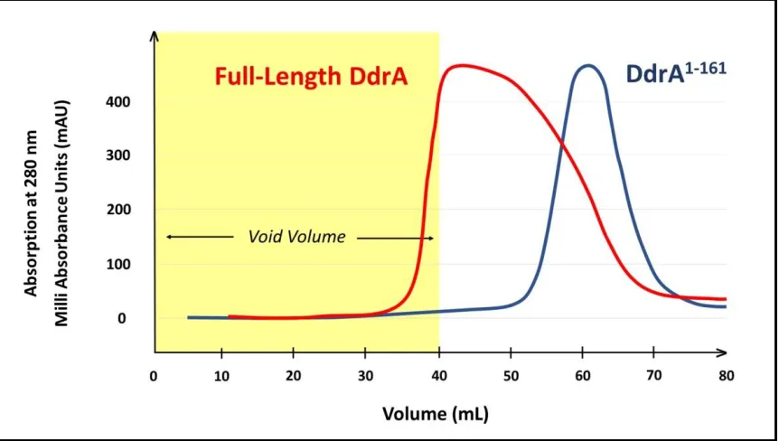
2.2 Analysis of Quaternary Structure of DdrA by SEC……….. 45
2.3 ssDNA Annealing Assessment
ix
viii
2.3.1 ssDNA Annealing Assessment of hRad52………... 49
2.3.2 ssDNA Annealing Assessment of Full-Length DdrA………. 50
2.3.3 ssDNA Annealing Assessment of DdrA1-161……… 51
2.3.4 Summary of ssDNA Annealing Data………... 52
2.4 Importance of DdrA ssDNA Annealing for DNA Damage Tolerance 2.4.1 Mutant Design: Multiple Sequence Alignment……… 54
2.4.2 Mutant Design: Thread-Based Homology Modelling………. 55
57 2.4.3 ssDNA Binding Assessment of DdrAK22A/K105A……… 2.4.4 ssDNA Annealing Assessment of DdrAK22A/K105A ……….. 58
2.4.5 Comparison of Wild-Type and Mutant ssDNA Annealing………. 59
2.4.6 DNA Damage Repair via ssDNA Annealing: Pictorial View……. 60
61 2.4.7 DNA Damage Repair via ssDNA Annealing: Graphical View…..
Chapter 3: Structural Characterization of DdrA
3.1 DdrA Construct Determination 3.1.1 Secondary Structure Predictions for Deinococcal DdrA………… 763.1.2 Homology Modelling of DdrA………. 77
83 3.2 Examples of Crystals Obtained……… 3.3 Analysis of Quaternary Structure of DdrA 3.3.1 Analysis of Quaternary Structure of DdrA by SEC-MALS………. 84
3.3.2 Analysis of Quaternary Structure of DdrA by AUC………. 85
3.4 Crystals Obtained with DdrA1-160 (D. radiodurans)………... 86
3.5 Preliminary X-Ray Diffraction of DdrA1-160 (D. geothermalis)………….. 87
ix
Tables
Chapter 3: Structural Characterization of DdrA
3.1 Summary of Protein Properties……… 79
3.2 List of Crystallographic Trials………... 81
Appendix
List of Materials Used
Proteins………..
DNA………
115
119
x
Abbreviations Used
6-4-PP: pyrimidine-(6-4)-pyrimidone photoproduct
APS: Advanced Photon Source
AUC: analytical ultracentrifugation
BER: base excision repair
BPP: bipyrimidine photoproduct
CPD: cyclobutyl pyrimidine dimer
D10: dose yielding 10% survival
D. radiodurans or D. rad: Deinococcus radiodurans
DAPI: 4′,6-diamidino-2-phenylindole
DdrA/B: DNA damage response A/B
DNA: deoxyribonucleic acid
DSB: double-strand break
dsDNA/ssDNA: double-stranded/single-stranded DNA
DTT: dithiothreitol
EDTA: ethylenediaminetetraacetic acid
EMSA: electrophoretic mobility shift assay
ESDSA: extended synthesis-dependent strand annealing
FPLC: fast protein liquid chromatography
FRET: Förster resonance energy transfer
H2O2: hydrogen peroxide
HR: homologous recombination
ICL: interstrand crosslink
IMAC: immobilized metal affinity chromatography
IPTG: isopropyl β-D-1-thiogalactopyranoside
IR: ionizing radiation
xi
LB: lysogeny broth
MMC: mitomycin C
MMR: mismatch repair
MWCO: molecular weight cut-off
NER: nucleotide excision repair
NHEJ: non-homologous end joining
O2-: superoxide 1O2: singlet oxygen
OD600: optical density 600
•OH: hydroxyl radical
PC: protein carbonylation
Phyre2: Protein Homology/Analogy Recognition Engine Version 2.0
Pol I/III: DNA polymerase I/III
PSIPRED: Position-Specific Iterative-Basic Local Alignment Search Tool Based Secondary Structure PREDiction
ROS: reactive oxygen species
SAD: single-wavelength anomalous dispersion
SEC: size-exclusion chromatography
SEC-MALS: size-exclusion chromatography with multi-angle light scattering
SeMet: selenomethionine
SSA: single-strand annealing
SSAP: single-strand annealing protein
SSB: single-stranded DNA-binding protein
TBE: tris/borate/EDTA
TEV: tobacco etch virus
TGY: tryptone, glucose and yeast
UV: ultraviolet
Chapter 1:
1.1 DNA Damage
Deoxyribonucleic acid (DNA) is the blueprint of life. DNA encodes all the
genetic information required for constructing an organism and maintaining
biological function. This information must therefore be faithfully protected from both
endogenous as well as exogenous sources of damage.
DNA is constantly under attack from various types of damage. Oxidative
damage is the most common type of stress that DNA encounters. This form of
damage typically occurs through exposure to reactive oxygen species (ROS),
which are a byproduct of many cellular metabolic processes. Reactive oxygen
species can also be formed in cells as a result of exposure to exogenous agents,
such as ionizing radiation (IR). Examples of ROS include, but are not limited to,
hydrogen peroxide (H2O2), superoxide (O2-), singlet oxygen (1O2) and the hydroxyl
radical (•OH). Base modification is the main form of oxidative damage induced by
ROS with over 80 different types of modified bases having been documented
(Bjelland & Seeberg, 2003). In addition, ROS can directly react with the sugar-
phosphate backbone of DNA, resulting in single-strand breaks. If such breaks
occur in close proximity, a DNA double-strand break (DSB) can be generated
(Kozmin et al., 2009). Since a single, unrepaired DSB is lethal to a cell, bacteria
have evolved elaborate mechanisms to mitigate these risks. For instance,
peroxidase, superoxide dismutase and catalase are all examples of enzymes
capable of detoxifying ROS, thereby preventing DNA damage from occurring (Tian
pathways have evolved to repair DSB’s. In lower organisms, such as bacteria, DNA
double strand breaks are typically repaired using the homologous recombination
(HR) pathway.
Middle wave ultraviolet (UV-B 290-320 nm) and, in particular, short wave
UV (UV-C 200-290 nm) radiation is further capable of inducing DNA damage,
chiefly through the introduction of covalent linkages between DNA bases. In this
manner, cyclobutyl pyrimidine dimers (CPD’s) and pyrimidine-(6-4)-pyrimidone
photoproducts (6-4-PP’s) may be formed. Both of these lesions could prove to be
lethal in bacteria if left unrepaired in large numbers, or mutagenic if bypassed
during replication or repaired incorrectly (Pfeifer, 1997). Cells have evolved an
arsenal of enzymes able to repair these lesions, including photolyases and DNA
glycosylases as well as a multi-protein repair pathway known as nucleotide
excision repair (NER). In addition to forming covalently linked DNA bases,
ultraviolet radiation may also produce ROS, which could then damage DNA
through strand breakage or base modification, as previously outlined.
Furthermore, chemical agents, produced in both the intra- and the extra-
cellular environments, may form covalent bonds with DNA bases, producing
adducts, which disrupt Watson-Crick base pairing. Disruption of base pairing may
result in incorrect incorporation of bases during replication (De Bont & van
Larebeke, 2004). Chemical adducts may also form interstrand crosslinks (ICL’s),
potentially inhibiting replication and transcription. Acrolein and malondialdehyde,
the formation of ICL adducts. ICL’s are also formed by clastogenic agents, most
notably mitomycin C (MMC) and cisplatin (Crooke & Bradner, 1976). In bacteria,
adducts and cross-links are most commonly repaired through the base excision
repair (BER), NER and HR pathways.
1.2 Deinococcus
Bacteria of the genus Deinococcus are notoriously resistant to all forms of
DNA damage and as such, the organism is of particular interest to the study of
DNA repair. Deinococcus radiodurans (D. radiodurans or D. rad) was the first strain
of the genus to be isolated. The bacteria were discovered in 1956 after surviving
on a can of meat that had been exposed to 4 kGy of gamma radiation (Anderson
et al., 1956). This is how the strain got its name, radiodurans, from the Latin
“radius”, meaning “ray of light”, and “durare”, meaning “to endure”, altogether
meaning “radiation resistant”. It took many more years for D. radiodurans to be
classified with other phylogenetically related bacteria into the Deinococcus genus
(Brooks & Murray, 1981). To date, the genus includes 47 strains, all of which are
remarkably resistant to a wide range of DNA damaging stimuli, including ionizing
radiation, UV-C radiation, mitomycin C and desiccation (Battista, 1997).
1.2.1 General Features
D. radiodurans is a red-pigmented, non-pathogenic, Gram-positive
optimally under moderate temperature conditions. D. radiodurans are easily
cultured at 30°C in tryptone, glucose and yeast (TGY) media with a doubling time
of approximately 2 hours. The complete genome of the bacterium is 3.28 Mb,
consisting of two chromosomes (2.6 Mb and 0.4 Mb) and two plasmids (177.5 kb
and 45.7 kb) (White et al., 1999). All four of these genetic elements are rich in
protein-coding regions. In fact, across these four elements, 80.9-93.5% of the
sequence encodes for protein. One third of the genes in D. radiodurans lack
identifiable matches, suggesting that the organism encodes a particularly high
amount of unique proteins, which, in part, explain the unique resistance of the
bacterium to DNA damaging stimuli. In addition to this collection of unique proteins,
genome analysis has identified homologues of proteins involved in
well-characterized DNA repair pathways, such as mismatch repair (MMR), NER, BER
and HR (Makarova et al., 2001). Interestingly, D. radiodurans continually maintains
at least 2 (and as many as 10) complete copies of its genome. Although there are
typically 4 copies, the exact number depends on the phase of growth and access to
nutrients (Hansen, 1978). Having several genome copies is thought to aid in repair
of DSB’s by HR, although it has been reported that genome number does not
appear to influence the degree of DNA damage resistance (Harsojo et al., 1981).
1.2.2 Ionizing Radiation
Perhaps the most impressive aspect of the DNA damage resistance profile
kGy of ionizing radiation is the dose yielding 10% survival (D10) in Escherichia coli
(E. coli), whereas the D10 for D. radiodurans is nearly 12 kGy. At 5 kGy, D.
radiodurans exhibits practically no loss of viability. Interestingly, D. radiodurans
does not resist exposure to DNA damage by protecting its genome
prophylactically. In fact, both D. radiodurans and E. coli accumulate DNA damage
to the same extent (Slade & Radman, 2011). A dose of 6 kGy results in the
formation of approximately 200 double-strand breaks, 3,000 single-strand breaks
and tens of thousands of altered bases in both organisms (Burrell et al., 1971).
Therefore, it would appear that the ability of D. radiodurans to resist exceptionally
large amounts of DNA damage is entirely due to the ability to restore its genome
rapidly (Zahradka et al., 2006) and faithfully (Repar et al., 2010). The underlying
DNA repair mechanisms responsible for this remarkable resistance to DNA
damage are poorly understood.
1.2.3 Ultraviolet-C Radiation
D. radiodurans exhibits extraordinary resistance toward the DNA damaging
properties of ultraviolet-C radiation. It is at least twenty times more resistant to
UV-C than E. coli. While only ~40 J/m2 of radiation are sufficient to kill 90% of E. coli,
it takes more than 900 J/m2 to achieve the same effect in D. radiodurans (Arrange
et al., 1993). Notably, photolyases, which are capable of directly reversing the
damage caused by UV-C, are absent in D. radiodurans. Likewise, the bacteria also
lack functional SOS response machinery, which routinely repair the damage in E.
repaired by a combination of NER (involving independent endonucleases, such as
uvrA and uvsE) and recombinational repair (involving proteins, such as RecA,
RecF and RecO) (Minton, 1994). It has been shown that a sub-lethal dose of
approximately 500 J/m2 results in the formation of tens of thousands of bipyrimidine
photoproducts (BPP’s), which are subsequently excised by uvrA and uvsE (Moeller
et al., 2010) and released into the medium (Boling & Setlow, 1966). Amazingly,
~9% of the total genomic content of cells is released into the medium following
exposure to this dose of radiation, corresponding to approximately 50 bases of
DNA per BPP (Varghese & Day, 1970).
Higher doses of UV-C have been shown to induce extensive genomic
fragmentation (Bonura & Smith, 1975). At high doses of radiation, large numbers
of BPP’s are formed. Subsequent excision of BPP’s leaves many gaps in the DNA,
which have the tendency to stall replication forks and lead to formation of DSB’s.
Furthermore, UV-C stimulates the production of ROS (Blaškovičová et al., 2017),
which, as previously outlined, have the potential to form DBS’s as well. It has been
shown that inactivation of proteins required for recombinational repair (eg., RecA,
RecO and RecF) renders D. radiodurans as sensitive to UV damage as mutations
in uvrA and uvsE (Tanaka et al., 2005; Xu et al., 2008; Chang et al., 2010). This
dependence on recombinational repair demonstrates the high degree of genomic
1.2.4 Mitomycin C
D. radiodurans is remarkably resistant to mitomycin C, a common
chemotherapeutic agent that has been utilized for the treatment of a wide variety
of cancers (Bradner, 2001). MMC is a cross-linking agent that forms deoxy- guanine
monoadducts, dG-dG intrastrand crosslinks and dG-dG interstrand crosslinks
(Weng et al., 2010). Excision of these adducts leads to the inhibition of transcription
and, similarly to the excision of BPP’s, the formation of double-strand breaks
(Kitayama et al., 1983). At an MMC concentration of 1 μg/mL, D. radiodurans
cultures experience no loss in viability after 40 minutes of exposure, whereas the
same dose decreases survival in E. coli by three orders of magnitude by that time.
Notably, D. radiodurans is also immune to the mutagenic effects of MMC that are
commonly observed when E. coli is treated with sub-lethal doses of the drug
(Sweet & Moseley, 1976). D. radiodurans is thought to respond to MMC- induced
damage in a similar fashion to UV-C induced damage as RecA (Gutman et al.,
1994) and uvrA (Moseley & Evans, 1983) mutant strains have been shown to
exhibit sensitivity to MMC in addition to UV-C. These findings are unsurprising
given the similarities in DNA damage induced by the two stimuli.
1.2.5 Desiccation
D. radiodurans is very resistant to extreme dryness, also referred to as
desiccation. While only 0.1% of E. coli survive following 2 days of desiccation at
<5% relative humidity, D. radiodurans remain fully viable after two weeks under
Given the scarcity of naturally occurring sources of ionizing radiation and
the prevalence of deserts throughout the history of the Earth, the radioresistance
of Deinococcus is believed to be a byproduct of adaption to periods of intense
dehydration. To demonstrate this, 41 strains of D. radiodurans identified as being
“radiation-sensitive” were found to be equivalently sensitive to desiccation
(Mattimore & Battista, 1996). Furthermore, the correlation between resistance to
IR and desiccation holds true in unrelated bacteria outside of the Deinococcus
genus (Shukla et al., 2007). At a cellular level, the forms of damage observed in
cells subjected to desiccation are similar to those observed in cells exposed to IR.
Transcriptome analyses of D. radiodurans recovering from exposure to IR or
desiccation revealed a subset of genes that respond similarly to both stimuli. Some
of these genes encode conserved hypothetical proteins of unknown function,
whereas other genes encode well characterized proteins involved in DNA
maintenance and ROS scavenging (Tanaka et al., 2004).
Desiccation is capable of inducing DNA damage in three ways. First, the
decrease in water availability leads to an increase in ROS production, which then
damages DNA in ways that have been previously outlined (Section 1.1). Second,
reduced water availability leads to protein denaturation. In this state, the function
of DNA repair proteins is compromised, allowing for accumulation of various types
of damage (Slade & Radman, 2011). Third, a dehydrated cell may enter cytostasis,
whereby cellular processes become stagnant, further allowing DNA damage to
accumulate until permissive growth conditions are restored (Potts, 1994).
numbers of differing types of DNA damage that must be repaired rapidly once
growth is reinitiated. D. radiodurans has evolved the mechanisms necessary to
meet this daunting challenge.
1.3 Factors Contributing to Resistance
D. radiodurans requires both protein and DNA synthesis for damage
recovery. Correlation between radiation dose and repair kinetics suggests the
existence of regulated checkpoints for DNA degradation, export, synthesis and
replication. None of these phenomena are unique to Deinococcus and as such,
are insufficient for explaining elevated radioresistance. Instead, three additional
factors have been suggested to underlie Deinococcal damage resistance: physical
scaffolding (Section 1.3.1), ROS scavenging (Section 1.3.2) and DNA repair
(Section 1.4).
1.3.1 Physical Scaffolding
Initially, it was hypothesized that a peculiar toroidal (doughnut-shaped)
arrangement of the genomic DNA, observed in stationary phase cells, may be
responsible for the radioresistance of D. radiodurans (Levin-Zaidman et al., 2003).
It was thought that this condensed arrangement of the genome might help maintain
proximity of broken ends through mechanical scaffolding. In this way, breaks could
be rapidly resealed ‘in-place’, reducing the risk of joining wrong pairs of broken
DNA segments. However, the absence of any genetic evidence for NHEJ being
1996) as well as the dependence on recombinational repair (Daly et al., 1994), the
increased radioresistance of cells cultivated in media inhibiting toroid formation
(Daly et al., 2004), and the fact that not all Deinococci store their genomic DNA in
a toroidal conformation (Zimmerman & Battista, 2005) led to the demise of this
hypothesis. Nevertheless, other methods of physical scaffolding, such as the
formation of DNA-membrane complexes (Burrell et al., 1971) and the
pre-alignment of homologous chromosomes (Minton & Daly, 1995) are still considered
important factors contributing to DNA damage resistance in Deinococci spp.
1.3.2 Protection of the Proteome via ROS Scavenging
In response to the damaging effects of ROS, Deinococcus spp. have
evolved a comprehensive array of ‘protective’ enzymes and free radical
scavengers. In addition to export from the cell (as discussed in Section 1.2.3),
damaged nucleotides are targeted for degradation by Nudix family hydrolases (Xu
et al., 2001) and nucleotidases (Kota et al., 2010). D. radiodurans contains 23
different Nudix hydrolases, twice the number found in E. coli. Five of these
hydrolases are upregulated following irradiation (Liu et al., 2003). In addition,
Deinococci spp. maintain an expanded set of subtilisin-like proteases that serve to
remove proteins that become modified, inactive or otherwise damaged during
exposure to DNA damaging stimuli. The frequent removal of damaged proteins
underscores the requirement for de novo protein synthesis prior to initiating DNA
While D. radiodurans accumulates DNA damage to the same degree as
non-radiation resistant bacteria, the same is not true for the proteome of the
bacterium, which is considerably better protected from oxidative damage (Daly et
al., 2007; Krisko & Radman, 2010). Protein carbonylation (PC) is a common
biomarker of oxidative stress that occurs when ROS react with amino acid side
chains to generate reactive aldehydes and ketones (Dalle-Donne et al., 2006).
Unlike DNA damage, which accumulates to the same degree in D. radiodurans
and E. coli, the rate of PC detected in D. radiodurans is 20 to 30 times lower than
that in E. coli at equivalent doses of radiation (Krisko & Radman, 2010). A similar
correlation between increased PC and decreased viability is, notably, observed in
both organisms. The differential effects of oxidative damage to DNA and protein
was inconsistent with the long-standing belief that ROS are capable of damaging
macromolecules in an indiscriminate fashion. As seen in Figure 1.1, radiolysis of
water leads to formation of three reactive oxygen species: •OH, O2- and H2O2.
While all three have potential to inflict DNA damage, each species has differing
downstream effects on protein and DNA. For instance, O2- is an inefficient oxidizing
agent due to its negative charge and as such, does not act directly on DNA or
amino acids (Imlay, 2003). Instead, O2- mainly targets iron-sulfur clusters without
oxidizing the coordinating residues (Flint et al., 1993; Imlay, 2003). This form of
oxidization could result in the termination of metabolic activity, since many of the
metallo-proteins containing iron-sulfur clusters play key roles in metabolism and
cellular respiration (Imlay et al., 2013). Likewise, H2O2 does not damage DNA, but
or iron containing ligands (Imlay, 2003). •OH may be generated from the radiolysis
of water or via the Fenton reaction and has the capacity to oxidize both protein and
DNA. Unlike O2- and H2O2, which persist for long periods of time unless scavenged,
•OH is short-lived and as such, can only react with molecules in the immediate
vicinity (Imlay, 2008).
D. radiodurans possesses a wide arsenal of catalases, peroxidases and
superoxide dismutases (see Section 1.1) for ROS neutralization (Makarova et al.,
2001). Even though the relative activity of these enzymes is elevated in
comparison to E. coli, D. radiodurans mutants deficient in catalase and superoxide
dismutase activity were found to be only marginally more sensitive to the effects
of ionizing radiation (Markillie et al., 1999). Furthermore, analysis of seven strains
of Deinococcus found no correlation between elevated enzymatic scavenging of
ROS and radioresistance (Shashidhar et al., 2010). These two findings suggest
that the enzymes in question are primarily involved in the neutralization of ROS
arising from ordinary cell activity and are of reduced importance in responding to
Figure 1.1: Generation of ROS. •OH may be formed either through the radiolysis of water
or via the Fenton reaction, which is subject to attenuation by elevated manganese:iron
ratios. •OH formed by Fenton chemistry primarily targets Fe metallo-proteins, whereas
•OH formed via the radiolysis of water is responsible for PC, DNA base modification as
well as single and double-strand break formation. The electron, which is generated as a
byproduct of the radiolysis of water, is then free to react with molecular oxygen to form O2
-, which chiefly targets iron-sulfur clusters. These superoxide radicals are then free to react
with protons to form H2O2, which in addition to targeting iron-sulfur clusters, also oxidizes
methionine, cysteine and residues coordinating either iron or iron containing ligands.
↓ Attenuated by Elevated Mn/Fe Ratios
The main contributor to anti-oxidant activity is believed to be the heightened
concentration of intracellular manganese (Mn) and manganese:iron ratio (Mn/Fe)
found in all Deinococci as well other radioresistant organisms (Daly et al., 2007;
Daly, 2009; Daly et al., 2010). D. radiodurans grown in TGY medium contains 0.36
nanomoles of manganese per milligram of protein and a Mn/Fe ratio of 0.24. In
contrast, radiosensitive organisms such as E. coli and Shewanella oneidensis (S.
oneidensis) have manganese concentrations of only 0.0197 nmol/mg and 0.0023
nmol/mg, respectively. Most significantly, when grown in media lacking Mn, the D10
of D. radiodurans drops from approximately 16 kGy of ionizing radiation to less than
2.5 kGy (Daly et al., 2004). An elevated Mn/Fe ratio is thought to be anti- oxidative
in two ways. First, manganese complexes formed with orthophosphate and
peptides act as efficient scavengers of H2O2 and O2- (Daly et al., 2010), the two
ROS species most responsible for protein oxidation. Second, as illustrated in
Figure 1.1, an elevated Mn/Fe ratio attenuates the Fenton reaction, thereby
decreasing the amount of •OH that can be produced in this manner. This
attenuation is protective of the proteome as •OH arising from Fenton chemistry is
short-lived and since it is only produced in proximity of iron, it can therefore only
target Fe metallo-proteins. In contrast, •OH formed by the radiolysis of water is
indiscriminate.
The selective neutralization of reactive oxygen species that primarily target
proteins explains the disproportionate protection of protein compared to DNA that
the proteome in optimal working condition allows Deinococcus to more efficiently
orchestrate the many protein-driven repair pathways necessary to respond to
excessive DNA damage. Nevertheless, the lack of direct protection of DNA from
ROS still necessitates an efficient mechanism for DNA repair. Since the work
presented in this thesis relates to a protein (DdrA) involved in double-strand break
repair, the following section will outline what is understood about these
mechanisms in Deinococcus.
1.4 Recombinational Repair in D. Radiodurans
Massive fragmentation of the Deinococcal genome in response to DNA
damage has been observed using pulsed-field gel electrophoresis (Grimsley et al.,
1991). When bacteria are exposed to 7 kGy of ionizing radiation, a dose at which
90% of cells survive, the average size of DNA fragments is 20-30 kb,
corresponding to approximately 100-200 double strand breaks per copy of the
genome (Zahradka et al., 2006). Depending on the phase of growth and exact
composition of the medium, Deinococcus may have up to 10 copies of the genome
present. Since strand breakage occurs stochastically, the probability of the same
locus being damaged in every single copy is thus negligible. Therefore, the cell
always has a template from which to repair (Harsojo et al., 1981).
1.4.1 The RecBCD Pathway
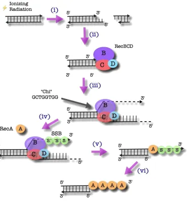
The RecBCD complex is responsible for the initiation of homologous
pathway has been derived from studies of E. coli (Dillingham & Kowalczykowski,
2008). As illustrated in Figure 1.2, a double-strand break results in a free DNA end
that is subsequently bound by the RecBCD complex. The RecBCD complex then
unwinds the double-stranded DNA (dsDNA) and digests the resulting exposed
single-stranded DNA (ssDNA) ends in a 3’ to 5’ fashion (Muskavitch & Linn, 1982).
Degradation continues until the complex reaches a chi (χ) sequence (5’-
GCTGGTGG-3’). At this point, the strand preference reverses (Anderson &
Kowalczykowski, 1997). Exonuclease activity in the 5’ to 3’ direction becomes
favoured and degradation of the 3’ end is terminated. The 3’ single-stranded DNA
product is then coated by single-stranded DNA-binding protein (SSB) to prevent
formation of secondary structures and further degradation (Muskavitch & Linn,
1982; Mackay & Linn, 1976). RecA is then recruited to this ssDNA/dsDNA junction
by the RecBCD complex and polymerizes in 5’ to 3’ fashion, displacing SSB in the
process. In this state, the resulting helical filament of RecA is primed for strand
invasion of homologous duplex DNA (Tsang et al., 1985).
Unlike E. coli, D. radiodurans has no homologues of RecB and RecC. While
it possesses a homologue of RecD, deletions of this gene do not result in
radiosensitivity (Zhou et al., 2007). Furthermore, when RecBC from E. coli are
expressed in D. radiodurans, radioresistance is not improved (Khairnar et al.,
2008), suggesting that the initiation of homologous recombination in D.
Figure 1.2: The Initiation of HR by the RecBCD Complex in E. coli
i) A double-strand break results from, in this instance, ionizing radiation.
ii) RecBCD complex binds the resulting DNA end.
iii) RecBCD complex unwinds the dsDNA and begins to digest the ssDNA, in a 3’ to 5’ fashion.
iv) When the complex reaches the χ region, degradation becomes favoured in the 5’ to 3’ direction, thus ending the breakdown of the 3’ end. SSB coats the 3’ strand to prevent secondary structure formation as well as further degradation.
v) RecA is recruited to the ss/dsDNA junction by the RecBCD complex.
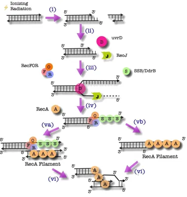
1.4.2 The RecFOR Pathway
In E. coli, RecBC-knockouts are capable of initiating the loading of RecA
using the alternative RecFOR pathway (Lloyd & Buckman, 1985). D. radiodurans
has homologues of the key proteins involved in the RecFOR alternative pathway
(White et al., 1999). Furthermore, when sbcB, an inhibitor of the RecFOR pathway
from E. coli, is expressed in D. radiodurans, a decrease in radioresistance is
observed (Misra et al., 2006). Additionally, in D. radiodurans, RecF, RecO, RecR
and RecA knockouts all exhibit radiosensitivity (Bentchikou et al., 2010),
suggesting that the RecFOR pathway is the principal method for initiation of
homologous recombination in D. radiodurans.
As illustrated in Figure 1.3, in the Deinococcal RecFOR pathway, uvrD
recognizes broken DNA ends and unwinds the dsDNA. RecJ then degrades the
ssDNA in a 5’ to 3’ fashion (Bentchikou et al., 2010). No sequence comparable to
the χ region has been identified and as such, the mechanism by which the activity
of RecJ is terminated remains uncertain. The other strand is then coated with either
SSB or DNA damage response B (DdrB), a protein unique to Deinococcus, with no
known homologues (Norais et al., 2009). RecFOR then binds the ss/dsDNA
junction (Timmins et al., 2007). From here, RecA ultimately promotes strand
exchange by homologous pairing of ssDNA and dsDNA by either coating a
Figure 1.3: The Initiation of HR by the RecFOR Complex in D. radiodurans
i) A double-strand break is generated, in this example, by IR.
ii) For D. radiodurans, the proteins subsequently involved are uvrD and RecJ, whereas in E. coli, RecQ replaces uvrD.
iii) UvrD recognizes the DNA end resulting from a double-strand break and unwinds the dsDNA so that RecJ may degrade the ssDNA in a 5’ to 3’ fashion.
iv) SSB or DdrB then coat the opposing strand to protect from secondary structure formation and further degradation.
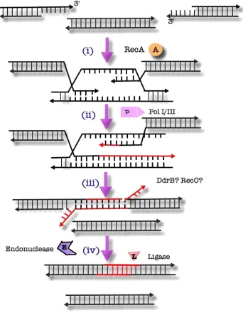
1.4.3. Extended Synthesis-Dependent Strand Annealing
Since complete and efficient recovery from extensive genomic
fragmentation requires significant DNA synthesis, conventional homologous
recombination cannot suffice as the primary mechanism of DNA repair in
Deinococcus (Zahradka et al., 2006). Instead, it is thought that a variation of
recombinational repair, termed “extended synthesis-dependent strand annealing”
(ESDSA) is used for repair of DSB’s.
According to this model (Figure 1.4), RecA-mediated strand invasion of
homologous duplex DNA initially occurs to form a D-loop. Extension of the invading
3’ strand is carried out by DNA polymerase III (pol III). DNA polymerase I (pol I) is
capable of facilitating, but not initiating, strand extension (Slade et al., 2009). The
extended invading strand then disassociates from the template strand and either
invades another homologous duplex to initiate a novel round of extension or
anneals with a complementary extension formed via the same mechanism using a
different template (Zahradka et al., 2006). Given the rapid speed by which long (up
to 20 kb) ssDNA overhangs are converted to duplex DNA, a model has been
proposed whereby extension occurs simultaneously along a single fragment that
further serves as a bridge or a scaffold (Figure 1.5) (Slade et al., 2009). Finally,
RecA-mediated recombination facilitates recircularization of the newly formed
Figure 1.4: ESDSA Repair of Double-Strand Breaks in D. Radiodurans
i) Hundreds of double-stranded fragments are formed following a DNA damaging stimulus, such as ionizing radiation, which is pictured above.
ii) UvrD and RecJ then process the fragments to generate long 3’ ssDNA strands.
iiia) Approximately one third of these fragments are then assembled into larger fragments via the mechanism of single-strand annealing.
Iiib) Fragments undergo RecA-mediated strand invasion.
iv) The invading 3’ strand is then extended by a combination of pol I and pol III. The strand is then free to participate in either:
via) another round of strand invasion or
vib) association with a complementary fragment.
vii) Any gaps in the annealed fragments are filled and nicks are sealed by a ligase.
Figure 1.5: Synthesis Along a Bridging Element
i) Two DNA fragments, missing a segment of sequence between them, simultaneously invade a fragment containing the missing sequence.
ii) Both fragments are extended by a combination of pol I and pol III.
iii) The two fragments then disassociate from the bridging fragment and associate with each other.
1.4.4 Single-Strand Annealing
DNA fragment assembly has been shown to occur in D. radiodurans in
RecA-knockouts, suggesting that the process can occur independently of RecA
and therefore, ESDSA (Daly & Minton, 1996; Slade et al., 2009). Following an
ionizing radiation dose of 10 kGy, one third of all double-strand breaks are rejoined
prior to RecA-mediated repair. Using this process, larger, partially repaired
fragments are formed that may be better suited for subsequent repair by ESDSA
(Daly & Minton, 1996). The RecA-independent repair occurs via a single-strand
annealing (SSA) mechanism, similar to the one observed in E. coli (Daly & Minton,
1996; Kowalczykowski et al., 1994). In this model (Figure 1.4, iiia), a 3’ ssDNA end
generated by uvrD and RecJ is annealed to a complementary 3’ ssDNA fragment
from a separate genomic copy that was processed in the same manner. Any 5’
flaps that remain after annealing are degraded and remaining gaps are filled in by
DNA polymerase (Daly & Minton, 1996).
Work reported in this thesis demonstrates that the DNA damage response
A (DdrA) protein possesses a novel ssDNA annealing activity that is required for
DNA damage resistance in D. radiodurans.
1.4.5 Novel Deinococcal Proteins Involved in Repair
A full understanding of the proteins required for ESDSA and SSA is
currently lacking. Only a few proteins have functionally assigned roles and most of
analogy to what is known from their study in other systems (Slade et al., 2009). In
addition to DdrA, two proteins (PprA and DdrB) have been shown to be critical for
repair. All three proteins appear unique to the bacterium and have been identified
in the genomes of all strains of Deinococcus that have been analyzed to date.
DdrA, PprA and DdrB are upregulated following exposure to ionizing radiation,
suggesting a role in repair. Furthermore, cells lacking DdrA, PprA or DdrB all exhibit
radiosensitivity. A combined knockout of DdrA and DdrB results in greater
radiosensitivity than knocking out either protein in isolation. Likewise, knocking out
DdrA or DdrB together with RecA yielded greater radiosensitivity than knocking out
RecA alone. Together, these findings suggest that DdrA and DdrB are epistatic to
one another and to RecA. One possible interpretation of the data is that, similar to
DdrA, DdrB may also enhance ssDNA annealing. If this hypothesis were correct,
and the proteins were indeed responsible for performing redundant functions, it
would explain why knocking out both proteins together leads to a greater reduction
in radioresistance than knocking out either protein alone (Tanaka et al., 2004).
Recent work in the Junop lab has demonstrated ssDNA annealing activity in DdrB
(Sugiman-Marangos et al., 2016), adding strength to the idea that DdrA and B may
share similar function in SSA repair.
In contrast, knocking out PprA and RecA together results in the same level
of radiosensitivity as knocking out RecA alone, suggesting that PprA and RecA act
in the same pathway. Knocking out RecA already eliminates the pathway that
PprA is involved in and the elimination of PprA function therefore results in no
1.5 DNA Damage Response A (DdrA)
1.5.1 Previous Research
When work on this thesis began, no evidence of the ability of DdrA to
enhance ssDNA annealing had been reported and no crystal structure of either the
protein alone or the protein in complex with DNA had been determined.
Nevertheless, the importance of DdrA for DNA damage resistance in Deinococcus
had been clearly demonstrated. As mentioned in Section 1.4.5, DdrA was shown
to be upregulated 23-fold following exposure to a sub-lethal dose of IR (3 kGy)
(Harris et al., 2004). As well, cells lacking DdrA were found to be highly sensitive
to IR and MMC (Harris et al., 2004). Work reported by Harris et al. (2004) further
indicated that DdrA is incapable of binding dsDNA unless a 3’ ssDNA extension is
present. Providing the protein with dsDNA containing a 5’ extension resulted in no
significant interaction. Together, these findings suggest that the preferred DNA
binding interaction of DdrA is with ssDNA containing a free 3’ end. DdrA was also
reported to lack ATPase, ssDNA annealing, helicase and recombinase activity
(Harris et al., 2004).
In an effort to further define DdrA structure-function relationships, limited
proteolysis was used to probe domain structure. These in vitro studies
demonstrated that the N-terminal 157 residues of DdrA form a stable domain with
full ssDNA binding activity. Deletion of the C-terminal protease sensitive region
(residues 158-208) resulted in a partial loss of binding preference for ssDNA
residues of DdrA form a stable, functional core domain. Surprisingly, when a gene
encoding DdrA1-157 was expressed in vivo, cells were as sensitive to IR as the
knockout, indicating that the C-terminal region of DdrA plays an important role in
vivo that is in addition to its interaction with ssDNA (Figure 1.6). The C-terminus of
DdrA may serve a regulatory role or be required for interaction with other proteins
and/or DNA structures. It is also possible that the C-terminal region may be
involved in an activity that remains to be characterized (Harris et al., 2008).
Determining a high-resolution structure, especially in complex with DNA, would
offer mechanistic insight that might help address these and other questions
surrounding DdrA function.
Although a crystal structure of DdrA has not been determined, a low-
resolution electron microscopy (negative stain) structure was reported for the N-
terminal domain (residues 1-160) of DdrA from D. deserti (Gutsche et al., 2008).
The final reconstruction was determined to 23 Å and revealed a surprisingly
complex quaternary structure. DdrA assembled into a 7-subunit heptameric ring
that further self-associated into a trimer of ring structures yielding a final complex
with 21 DdrA subunits (Gutsche et al., 2008). Since the interaction surface
observed between ring structures was relatively small, it was suggested that DdrA
would most likely exist as a heptameric ring in its biologically relevant state.
Unfortunately, the low-resolution precluded further insight into structure-function
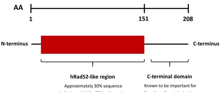
Figure 1.6: Domain Organization of DdrA. The first 151 residues of DdrA constitute a
Rad52-like domain, whereby approximately 30% of the sequence is shared with Rad52.
The thread-based homology modelling, which yielded these results, is detailed in Chapter
3. The remainder of the protein constitutes a C-terminal domain, which is important for
radioresistance in vivo (Harris et al., 2008). The first 157 residues have been shown to be
sufficient in forming a stable, functional core as DdrA1-157 (D. radiodurans) displayed a
nearly identical biochemical profile in vitro as the full-length protein. DdrA1-160 (D. deserti)
was useful in determining a low-resolution heptameric structure of the protein by negative
stain EM. DdrA1-160 (D. geothermalis) was useful in obtaining a crystal that diffracted to 2.4
Å (see Section 3.4.4). The work reported in Chapter 2 has shown that DdrA1-161 (D.
1.5.2 Current Research Outline
Given the importance of DdrA for DNA damage tolerance in Deinococcus,
a primary objective of this thesis was to further characterize the structure-function
relationships of DdrA using a combination of biophysical and cell-based
techniques. The primary goal for structural characterization was to determine an
X-ray crystal structure of DdrA in complex with DNA. Such information would not
only inform on potential mechanisms for the protein, but also provide a framework
for further hypothesis-based studies. In Chapter 3, we outline steps taken to obtain
optimal constructs of DdrA suitable for crystallization. In addition, conditions for the
successful crystallization of DdrA are reported.
Although DdrA appears to be unique to Deinococcus, it does share some
weak sequence similarity with a domain of eukaryotic Rad52 (residues 1-209)
responsible for binding ssDNA and enhancing strand annealing (Singleton et al.,
2002). We therefore sought to determine if DdrA might also be capable of
annealing ssDNA. In Chapter 2, we report the identification of robust annealing
activity within the first 161 residues of DdrA, comparable to human Rad52. This
finding is particularly significant as it contradicts a previous report that suggested
DdrA is incapable of annealing ssDNA (Harris et al., 2004). Importantly, we further
establish, through mutational studies, that this novel annealing activity is required
for the ability of DdrA to function in its role of promoting extreme DNA damage
Chapter 2: Functional
2.1 Abstract
Deinococcus radiodurans has several unique proteins required for its
extraordinary resistance toward a wide range of DNA damaging stimuli. DdrA
represents one of these proteins and is thought to be directly involved in DNA
double-strand break repair. Although DdrA shares weak sequence similarity with
human Rad52 (<10% identity), no prokaryotic Rad52-like homologue has been
shown to possess ssDNA annealing activity, suggesting that DdrA may perform a
different role in Deinococcus. To further characterize DdrA, we tested the
possibility that DdrA might function as a ssDNA annealing protein. Contrary to prior
reports, DdrA was found to possess robust ssDNA annealing activity. This activity
was localized to an N-terminal domain (residues 1-161) that appears to be partially
regulated by elements in the less structured C-terminal region (residues 161-208).
Two residues (K22 and K105) necessary for ssDNA annealing were identified and
used to demonstrate a requirement for DdrA annealing activity in Deinococcus
following exposure to extreme levels of DNA damage. Taken together, this work
not only suggests that DdrA functions as a Rad52 homologue for annealing of
ssDNA, but also represents the first demonstration that any prokaryote contains
both a functional and structural Rad52 homologue.
2.2 Introduction
Double-strand breaks are perhaps the most lethal form of damage
sustained by DNA. In lower organisms, a single DSB may prove to be lethal if left
associated with mutagenic events that can lead to a variety of deleterious
outcomes, including cancer (Pardo et al., 2009).
The bacteria of the genus Deinococcus possess a remarkable capacity to
recover from exposure to high levels of DNA damage. The model organism
Deinococcus radiodurans is capable of surviving approximately 15 kGy of gamma
radiation, which effectively shatters the genome into hundreds of fragments, each
approximately 20 to 30 kb in length (Zahradka et al., 2006). Remarkably, the
complete genome of the bacterium is faithfully reassembled from these fragments
in a matter of hours. In contrast, humans are several thousand times more
sensitive to gamma radiation with a lethal dose in the range of 2-10 Gy
(Mihandoost et al., 2014).
Since the discovery of D. radiodurans in 1956 (Anderson et al., 1956),
numerous hypotheses have been proposed in an effort to explain this remarkable
degree of survival. It has been demonstrated that D. radiodurans accumulates
DNA damage to the same extent as radiosensitive organisms, such as E. coli,
meaning that the bacterium does not protect its genome prophylactically. Instead,
radioresistance is thought to result from a combination of 1) protection of repair
proteins by elevated intracellular concentrations of manganese and 2) robust
repair pathways reliant on a collection of seemingly unique proteins (Zahradka et
al., 2006; Tanaka et al., 2004; Slade et al., 2009; Makarova et al., 2007).
The restoration of the Deinococcal genome in response to severe irradiation
extensions greater than 20 kb in length, which result from successive rounds of
strand invasion of homologous fragments followed by Pol I and Pol III-mediated
strand extension (Zahradka et al., 2006; Slade et al., 2009). In the second stage
of repair, complete circular chromosomes are generated by the joining of long
linear DNA molecules via RecA-dependent homologous recombination.
Interestingly, fragment assembly has also been observed in Deinococcal cells
lacking RecA function, indicating that the process is capable of occurring
independently of ESDSA (Daly & Minton, 1996; Slade et al., 2009). In fact,
approximately one third of all double-strand breaks resulting from a dose of ionizing
radiation of 10 kGy are repaired in a RecA-independent manner. It is thought that
larger DNA fragments, generated by RecA-independent repair, serve as more
ideal substrates for ESDSA (Daly & Minton, 1996). This RecA-independent
process is believed to occur through a single-strand annealing mechanism (Daly &
Minton, 1996; Kowalczykowski et al., 1994), similar to the one observed in E. coli.
Despite knowing that ESDSA and SSA are essential for extreme DNA
damage tolerance, a complete list of proteins responsible for carrying out various
functions in these pathways is not yet available. It is already clear, however, that
some functions are fulfilled by ‘house-keeping’ repair proteins (i.e. RecFOR) while
other functions are completed by proteins (such as DdrA, DdrB and PprA) that are
only needed for repair of extreme amounts of DNA damage. This latter class of
proteins is of particular interest since they are likely to be directly responsible for
DdrA has been shown to be essential for extreme DNA damage resistance,
but its mechanism of action has remained elusive. Although DdrA shares weak
sequence similarity (<10% identity) with human Rad52, it was thought that DdrA
does not function as a Rad52 homologue for annealing of ssDNA. This thinking
was based on the fact that very few Rad52-like homologues have been identified
in prokaryotes and that there already exists a well characterized functional
homologue (RecO, not structurally related to Rad52) of Rad52 sufficient for
mediating strand annealing during RecA-directed repair. Nevertheless, no firm
data have been reported to conclusively rule out the possibility that DdrA functions
as a ssDNA annealing factor uniquely required for extreme DNA damage
tolerance.
In this chapter, we demonstrate robust ssDNA annealing activity in DdrA.
This represents the first report of such activity for any Rad52-like protein in any
prokaryote. Annealing activity was further localized to an N-terminal (residues 1-
161) domain that appears to be partially regulated by elements in the less
structured C-terminal region (residues 161-208). Furthermore, we identify amino
acid residues of DdrA (K22 and K105) required for binding and annealing of
ssDNA. Most significantly, we present evidence suggesting that DdrA annealing
activity is essential for Deinococcus to achieve extreme levels of DNA damage
2.3 Materials & Methods
2.3.1 Protein Preparation
Gateway Cloning
The gene encoding full-length DdrA (D. radiodurans) was synthesized
(GenScript) and codon optimized for expression in E. coli. DdrA was Gateway
cloned using the pUC57 entry vector obtained from GenScript into a pDEST527
expression vector encoding an N-terminal polyhistidine-tag and tobacco etch virus
(TEV) protease cleavage site. To determine whether the C-terminus is required in
vivo due to reasons related to ssDNA annealing, we also prepared DdrA1-161 (D.
radiodurans). This protein was similarly Gateway cloned but placed into a
pDEST14 expression vector. Unlike the pDEST527 expression vector, the
pDEST14 expression vector does not add any fusions. Instead, a C-terminal
polyhistidine-tag was engineered during gene synthesis. The integrity of the two
expression vectors was verified by Sanger sequencing. The bacterial expression
vector (pSF2285) encoding full-length human Rad52 (with an N-terminal His6x
fusion and SUMO protease cleavage site) was obtained as a kind gift from Dr.
Mauro Modesti (Centre de Recherche en Cancérologie de Marseille).
Protein Expression and Purification
All proteins were expressed in E. coli BL21 (DE3)-T1R cells (InvitrogenTM).
Cultures were grown in lysogeny broth (LB) supplemented with ampicillin (100
with 1 mM of isopropyl β-D-thiogalactopyranoside (IPTG) for 16 hours at 16°C.
Cells were harvested by centrifugation at 6000 x g for 15 min at 4°C. Cell pellets
were resuspended in lysis buffer (1 M NaCl, 20 mM Tris-HCl pH 8.0 and 5 mM
imidazole) using 10 mL buffer per gram of cell pellet and lysed by 4 sequential
French press passages at 10,000 psi. Following clarification by centrifugation at
48,384 x g for 40 min, soluble lysate was loaded onto a Ni-charged HisTrapTM Fast
Flow (FF) 5 mL column (GE Healthcare) at 1 mL/min using an ÄKTA Fast Protein
Liquid Chromatography (FPLC) system. The column was washed with 15 column
volumes each of buffer (1 M NaCl and 20 mM Tris-HCl pH 8.0) containing
increasing amounts of imidazole (0, 5, 15, 30, 45 and 60 mM imidazole) prior to
elution (600 mM imidazole). Eluted protein was buffer exchanged using a HiPrep
16/10 desalting column (GE Healthcare), equilibrated with TEV protease buffer,
composed of 150 mM NaCl, 50 mM Tris-HCl pH 8.0, 5 mM dithiothreitol (DTT) and
0.5 mM ethylenediaminetetraacetic acid (EDTA).
While the C-terminal polyhistidine-tag of DdrA1-161 was designed to be
uncleavable, full-length DdrA and Rad52 were digested using TEV and SUMO
proteases, respectively. Small-scale assays were first performed to determine
optimal conditions for cleavage. Large-scale cleavage reactions were performed
with a 10:1 ratio of fusion protein to protease. Following digestion with TEV or
SUMO protease, samples were exchanged into lysis buffer (1 M NaCl, 20 mM Tris-
HCl pH 8.0 and 5 mM imidazole) and passed over a 5 mL Ni-charged HisTrapTM
column to isolate digested protein. Proteins were concentrated by ultrafiltration
2.3.2 Analysis of Quaternary Structure
To assess monodispersity of purified DdrA and estimate quaternary
structure, size-exclusion chromatography (SEC) was performed. DdrA (10 mg/mL)
was resolved on a HiLoadTM 16/60 SuperdexTM 200 prep grade column (GE
Healthcare) using an ÄKTA Pure system (GE Healthcare) housed at 10°C. The
column was equilibrated and run with buffer containing 20 mM Tris-HCl pH 8.0, 1
M NaCl and 15% glycerol (v/v). Molecular weight standards were run under the
same conditions to calibrate the column for size estimation of DdrA.
Size-exclusion chromatography with multi-angle light scattering (SEC-
MALS) was also performed under the same conditions with the SEC column
connected in-line to a Dawn HELEOS II MALS detector equipped with a 662 nm
laser source and an Optilab T-rEX differential refractometer equipped with a 658
nm LED source (Wyatt Technology, Santa Barbara, CA, USA). Molecular weights
were calculated by Zimm plot analysis using ASTRA software (v6.1.5.22; Wyatt
Technology).
2.3.3 ssDNA Binding Assessment
ssDNA Synthesis
As the 3’ end of ssDNA has been shown to be important for ssDNA-DdrA
interactions, oligonucleotides were labelled (BioBasic) at the 5’ end with an Alexa
Fluor488 for the purpose of visualization. Since SEC analysis suggested that DdrA