University of Pennsylvania
ScholarlyCommons
Publicly Accessible Penn Dissertations
Spring 5-17-2010
Functions of DNA Damage Response Factors in
Lymphocyte Development and Transformation
Bu YinUniversity of Pennsylvania School of Medicine, [email protected]
Follow this and additional works at:http://repository.upenn.edu/edissertations
Part of theCancer Biology Commons,Immunity Commons, and theMolecular Biology Commons
This paper is posted at ScholarlyCommons.http://repository.upenn.edu/edissertations/410 Recommended Citation
Yin, Bu, "Functions of DNA Damage Response Factors in Lymphocyte Development and Transformation" (2010).Publicly Accessible Penn Dissertations. 410.
Functions of DNA Damage Response Factors in Lymphocyte
Development and Transformation
Abstract
DNA double strand breaks (DSBs) can activate cell cycle checkpoints or apoptosis, and lead to genomic alterations that drive malignant transformation. The H2AX core histone variant is phosphorylated in chromatin around DSBs by kinases such as ATM and DNA-PKcs. However, how H2AX suppresses chromosome breaks and translocations in cells and prevents tumorigenesis in mice and humans is not well understood. V(D)J recombination is a genetically programmed DNA damage and repair process that assembles the variable region exons of antigen receptor genes in developing lymphocytes. Using an inducible V(D)J recombination system, I found that H2AX is phosphorylated along cleaved antigen receptor loci DNA strands, prevents their irreversible separation in G1 phase, and reduces chromosome breaks and
translocations in subsequent cell cycles. Consistent with H2AX functions in DSB repair, I also demonstrated that conditional H2AX deletion results in accumulation of genomic instability in cells, but delays tumor onset in a mouse thymic lymphoma model, presumably due to increased death of cells with synthetic loss of multiple repair factors. To further test this possibility, I generated cells and mice deficient in both H2AX and ATM to examine whether ATM-independent H2AX functions downstream of other kinases are essential for proper DSB repair. I found that thymocyte-specific ablation of H2AX in ATM-deficient mice results in a 50% reduction in thymus cellularity but does not accelerate or delay tumorigenesis. My results suggest that the outcomes of functional interactions between DNA damage response factors likely depend on the cellular context. Additionally, I discovered a novel function of ATM in regulating mono-allelic recombination at the immunoglobulin light chain locus. ATM could orchestrate the signaling pathways enforcing allelic exclusion to provide a time window to test whether the rearrangement on the first allele is productive. In summary, my work has provided novel mechanistic insights into how DNA damage and repair factors coordinately regulate V(D)J recombination, lymphocyte development and neoplastic transformation.
Degree Type Dissertation
Degree Name
Doctor of Philosophy (PhD)
Graduate Group
Cell & Molecular Biology
First Advisor
Craig H. Bassing, PhD
Keywords
DNA Damage and Repair, V(D)J Recombination, H2AX, ATM, Lymphoma, Allelic Exclusion
Subject Categories
FUNCTIONS OF DNA DAMAGE RESPONSE FACTORS IN LYMPHOCYTE
DEVELOPMENT AND TRANSFORMATION
Bu Yin
A DISSERTATION
in
Cell and Molecular Biology
Presented to the Faculties of the University of Pennsylvania
in Partial Fulfillment of the Requirements for the Degree of Doctor of Philosophy
2010
Supervisor of Dissertation
_______________________________
Craig H. Bassing, PhD, Assistant Professor, Pathology and Laboratory Medicine
Graduate Group Chairperson
______________________________
Daniel S. Kessler, PhD, Associate Professor, Cell and Developmental Biology
Dissertation Committee
Shelley L. Berger, PhD, University Professor, Cell and Developmental Biology Tom Curran, PhD, FRS, Professor, Pathology and Laboratory Medicine
Zissimos Mourelatos, MD, Associate Professor, Pathology and Laboratory Medicine Warren S. Pear, MD, PhD, Professor, Pathology and Laboratory Medicine
DEDICATION
I dedicate this thesis to my parents, who raised me, cared for me, and had to get
used to living without me being around, just because they love me and have always had
ACKNOWLEDGEMENTS
I would like to thank my advisor Dr. Craig H. Bassing for his generous support,
without which I would not have achieved what I have now. Thank you for your
encouragement in my research, your guidance at and beyond the bench, and your efforts
in helping me to become a better scientist and a better person in general. I am indebted to
all current and past Bassing lab members, especially Andrea Carpenter, Velibor Savić
and Beth Nuskey, for teaching me the essential techniques, Brenna Brady, Natalie
Steinel, Marta Rowh and Angela Fusello for discussions, and Katherine Yang-Iott for
making the lab feel like home. A lot of thanks also go to the Cancer Biology/Cell and
Molecular Biology graduate group at Penn. The excellent faculty members and many
exceptional graduate students have been tremendous role models. Among them I would
like to acknowledge my thesis committee, Dr. Tom Curran, Dr. Shelley L. Berger, Dr.
Warren S. Pear, Dr. Zissimos Mourelatos, and my chair Dr. M. Celeste Simon, for their
support and scientific advice. Marisa Juntilla from Dr. Gary Koretzky’s lab was
instrumental in the work described in Chapter II. Dr. Aaron Gitler has been very
supportive in my job search. I would also like to acknowledge my collaborators, Dr.
Barry P. Sleckman together with Grace Mahowald, Beth Helmink, Eric Gapud and
Andrea Bredemeyer from Washington University at St. Louis. Without their pioneering
work and sharing of expertise and reagents, most of my work would not have been
possible. I’ve also enjoyed collaborations with Dr. Jane Skok’s lab at New York
University, and with Dr. Larry Turka’s lab at Penn. I thank Xiaohe Liu from the Turka
ABSTRACT
FUNCTIONS OF DNA DAMAGE RESPONSE FACTORS IN LYMPHOCYTE
DEVELOPMENT AND TRANSFORMATION
Bu Yin
Advisor: Craig H. Bassing
DNA double strand breaks (DSBs) can activate cell cycle checkpoints or apoptosis,
and lead to genomic alterations that drive malignant transformation. The H2AX core
histone variant is phosphorylated in chromatin around DSBs by kinases such as ATM and
DNA-PKcs. However, how H2AX suppresses chromosome breaks and translocations in
cells and prevents tumorigenesis in mice and humans is not well understood. V(D)J
recombination is a genetically programmed DNA damage and repair process that
assembles the variable region exons of antigen receptor genes in developing
lymphocytes. Using an inducible V(D)J recombination system, I found that H2AX is
phosphorylated along cleaved antigen receptor loci DNA strands, prevents their
irreversible separation in G1 phase, and reduces chromosome breaks and translocations in
subsequent cell cycles. Consistent with H2AX functions in DSB repair, I also
demonstrated that conditional H2AX deletion results in accumulation of genomic
instability in cells, but delays tumor onset in a mouse thymic lymphoma model,
presumably due to increased death of cells with synthetic loss of multiple repair factors.
To further test this possibility, I generated cells and mice deficient in both H2AX and
kinases are essential for proper DSB repair. I found that thymocyte-specific ablation of
H2AX in ATM-deficient mice results in a 50% reduction in thymus cellularity but does
not accelerate or delay tumorigenesis. My results suggest that the outcomes of functional
interactions between DNA damage response factors likely depend on the cellular context.
Additionally, I discovered a novel function of ATM in regulating mono-allelic
recombination at the immunoglobulin light chain locus. ATM could orchestrate the
signaling pathways enforcing allelic exclusion to provide a time window to test whether
the rearrangement on the first allele is productive. In summary, my work has provided
novel mechanistic insights into how DNA damage and repair factors coordinately
TABLE OF CONTENTS
Title Page i
Dedication ii
Acknowledgements iii
Abstract iv
Table of Contents vi
List of Figures viii
List of Tables x
Chapter I – Introduction 1
DNA Damage and Repair Mechanisms Are Essential for Tumor Suppression 2 DNA Double Strand Break Response and Repair within Chromatin 3 H2AX Phosphorylation Is a Hallmark of Chromosomal DSB Response 9 V(D)J Recombination is a Physiological DNA Damage and Repair Process 15 Novel Cell Line Systems and Mouse Models to Study H2AX Functions 20 DNA Damage Signals and Mono-allelic V(D)J Recombination 26
Chapter II – Histone H2AX Stabilizes Broken DNA Strands to Suppress Chromosome Breaks and Translocations in V(D)J Recombination
35
ABSTRACT 36
INTRODUCTION 37
RESULTS 40
DISCUSSION 54
FIGURES AND FIGURE LEGENDS 59
Chapter III – Cellular Context Dependent Effects of H2ax and p53 Deletion Upon Development of Thymic Lymphoma
74
ABSTRACT 75
INTRODUCTION 76
RESULTS 80
DISCUSSION 91
FIGURES, FIGURE LEGENDS AND TABLES 96
Chapter IV – Functional Interactions Between ATM and H2AX in
Lymphocyte Development, Recombination and Tumorigenesis
115
ABSTRACT 116
RESULTS 119
DISCUSSION 126
FIGURES, FIGURE LEGENDS AND TABLES 130
Chapter V – ATM-dependent DNA Damage Signals Prevents Biallelic Igκ
Chromosome Breaks
137
ABSTRACT 138
INTRODUCTION 140
RESULTS 143
DISCUSSION 147
FIGURES AND FIGURE LEGENDS 150
Chapter VI – Summary and Discussion 154
Chapter VII – Materials and Methods 164
LIST OF FIGURES
Figure 1. Model for signal propagation and amplification in DDR 30 Figure 2. Model for the chromosomal V(D)J recombination reaction 31 Figure 3. Defective repair of DSB intermediates in V(D)J recombination can lead to
chromosome breaks/translocations 32
Figure 4. Inducible V(D)J recombination in the Abelson cell lines 34 Figure 5. H2AX is not required for normal resolution of V(D)J recombination
intermediates of chromosomally integrated pMX-INV in G1 abl cells
59
Figure 6. H2AX deficient cells exhibit normal coding join formation within chromosomal substrates
61 Figure 7. No accumulation of Jκ coding ends in the absence of H2AX
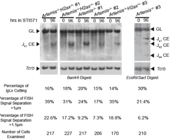
phosphorylation along RAG cleaved Igκ DNA strands 63 Figure 8. H2AX suppresses separation of RAG cleaved Igκ locus DNA strands 65 Figure 9. Quantification of Igκ cutting following STI571 induction of the cell lines
used in 2D-2C-FISH 67
Figure 10. H2AX prevents transition of RAG cleaved Igκ DNA strands into chromosome breaks and translocations during cellular proliferation
68 Figure 11. OP9-DL1 feeder cell metaphases can be separated from lymphocyte
spreads 70
Figure 12. H2AX prevents chromosome breaks emanating from un-repaired TCRα/δ locus coding ends in primary thymocytes
71 Figure 13. Models of H2AX function during end-joining repair of chromosomal
DSBs
73 Figure 14. Mice with somatic inactivation of H2ax and p53 in thymocytes succumb to clonal thymic lymphomas
96 Figure 15. LHP thymic lymphomas harbor translocations that arise from
spontaneous and programmed DNA breakage
100 Figure 16. Combined inactivation of H2ax and Artemis in p53-deficient thymocytes reduces the rate of mortality of mice from thymic lymphoma
103 Figure 17. LAP and LAHP thymic lymphomas exhibit distinct patterns of genomic
instability
110 Figure 18. LAHP thymic lymphomas exhibit increased genomic instability as
compared to LAP and LHP tumors
114 Figure 19. Characterization of thymic and splenic T cell development in LH, LA and
LAH mice
130 Figure 20. Genomic instability of stimulated splenic T cells from one LAH mice
analyzed by SKY
Figure 21. Conditional inactivation of H2ax in Atm-deficient thymocytes does not alter thymic lymphoma development or pattern of genomic instability
133 Figure 22. ATM prevents the biallelic cleavage and dissociation of Igκ coding ends and biallelic chromosome breaks or translocations
150 Figure 23. No single ATM substrates tested fully recapitulates the Atm–/– phenotype 153 Figure 24. Proposed model to explain delayed tumor development in LAHP mice
LIST OF TABLES
Table 1. Characterization of LHP Cohort Mice 98
Table 2. SKY Analysis of LHP Tumors 102
Table 3. Characterization of LAP Cohort Mice 106
Table 4. Characterization of LAHP Cohort Mice 108
Table 5. SKY Analysis of LAP Tumors 112
Table 6. SKY Analysis of LAHP Tumors 113
Chapter I
DNA Damage and Repair Mechanisms Are Essential for Tumor Suppression
The existence of most life forms depends on the stability of the genomes and the
fidelity of genomic replication, despite constant assaults to the genetic materials by
endogenous and exogenous agents (Lindahl and Barnes, 2000). To counter these threats,
multiple layers of DNA damage recognition and repair mechanisms have evolved, and
defects in these pathways are closely related to cellular and organism survival and aging,
and development of human diseases including cancers (Rouse and Jackson, 2002;
Harrison and Haber, 2006; Harper and Elledge, 2007; Jackson and Bartek, 2009).
DNA damage is common and constantly occurring. It is estimated that each of the
cells in the human body (approximately 1013 cells in total) receives thousands of DNA
lesions every day (Lindahl and Barnes, 2000). These lesions can arise during DNA
replication in S phase, in which abortive topoisomerase activities can lead to DNA strand
breaks. They can also arise in any cell cycle phases from exogenous factors like ionizing
radiation (IR) and ultraviolet light (UV), or endogenous processes such as oxidative
stress and DNA hydrolysis. Attacks on DNA can generate base losses, adducts that
impair base pairing, or DNA single-strand breaks (SSBs). In addition, double-strand
breaks (DSBs) can result from two SSBs in close proximity, replication through SSBs or
other DNA lesions, collapsed replication forks, or endonuclease cleavage. If left
un-repaired or erroneously un-repaired, DNA lesions can block transcription and genome
replication, leading to impaired celllar functions, cell cycle arrest or cell death (Figure 1).
Such persistent DNA lesions may also result in mutations or large-scale genome
advantages to incipient cells, leading to their malignant transformation. Indeed,
chromosome translocations that activate oncogenes and chromosome deletions that
inactivate tumor suppressor genes are frequently found in many human tumors (van Gent
et al., 2001; Hoeijmakers, 2001; Thompson and Schild, 2002; Mills et al., 2003; Jackson
and Bartek, 2009).
DSBs are among the most hazardous forms of DNA lesions, since liberated DNA
ends are difficult to repair and can separate irreversibly or join promiscuously (Khanna
and Jackson, 2001). Mutations of DSB repair factors result in increased cellular
sensitivity to DSBs, elevated levels of spontaneous genomic instability, and are closely
associated with an increased predisposition to cancers in both mice and humans (Mills et
al., 2003). In addition, many cancer cells also have subverted the cell cycle checkpoint
activation and cell death mechanisms to allow survival and proliferation of premalignant
cells and further neoplastic transformation (Dasika et al., 1999). Thus, understanding how
the DNA damage response (DDR) coordinates DNA repair with cell fate determination is
crucial to better prevent, diagnose and treat human malignancies.
DNA Bouble Strand Break Response and Repair within Chromatin
DNA in eukaryotic cells is packaged into chromatin, a highly organized and
dynamic DNA/protein polymer. Since chromatin is the native environment in which
DNA damage and repair must occur, how chromatin dynamics contributes to the
initiation, propagation and termination of the DDR has been the focus of recent studies
The most basic pattern of chromatin organization is ~147 base pairs of DNA
wrapped around a histone octomer core, which is composed of two of each core histones
H2A, H2B, H3 and H4, to form a nucleosome (Downs et al., 2007). Each of these
histones has a globular core domain, as well as an unstructured N-terminal tail domain
that protrude from the nucleosome core. Histones H2A and H2B also possess
functionally important C-terminal tails. In higher eukaryotes, each histone subtype, with
the possible exception of histone H4, contains a family of genes encoding multiple
variants that have altered amino acid sequences that may confer unique functions
(Brown, 2001). Core histones, including their variants, can be covalently modified by a
myriad of histone marks, including phosphorylation, acetylation, methylation,
ubiquitylation and sumoylation (Strahl and Allis, 2000). These covalent modifications
can alter chromatin structure via selectively allowing binding of non-histone effector
proteins. The combination of the covalent histone marks and the non-covalently bound
effector proteins constitute a code that regulates diverse processes such as transcription,
DNA replication, and DNA damage and repair (Strahl and Allis, 2000; Downs et al.,
2007). Between nucleosomes there are nucleosome-free regions where linker histones
and high-mobility group proteins can bind to regulate chromatin configuration and
compaction. In addition, a number of chromatin-remodeling complexes can alter
histone-DNA contacts by histone eviction, exchange of histone variants, nucleosome sliding and
changing the accessibility of particular DNA elements (Downs et al., 2007). Thus, DNA
damage within chromatin may elicit chromatin remodeling, unmask constitutive histone
modifications, making damaged DNA more accessible and coordinating DNA damage
response and repair.
In all mammalian cells, DSBs can arise accidentally following collapse of stalled
replication forks during DNA replication, or by exogenous factors such as IR. During
lymphocyte development, DSBs are also imperative intermediates during antigen
receptor gene rearrangement reactions such as V(D)J recombination (Figure 2) and B cell
class switch recombination (CSR), which are essential processes for the generation of
fully functional adaptive immune systems (Bassing et al., 2002a; Chaudhuri et al., 2007).
Whichever source they are from, DSBs pose a major threat to genome integrity since they
can lead to mutations, chromosome deletions and oncogenic translocations. Thus,
eukaryotic cells have evolved sophisticated and redundant genome surveillance
mechanisms to rapidly signal the presence of DSBs, initiate DNA repair, and coordinate
repair with cell cycle progression and cell survival decisions (Figure 1) (Rouse and
Jackson, 2002; Harrison and Haber, 2006; Harper and Elledge, 2007; Jackson and Bartek,
2009).
The DDR is executed coordinately by a constellation of sensor, transducer and
mediator/effector proteins (Figure 1). While the mechanisms by which DNA breaks are
initially sensed remain elusive and may vary depending on the context, the recognition of
DNA damages generally leads to rapid activation of the transducer kinases Ataxia
Telangiectasia Mutated (ATM) (Andegeko et al., 2001; Burma et al., 2001),
ATM/Rad3-like (ATR) (Furuta et al., 2003; Zou and Elledge, 2003). These three kinases
belong to the phosphoinositide 3-kinase (PI3K) superfamily, and optimal kinase
activation/function following DNA damage critically depends on their interactions with
the DNA ends and/or chromatin and associated factors. Active ATM/ATR/DNA-PKcs
kinases can then activate, among others, effector kinases such as Chkl or Chk2 (Dasika et
al., 1999; Matsuoka et al., 2007). Together with ATM and ATR, Chk1 and Chk2 act to
reduce cyclin-dependent kinase (CDK) activity by various mechanisms, slowing down or
arresting cell cycle progression (Jackson and Bartek, 2009). In mammalian cells, Chk1 is
the primary effector to activate intra-S and G2/M phase checkpoints, while Chk2
contributes to intra-S and G1/S checkpoint activation. ATM/Chk2-mediated stabilization
of the p53 transcription factor is essential for effective G1/S checkpoint activation
(Bartek and Lucas, 2007). The consequences of DDR signaling include cell cycle arrest
to allow time for repair of such breaks, or cell death in cases where DSB repair cannot be
easily accomplished.
Mammalian cells have evolved distinct yet complementary DSB repair pathways
that are conserved among species to ensure genomic stability. DSBs generated in
post-replicative cell cycle phases are primarily repaired by homologous recombination (HR),
whereas DSBs generated in G1-phase cells are repaired through non-homologous end
joining (NHEJ) (Lieber et al., 2003; Rooney et al., 2004b; Wyman et al., 2004). HR
involves two major sub-pathways, sister chromatid recombination (SCR) and single
strand annealing (SSA). SCR is generally limited to late S and G2 phases of the cell cycle
templates, to accurately repair breaks (Wyman et al., 2004). In contrast, NHEJ mediates
the joining of DSB ends independent of extensive homology, and is the principle
mechanism used in G1 phase although it is functional throughout the cell cycle (Lieber et
al., 2003; Rooney et al., 2004b). SSA also involves ssDNA intermediates, but repair
proceeds through the annealing of complementary sequences on the same strand with
deletion of intervening sequences. Thus, SSA is error-prone; and, as it often utilizes
repetitive elements dispersed along a chromosome, SSA deletes much larger genomic
sequences than NHEJ. Although both HR and NHEJ are important for the well-being of
cells and organisms, only NHEJ is discussed in detail due to the focus of my thesis.
The NHEJ machinery is conversed from yeast to human and consists of factors
with functions such as end stabilization, end processing and end ligation. In yeast, the
Mre11/Rad50/Xrs2 (MRX) and Ku complexes are among the first to bind DSB ends,
where they serve to protect the ends from degradation and bridge the two termini to
prevent their irreversible separation (Daley et al., 2005). In vertebrates Ku70/80 forms a
trimeric complex with the catalytic subunit DNA-PKcs, and the holoenzyme DNA-PK is
activated upon loading onto DSB ends. Besides its DNA end-bridging role, DNA-PK
may also recruit, stabilize and stimulate the ligase complex LigIV/XRCC4/XLF and
allow end re-joining to occur (Mahaney et al., 2009). It is estimated that approximately
10-15% of IR-induced DSBs are dependent on the exonuclease Artemis, whose
endonuclease activity is activated through phosphorylation by DNA-PKcs, to process
hairpin-like structures that are incompatible with end-joining (Riballo et al., 2004). Other
before ends are ligated, making NHEJ an inherently imprecise way of DNA repair
(Lieber et al., 2003).
One of the earliest events and an important component of eukaryotic DSB response
is the phosphorylation of the C-terminal SQE motif (phosphorylation site Ser139) on the
histone variant H2AX within chromatin around DSB sites, forming γH2AX that can be
visualized microscopically as foci with phospho-specific antibodies or detected by
chromatin immunoprecipitation (ChIP) around site specific DSBs (Rogakou et al., 1998;
Rogakou et al., 1999; Bonner et al., 2008; Savić et al., 2009). H2AX represents 2-25% of
the cellular H2A pool and can be incorporated into nucleosomes together with other core
histones (Rogakou et al., 1998; Bewersdorf et al., 2006). H2AX phosphorylation and
γH2AX foci formation is of fundamental importance to further signaling at the break
(Figure 1) (Downs et al., 2007; Bonner et al., 2008). An attractive model has been
proposed to emphasize the importance of this phosphorylation event. Upon DNA
damage, sensors such as the MRE11/RAD50/NBS1 (MRN, homolog of the yeast MRX
complex) complex can recognize un-repaired DSBs (Paull et al., 2000; Carson et al.,
2003; Difilippantonio et al., 2005; Horejsí et al., 2004; Lee and Paull, 2005; Uziel et al.,
2003), to which ATM is recruitmented and activated within chromatin (Bakkenist and
Kastan, 2003). Phosphorylation of H2AX by ATM then provides a binding site for DDR
factors including MRN, MDC1 and 53BP1, all of which can be phosphorylated and
activated by ATM (Downs et al., 2007; Bonner et al., 2008). The accumulation of these
DDR factors and chromatin conformation changes around the breaks may potentiate
leading to a propagation of γH2AX signal into chromatin around the breaks and
amplification of DDR signals (Figure 1) (Lou et al., 2006).
H2AX Phosphorylation Is a Hallmark of Chromosomal DSB Response
Formation of γH2AX foci after DNA damage has been widely used as an indicator
of DSB induction. Recent studies of protein-protein/protein-DNA interactions have
started to unravel the molecular architectures of the multi-component complexes within
the DNA damage foci. Among the many molecular interactions with γH2AX, the tandem
BRCA1 carboxyl-terminal (BRCT) domain of MDC1 appears to be the primary γH2AX
recognition module in higher eukaryotes (Stucki and Jackson, 2006). MDC1 mediates
effcient foci formation by other DDR factors including the MRN complex, BRCA1, PITP
and likely also 53BP1 (Stucki and Jackson, 2006; Gong et al., 2009). However, there are
factors, such as MCPH1/BRIT1, that form DNA damage-induced foci in an
H2AX-dependent but MDC1-inH2AX-dependent manner (Wood et al., 2007). In this regard, although
H2AX is not required for the initial recruitment of DDR factors, γH2AX can facilitate
DSB repair by retention of repair factors at the breaks sites (Celeste et al., 2003a). The
chromatin changes conferred by the γH2AX-recruited DDR complexes have been
proposed to anchor broken DNA ends in proximity, thus function more directly in end
joining (Bassing and Alt, 2004). When repair is completed, removal of γH2AX might be
crucial for restarting the cell cycle, and this could be accomplished by nucleosome
exchange or eviction from chromatin, or dephosphorylation via phosphatases such as
The MRN complex is required for the initial detection of DSBs and several effector
pathways, in agreement with its role as an upstream activator and a downstream target of
ATM (Uziel et al., 2003; Lee and Paull, 2004; Lee and Paull, 2005; Difilippantonio et al.,
2005). Although a direct function of ATM in HR remains controversial, the involvement
of MRN compents in HR has been documented. First, the endonuclease and exonuclease
activities of Mre11 might be involved in DNA end processing (D'Amours and Jackson,
2002; Stracker et al., 2004). Second, Rad50 has ATPase and adenylate kinase activities
that may be required for DNA tethering (D'Amours and Jackson, 2002; Stracker et al.,
2004; Bhaskara et al., 2007). Besides, Rad50 has N- and C-terminal Walker A and B
nucleotide binding motifs, respectively, that form a DNA binding domain upon their
intra-molecular association. This association also leads to the formation of a central
Rad50 hook domain (D'Amours and Jackson, 2002; Stracker et al., 2004), which can
facilitate the tethering of two distinct MRN-bound DNA molecules through an
inter-molecular interaction of two Rad50 hook domains (Anderson et al., 2001; de Jager et al.,
2001; Hopfner et al., 2002; Wiltzius et al., 2005; Moreno-Herrero et al., 2005). Since
ATM phosphorylates Mre11, RAD50 and NBS1 in response to DNA DSBs, ATM may
modulate HR repair by regulating MRN effector functions (Gatei et al., 2000; Wu et al.,
2000; Lim et al., 2000; Matsuoka et al., 2007). So far no enzymatic activities have been
attributed to NBS1; nevertheless, it’s an essential DDR mediator due to its interactions
with γH2AX, ATM and MDC1, among others. In light of the strikingly different
phenotypes of several NBS1 mutants, MRN might mediate checkpoint activation and
apoptosis induction in response to DSBs through distinct molecular domains on NBS1
complex was recently shown to also play an important role in NHEJ, likely by bridging
and stabilizing DSB ends (Helmink et al., 2009; Deriano et al., 2009; Xie et al., 2009;
Dinkelmann et al., 2009; Rass et al., 2009).
MDC1 is a major γH2AX recognition module in higher eukaryotes. MDC1
modulates S-phase and G2/M checkpoint activation after DNA damage and mediates
efficient foci formation by other DDR factors (Stucki and Jackson, 2006). Importantly,
docking of MDC1 on γH2AX has been postulated to facilitate the local accumulation of
ATM in damaged chromatin, where activated ATM phosphorylates adjacent H2AX,
generating a positive feedback loop that leads to γH2AX spreading into surrounding
chromatin and amplification of DNA damage signals (Stucki and Jackson, 2006; Lou et
al., 2006). Recent work using an inducible V(D)J recombination system in mammalian
cells has show that the γH2AX densities equilibrate within a fixed distance from break
sites, and MDC1 is essential for γH2AX formation at high densities near DSBs (Savić et
al., 2009). This in part could be due to that MDC1 bound to γH2AX might prevent
phosphatase access to the phosphoepitope. Most recently the phosphorylated SDT repeats
of MDC1 have been shown to dynamically associate with NBS1 in undamaged cells, and
mediate chromatin retention of MRN after concentration of MDC1 around DSB sites
(Melander et al., 2008; Spycher et al., 2008; Chapman and Jackson, 2008). These results
confirmed a pivotal role of MDC1 in the relay and amplification of DDR signals.
γH2AX-dependent MDC1 accumulation at the breaks site also orchestrates the
phosphorylation with ubiquitylation (Stewart, 2009). RNF8 is an E3 ubiquitin ligase and
through its FHA domain, interacts with a conserved TQXF motif on MDC1 following its
phosphorylation by ATM in chromatin. Recruitment of RNF8 to phosphorylated MDC1,
in conjunction with the E2 ubiquitin conjugating enzyme, UBC13, catalyses
poly-ubiquitylation of H2AX and other H2A histones surrounding DSBs (Huen et al., 2007;
Kolas et al., 2007; Mailand et al., 2007; Wang et al., 2007). Another E3 ubiquitin ligase
RNF168 was recently found to function downstream of RNF8 to ubiquitylate more H2A
molecules (Stewart et al., 2009; Doil et al., 2009). The ubiquitylation events around DSB
sites may provide binding sites for additional factors, or configure the chromatin into a
more accessible conformation, allowing concealed epigenetic marks such as histone
methylation to recruit additional, albeit kinetically slower, component of the DDR
cascade (Stewart, 2009).
One such component of the DDR response is 53BP1, which is phosphorylated by
ATM and contains a potential γH2AX-binding motif. Curiously, this tandem BRCT
domain is not required for its repair function; instead, its tandem Tudor motifs bind to
methylated H3K79 or H4K20 and target 53BP1 to sites of DSBs. The chromatin
conformational changes following DSB induction, as discussed above, can expose these
constitutively methylated histone marks, providing sites for the initial recruitment of
53BP1 (van Attikum and Gasser, 2009). However, 53BP1 foci form at a slower kinetics
than those of γH2AX, MDC1 or NBS1, and absence of any of these factors impairs
53BP1 foci formation (van Attikum and Gasser, 2009), presumably because these factors
interaction or exposure of large numbers of methylated histones, or both.
Recently, it was discovered that in addition to the Ser139 phosphorylation, H2AX
is also phosphorylated at residue Tyr142. In unstressed cells, Tyr142 is already
phosphorylated by the kinase WSTF; upon DNA damage, Tyr142 becomes gradually
dephosphorylated through actions of the phosphatases Eya1 and Eya3. Tyr142
dephosphorylation may be required for Ser139 phosphorylation, since the dually
phosphorylated H2AX is incapable to retain MDC1 and elicit further signaling. Thus,
Tyr142 phosphorylation status determines the relative recruitment of either DNA repair
or pro-apoptotic factors to γH2AX tails, and functions as a switch of repair versus
apoptotic responses to DNA damage (Xiao et al., 2009; Cook et al., 2009).
In summary, H2AX phosphorylation, H2A(X) ubiquitylation and other histone
modifications surrounding the DNA breaks may be required to cooperatively promote
chromatin changes around the DSB sites, efficient DNA repair, effective checkpoint
activation, and/or apoptosis decisions.
Consistent with the important roles of H2AX in response to and/or repair of DSBs,
cells deficient in H2AX have been shown to display significant growth defects, IR
sensitivity, and increased levels of genomic instability (Bassing et al., 2002b; Celeste et
al., 2002). The genomic instability of H2ax–/– cells could be due to impaired HR and/or
NHEJ repair (Bassing et al., 2002b; Celeste et al., 2002; Xie et al., 2004; Franco et al.,
efficient HR and prevent the error-prone deletional repair through SSA (Xie et al., 2004).
One potential mechanism is provided by studies in yeast post-replicative DSB repair,
where cohesin binding to the γH2AX domains around the break sites ensures sister
chromatid cohesion, promoting proper HR that uses the adjacent intact sister chromatid
as a template (Unal et al., 2004). This role of H2AX in HR is consistent with the
observation of chromatid breaks that accumulate in H2ax–/– cells (Bassing et al., 2002b;
Celeste et al., 2002). Although H2AX does not seem to be essential for the joining step of
NHEJ using an extra-chromosomal V(D)J recombination assay (Bassing et al., 2002b),
H2ax–/– cells accumulate chromosome breaks either during normal proliferation in culture
or following treatment of DNA damaging agents, indicative of compromised
pre-replicative DSBs repair that does not depend on extensive homology (Bassing et al.,
2002b; Celeste et al., 2002). Futhermore, in CSR where DSB intermediates are joined
through the NHEJ pathways, H2ax–/– B cells show reduced CSR efficency and harbor
elevated levels of CSR-associated chromosome breaks/translocations independent of the
p53-mediated checkpoint, consistent with a role of H2AX in NHEJ (Reina-San-Martin et
al., 2003; Franco et al., 2006; Ramiro et al., 2006). In addition, H2AX-deficient cells
exhibit impaired G2/M checkpoint activation in response to low, but not high, doses of IR
(Fernandez-Capetillo et al., 2002), suggesting checkpoint signaling defects in response to
physiological levels of DNA breaks may also have contributed to the accumulation of
genomic aberrations in the absence of H2AX. Thus, the delineation of H2AX functions in
DSB repair would be facilitated by experimental systems in which break repair could be
V(D)J Recombination is a Physiological DNA Damage and Repair Process
Although protocols that induce large amouts of cellular DNA damage have
provided important insights into the DDR mechanisms and the rationales of current
cancer therapies (chemotherapies and radiotherapies), cell responses to low,
physiologically relevant doses of DNA damage and mechanisms of DNA repair may be
different and are poorly understood. This knowledge is necessary to understand tumor
initiation during which somatic cells rarely receive large, but rather accumulate mutations
due to low, amounts of DNA damage. This knowledge may also form the basis for
improved cancer prevention strategies. Thus, systems where a few DSBs can be induced
and tracked in each cell would be extremely valuable for this purpose. For my thesis, I
have been using V(D)J recombination as a model to elucidate the mechanisms of
physiological DNA damage and repair.
The ability of adaptive immune systems to recognize and destroy a plethora of
pathogens depends on the immense repertoire of antigen receptor specificities of B and T
cells. Each antigen receptor is composed of a constant (C) region and a variable region,
whose extreme variability allows the antigen-binding site to recognize antigens with
specificity. In developing immature lymphocytes, the variable region exons of either
T-cell receptor (TCR) or B-T-cell receptor (BCR) genes are assembled through cutting and
pasting of germ line variable (V), diversity (D) and joining (J) gene segments, in a
genetically programmed DNA recombination process termed V(D)J recombination
In humans and mice, there are seven different antigen receptor gene loci that can
undergo V(D)J recombination. In B cells, the BCRs or immunoglobulins (Ig) are encoded
by the heavy chain (IgH) and light chain κ and λ (Igκ and Igλ) loci; in T cells, the TCRs
are encoded by the α and β chain loci in αβ T cells, or the γ and δ chain loci in γδ T cells.
Mature B cell and αβ T lymphocytes develop through a highly regulated stepwise
differentiation program that includes V(D)J recombination and periods of rapid cell
proliferation (Bassing et al., 2002a). V(D)J recombination is initiated by the lymphoid
specific endonuclease complex RAG1/2 (recombinase activating genes 1 and 2) in the
G0/G1 phase of slowly cycling immature lymphocytes. In the bone marrow, for example,
pro-B cells assemble IgH V genes and express the IgH µ chains together with the
surrogate light chains (VpreB and λ5) to form the pre-BCR, resulting in cell proliferation
and maturation into the pre-B cells stage. Early large pre-B cells turn off the
recombination machinery, and exit the cell cycle following several cell divisions. These
G0/G1 phase late small pre-B cells can now undergo IgL (IgLκ or λ) rearrangement,
becoming surface IgM+ immature B cells. The initial un-selected B cell repertoire
contains large numbers of cells with self-reactive antibodies or receptors. Further IgL
rearrangements such as secondary V(D)J recombination or receptor editing can occur in
these self-reactive B cells to rescue them from being selected against (Jankovic et al.,
2007). In parallel, in-frame VDJβ rearrangements in CD4–/CD8– (double negative, DN)
thymocytes generate TCRβ chains that, when expressed and paired up with pre-Tα
chains to form the pre-TCR on the cell surface, provide survival signals for DN
thymocytes and drive rapid cell proliferation and differentiation to the CD4+/CD8+
thymocytes. Successful VJα rearrangements generate TCRα chains that, when expressed
on the cell surface with TCRβ chains to form the αβ TCR, rescue DP cells from
apoptosis and signal differentiation to CD4+ or CD8+ (single positive, SP) thymocytes.
V(D)J recombination is an essential component of lymphocyte development, as
evidenced by the observations that Rag1–/– or Rag2–/– mice completely lack mature B and
T lymphocytes.
The combinations of joining between different V, J, and sometimes D, gene
segments generate the combinatorial diversity, which contributes to the final repertoire of
receptor specificities but by itself is not sufficient to account for the immense pool size.
Additional diversity is determined by the repair mechanisms of DSB intermediates during
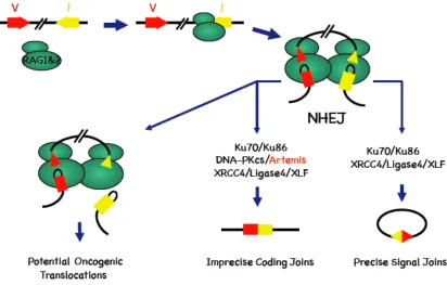
V(D)J recombination. As shown in Figure 2, The RAG1/2 complex introduces DSBs at
recombination signal sequences (RSSs) that flank V, D and J gene segments. Upon
binding to the RSSs RAG proteins introduce a single stranded nick in the DNA and
trans-esterification leads to two types of double strand breaks: blunt, 5’ phosphorylated signal
ends (SE) and covalently sealed hairpin coding ends (CE) (Fugmann et al., 2000). RAG
proteins hold V(D)J intermediates together in a synaptic complex (Agrawal and Schatz,
1997; Lee et al., 2004), facilitating their repair by seven known NHEJ factors, Ku70,
Ku86, XRCC4, ligase IV, DNA-PKcs, Artemis and XLF/Cernunnos (Lieber, 2008)
(Figure 2). All seven factors are required to process and join CEs together to form coding
joins (CJs) that encode antigen receptor exons, while all seven except DNA-PKcs and
Artemis are required to join SEs to form signal joins (SJs). In CJ formation, DNA-PKcs
CE hairpins for repair (Ma et al., 2002). Hairpin opening often generates staggered DNA
ends from which nucleotides can be trimmed by the DNA-PKcs/Artemis complex (Figure
2). Before ligation of coding ends by the XLF/XRCC4/ligase IV complex, polymerase µ
and/or polymerase λ can fill in the gaps in a template-dependent manner (Nick
McElhinny and Ramsden, 2004). In addition, terminal transferase (TdT), a
lymphocyte-specific template-independent polymerase, can add nucleotides to the CEs. The imprecise
nature of NHEJ thus contributes to the junctional diversity.
NHEJ is essential for functional V(D)J recombination in both mice and humans.
Most NHEJ-deficient mice lack mature B and T cells due to their inability to form coding
joins, which are prerequisites for clonal expansion of B and T cell precursors (Rooney et
al., 2004b). In humans, nearly 30% of all T–B– SCID (severe combined
immunodeficiency) patients show hypersensitivity to IR, because the V(D)J
recombination deficiency results from defective NHEJ, rather than mutations in their
RAG1 and/or RAG2 genes (Revy et al., 2005). The majority of these patients harbor
mutations in ARTEMIS. Mutations of other NHEJ components are also associated with
human immunodeficiencies, including those of the DNA ligase IV gene (LIG4
Syndrome) and XLF/Cernunnos gene (O’Driscoll et al., 2004; Ahnesorg et al., 2006;
Buck et al., 2006). Phenotypes of knockout/mutant mice for Ku70, Ku80, XRCC4 and
DNA-PKcs genes suggest their mutations might also lead to SCID in humans (Rooney et
al., 2004b). Indeed, van der Burg et al. recently identified a DNA-PKcs mutation that
inhibits Artemis activation and NHEJ in a radiosensitive T–B– SCID patient (van der
DSBs but are defective in the joining reaction suggests that RAG may play roles in repair
itself (such as holding the DNA ends in proximity) or in recruitment of NHEJ DNA
repair factors (Agrawal and Schatz, 1997; Lee et al., 2004). In contrast to RAGs in V(D)J
recombination, the enzyme that introduces the DSBs in CSR, and the restriction enzymes
used in a number of in vitro studies, are not known to hold DSB ends with high affinity.
Not unlike general DSBs, lymphocytes are challenged to properly activate DDR in
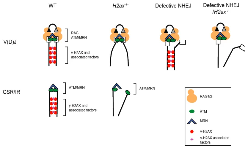
response to programmed DSBs during V(D)J recombination and CSR (Figure 3). First,
γH2AX and NBS1 form RAG-dependent foci at the Tcrα locus in developing
thymocytes, indicative of ongoing V(D)J DSB induction and/or repair (Chen et al., 2000).
Second, RAG-induced breaks are restricted to the G1 cell cycle phase, since the
Skp1-Cul1-Skp2 (Skp2-SCF) polyubiquitylates RAG2 for the proteosome-dependent
degradation at G1/S transition (Lee and Desiderio, 1999) (Figure 3). Most DSB
intermediates are believed to be repaired before S-phase entry, since un-repaired DSBs
can activate the G1/S checkpoint, although it has been reported that in at least a small
percentage of lymphocytes with un-repaired V(D)J breaks can persist into S phase even
in wild type mice (Pedraza-Alva et al., 2006). An even higher percentage of cells with
un-repaired V(D)J breaks may be able to start DNA replication in NHEJ-deficient mice,
or in the absence of effective G1/S checkpoint activation, such as in p53–/– and Atm–/–
cells (Dujka et al., 2010) (Figure 3). This fraction of DNA breaks can be replicated to
form chromosome breaks, or participate in chromosome translocations after illegitimate
joining with other breaks such as those generated during collapsed replication forks.
result in dramatically elevated genomic instability due to faulty V(D)J recombination
(Figure 3).
Consistence with the aforementioned idea, NHEJ-deficient mice rarely develop
lypmphomas because p53-mediated apoptosis can eliminate DN or pro-B cells with
un-repaired RAG-generated DSBs (Rooney et al., 2004b). NHEJ/p53 compound deficiency
allows these cells to survive but does not rescue lymphocyte development. Furthermore,
most NHEJ/p53-deficient mice rapidly succumb to pro-B lymphomas with IgH
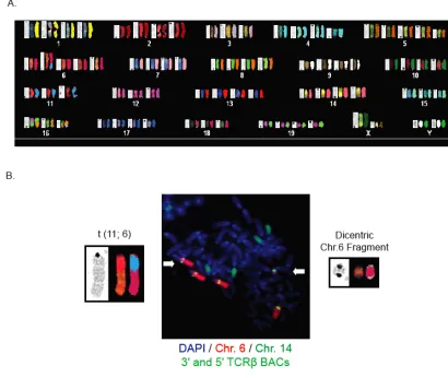
translocations, and some Artemis–/–p53–/– mice also develop thymic lymphomas with
potential TCRα/δ translocations (Rooney et al., 2004a, 2004b). Interestingly, over 10%
of peripheral T cells from Atm–/– mice contain Chr. 14 breaks or translocations that likely
involve the TCRα/δ locus, and Atm–/– mice invariably die from thymic lymphomas with
clonal TCRα/δ translocations (Liyanage et al., 2000). These abnormalities cannot be
fully explained by the checkpoint activation or apoptotic functions of ATM, suggesting
ATM may in addition function in the joining step of NHEJ, which is discussed later in
this chapter.
In summary, V(D)J recombination is highly regulated in a lineage-, developmental
stage-and cell cycle-specific manner. This V(D)J recombination program must
orchestrate chromatin conformation and DNA damage and repair proteins, in addition to
cis-acting DNA elements and trans-acting transcription factors.
Whether and how H2AX functions in NHEJ and V(D)J recombination is poorly
understood and existing results are contradictory. Similar to Atm–/– mice, H2ax–/– mice
are mildly lymphopenic and their primary αβ T cells harbor potential TCR translocations,
although the H2ax–/– defects are generally less severe than that of Atm–/– mice (Celeste et
al., 2002; Liyanage et al., 2000). Despite the dramatic genomic instability of H2ax–/–
cells, H2ax–/– mice are only slightly predisposed to thymic lymphomas, whereas H2ax–/–
p53–/– mice rapidly succumb to immature T and B lineage lymphomas and solid tumors
(Celeste et al., 2003b; Bassing et al., 2003). H2ax–/–p53–/– thymic lymphomas harbor
clonal translocations that predominantly do not involve TCR loci and, which, therefore
have been proposed to arise through the mis-repair of spontaneous DSBs. Nevertheless,
at very low frequencies, H2ax–/– and H2ax–/–p53–/– mice do develop thymic lymphomas
with clonal TCR translocations, which are not observed in p53–/– mice (Celeste et al.,
2003b; Bassing et al., 2003). Moreover, the majority of H2ax–/–p53–/– pro-B lymphomas
contain Igh/c-myc translocations, similar to those in NHEJ/p53-deficient pro-B
lymphomas (Mills et al., 2003), suggesting a role of H2AX in resolution of DSB
intermediates during V(D)J recombination. Indeed, the subset of thymic lymphomas and
pro-B lymphomas with clonal antigen receptor translocations are absent in H2ax–/–p53–/–
Rag2–/– mice, though RAG2-deficiency does not significantly alter lymphoma
development of H2ax–/–p53–/– mice (Bassing et al., 2008). Thus, among its more general
functions as a genome caretaker, H2AX may play a role in suppressing aberrant
processing of RAG-initiated DSBs, though compelling experimental evidence assaying
On the other hand, H2AX-deficient cells support normal signal and coding end
formation on transiently introduced extra-chromosomal V(D)J recombination substrates
(Bassing et al., 2002b), suggesting H2AX might not be required for NHEJ per se,
contradicting aforementioned phenotypes of H2ax–/– mice. Such discrepancies also
existed for the upstream kinase ATM until a novel cell line base system was developed to
assay chromosomal V(D)J recombination. Treatment of v-Abl transformed (Abelson)
pre-B cells with STI571, a small molecule Abl kinase inhibitor, results in rapidly
up-regulation of RAG expression and induction of DSBs at the endogenous Jκ locus and
within chromosomally-integrated recombination substrates (Figure 4) (Muljo and
Schlissel, 2003; Bredemeyer et al., 2006). A concomitant G1 arrest independent of V(D)J
recombination provides large populations of synchronized cells. To prevent elimination
of DSB-bearing cells through apoptosis, these Abelson cells were also engineered to
express the anti-apoptotic Bcl2 protein (Bredemeyer et al., 2006). Thus, RAG-generated
chromatin DSBs can be induced with high efficiency, and their repair tracked, in large
populations of G1-arrested Abelson cells. Using this system, a previously unappreciated
role of ATM was unraveled: dependent on its kinase activity, ATM stabilizes
chromosomal V(D)J recombination DSB intermediates, facilitates coding join formation,
and prevents broken DNA ends from participating in chromosome deletions, inversions,
and translocations (Bredemeyer et al., 2006). This is consistent with both the lymphoid
tumor-prone phenotype in ataxia telangiectasia (A-T) patients and the development of
thymic lymphomas harboring RAG-dependent Tcrα/δ translocations in Atm–/– mice
(Liyanage et al., 2000), neither of which could be explained solely by ATM’s checkpoint
allows immature lymphocytes with un-repaired chromosome breaks to develop and
persist into the periphery, where in the case of B cells, these RAG-initiated breaks can
join to AID-dependent breaks during CSR (Callén et al., 2007).
Definite evidence that the MRN complex components play a role in NHEJ and
V(D)J recombination was also obstained for the first time using the Abelson cells. In
humans, mutations of the ATM gene cause A-T, mutations of NBS1 cause Nijmegen
breakage syndrome (NBS) and mutations of MRE11 cause ataxia-telangiectasia-like
disease (ATLD), and all three syndromes are genetic disorders exhibiting multiple
phenotypes that include genomic instability, immunodeficiency, and increased
predisposition to lymphoid malignancies associated with antigen receptor locus
translocations. Consistent with the similarities between A-T and NBS/ATLD patient
symptoms, MRN also functions in the NHEJ-mediated repair of DNA DSBs generated
during V(D)J recombination (Helmink et al., 2009). The defects observed in MRN
mutant lymphcoytes are strikingly similar to those observed in ATM-deficient
lymphocytes, suggesting that MRN and ATM function in the same pathway in
NHEJ-mediated repair of RAG-NHEJ-mediated DSBs in G1-phase cells. Both ATM and NBS1
localize to V(D)J DSB ends and may function to tether ends in proximity in addition to
the RAG complex (Chen et al., 2000; Perkins et al., 2002). Normally, the RAG
post-cleavage complex prevents V(D)J DSBs from being repaired through alternative NHEJ,
an error-prone mechanisms for joining DSBs in mammalian cells (Corneo et al., 2007).
Using certain RAG mutants that allow destabilization of the post-cleavage complex and
pathway choice by enhancing the stability of DNA end complexes and participate in
alternative NHEJ of coding ends (Deriano et al., 2009). Thus, I reasoned that the Abelson
inducible V(D)J recombination system would allow me to uncover novel H2AX
functions.
Based on the model of γH2AX nucleating foci formation in response to general
DSBs (Bonner et al., 2008), Chapter II of my thesis focuses on testing the hypothesize
that one specific function of γH2AX is to serve as an anchor for the assembly of multiple
protein-protein/protein-DNA interactions involving MDC1, MRN and 53BP1, among
others, around chromosomal V(D) breaks. These complexes would prevent irreversible
dissociation and subsequent mis-repair of broken V, D, and J segments. Thus, by
inducing V(D)J recombination in H2ax–⁄– Abelson cells, I speculate there might be
accumulation of un-repaired DSB ends that have escaped from the post-synaptic
complex, and they are capable of participating in illegitimate end joining either in G1 or
subsequent cell cycles. Indeed, H2AX has been shown to prevent AID-dependent DNA
breaks from progressing into chromosome breaks/translocations in cells undergoing CSR,
where H2AX and ATM are required for long-range switch region synapsis (Franco et al.,
2006; Ramiro et al., 2006). However, it is equally possible that no V(D)J end joining
defects would be observed within G1-arrested H2ax–⁄– Abelson cells, either due to high
affinity of RAG proteins to DNA ends evidenced at least in vitro, or the rapid kinetics of
NHEJ as soon as breaks are induced, or a combination of both. If this is the case,
H2AX-deficiency might need to be coupled with continued cell cycle progression in order to
Abelson cells are released from G1-arrest, allow RAG2 degradation upon S-phase entry,
and monitor fates of RAG-generated DSBs (Figure 4). Since in the context of rapid
NHEJ the level of RAG-initiated breaks persisting into S-phase might be below detection
even in the absence of p53-mediated checkpoint activation, I’ve also generated and
utilized Artemis–⁄–H2ax–⁄– and Artemis–⁄–H2ax–⁄–p53–⁄– Abelson cells, where NHEJ kinetics
of coding ends is nearly down to zero due to inability to open hairpins. In these
NHEJ/p53-deficeint cells, H2AX deficiency might severely compromise positional
stability of persistent broken DNA ends even in G1 phase, and after G1/S transition, lead
to enhanced chromosome breaks/translocations involving ends that are permanently
separated from their legitimate partners. Thus, when Artemis–⁄–H2ax–⁄– and Artemis–⁄–
H2ax–⁄–p53–⁄– Abelson cells are released from cell cycle arrest, H2AX deficiency may
lead to more un-repaired V(D)J DSBs being replicated to form chromosome breaks or
translocations (Figure 4).
Previous comparative genomic hybridization analysis of NHEJ/p53-deficient mice
pro-B cell tumors show that the majority of these tumors carry a mono-allelic deletion of
a region spanning the H2ax gene on chromosome 9, suggesting H2ax haploinsufficiency
may have contributed to genomic instability and tumor development in NHEJ/p53 mice
(Bassing et al., 2003). Artemis–⁄–p53–⁄– mice can develop thymic lymphomas with clonal
TCRα/δ locus translocations (Rooney et al., 2004a), and provide a model to test the roles
of H2AX during V(D)J recombination in vivo and in subsequent lymphomagenesis. To
characterize the tumor phenotype of Artemis–⁄–p53–⁄– and Artemis–⁄–H2ax–⁄–p53–⁄– mice is
Extensive studies summarized above have placed H2AX, ATM and MRN into
highly cooperative yet non-overlapping regulatory networks during DDR. ATM and
MRN clearly have H2AX-independent functions, including roles in cell cycle
checkpoints and p53-mediated apoptosis, the impairment of ATM or MRN components
results in unique phenotypes associated with each disorder. On the other hand, H2AX is
downstream of other kinases such as DNA-PKcs and ATR, and is shown to be critical for
HR. The organization of H2AX and ATM genes in a syntenic group across species
suggests their functional interactions could be particularly relevant in tumor suppression.
The human H2AX gene (H2AFX) maps 11 Mb telomeric of ATM on Chr. 11 at 11q23,
which frequently exhibits LOH or deletion spanning both geneloci in a number of human
cancers, including immature T cell and mature B cell lymphomas (Stilgenbauer et al.,
1996, 1999). By comparing G1-arrest H2ax–/–, Atm–/– and H2ax–/–Atm–/– Abelsons cells
undergoing V(D)J recombination, I can test the hypothesis that H2AX and ATM occupy
distinct chromatin regions around break sites to promote DNA ends stability. Moreover,
haploinsufficient H2AX expression dramatically predisposes mice to genomic instability
and cancer in a p53-deficient background (Bassing et al., 2003). In this context, it will be
of particular interest to determine whether loss of one or both H2AX genes modifies the
ATM-deficient phenotype in mice. In Chapter IV of my thesis, I present some
preliminary results examining whether there are functional interplays between H2AX and
ATM in V(D)J recombination, lymphocyte development and transformation.
In the absence of any regulatory control mechanisms, both alleles of each antigen
receptor locus in diploid human and mouse lymphocytes would be available for V(D)J
recombination. However, most B and T cells express a unique antigen receptor encoded
by V-to-(D)J rearrangements of only one of the two alleles for each individual TCR/Ig
gene (Cedar and Bergman, 2008). The functional exclusion of the non-expressed alleles,
also known as allelic exclusion, is observed at the TCRβ locus in T cells and the IgH,
IgLκ and IgLλ light chain loci in B cells. Despite extensive efforts, the precise
mechanisms of how allelic exclusion is initiated and maintained remain largely unknown,
partly due to asynchronous V(D)J recombination in populations of cells in vivo.
Allelic exclusion can be achieved at multiple levels, such as locus accessibility,
DNA rearrangement, differential expression and cellular selection. For example, both
IgH and TCRβ loci exhibit allelic exclusion at the V-to-DJ step but not at the D-J step.
First, due to the imprecise nature of V(D)J end joining and 2/3 of the time each joining is
out of frame, it is suggested that the chances of both alleles undergoing productive
V-to-DJ recombination simultaneously are small, assuming the choice of which allele to
rearrange is stochastic. Second, once a productive rearrangement on one allele occurs, the
pre-BCR/TCR can send a feedback signal to suppress further V-to-DJ recombination on
the remaining allele. The pre-BCR/TCR signaling also drives proliferation and
differentiation until the IgL or TCRα rearrangements, during which further IgH or TCRβ
rearrangements need to be suppressed. This suppression could be achieved in a number of
ways, which are not mutually exclusive: down-regulation of the recombination
chromatin between V and (D)J segments, and association of alleles with nuclear
periphery or pericentromeric heterochromatic regions (Cedar and Bergman, 2008). In
contrast to the stochastic model for the initiation if allelic exclusion, it has been suggested
at least for Igκ, the two alleles are differentially marked as early and late replicating
alleles. This regulated or deterministic model argue that in pre-B cells the early
replicating allele becomes more transcriptionally accessible through DNA demethylation
and thus is predisposed to rearrange first. However, this model still cannot explain why
D-to-J recombination occurs on both TCRβ and IgH alleles. Since both TCRβ alleles are
accessible by RAGs (Carpenter et al., 2009) and germ line transcription occurs
bi-allelically (Jia et al., 2007), allelic exclusion must be initiated through other mechanisms.
Although V(D)J recombination is widely recognized as a physiological DNA
damage response, surprisingly, the possibility of DNA damage signals in coordinating
bi-allelic V(D)J recombination has never been explored. This lack of interest presumably
comes from the observation that not all antigen receptor loci exhibit allelic exclusion, and
for those that do, for example, IgH and TCRβ, D-J rearrangements can happen on both
alleles in the same cell, as evidenced by Southern blot analysis of mature T cell
hybridomas. However, it was never shown that the bi-allelic D-J rearrangements take
place simultaneously. Thus it remains possible that DNA damage signals emanating from
one allele can repress simultaneous break induction on the other allele. Intuitively, this
intra-nucleus suppressive signal would work faster than the feedback signal following
In Chapter V of my thesis, I attempt to uncover roles of the DDR machinery in
allelic exclusion. In Artemis–/– Abelson cells un-repaired Igκ locus DNA ends
accumulate, and mono-allelic recombination is not violated. On the contrary, both Igκ
alleles undergo rearrangement in a substantial portion of Artemis–/–Atm–/– cells. DSBs
generated on both alleles can persist and separate in G1 phase nuclei, and form
chromosome breaks and/or translocations upon cell cycle re-entry. These data are
consistent with the notion that ATM-mediated DNA damage signals emanating from one
rearranging Igκ allele can prevent initiation of V(D)J recombination on the second allele,
Figure 1.Model for signal propagation and amplification in DDR. Presence of DNA
lesions are detected by sensors, and H2AX can be rapidly phosphorylated by kinases such
as ATM. Recruitment of mediators through γH2AX and/or chromatin conformation
changes around the breaks can potentiate ATM activation, which leads to more H2AX
phosphorylation. DDR signals are relayed and amplified through activation of
Figure 2.Model for the chromosomal V(D)J recombination reaction. RAG1/2
(green) binds to the RSSs (triangles) of the two segments that are to be joined, in this case
a V and a J, and brings the two segments into a synaptic complex, generating two types
of DNA DSBs. The blund signal ends a re joined through NHEJ with minimal processing
to form an extra-chromosomal circle. The hairpin coding ends need to be opened by
DNA-PKcs/Artemis, before they are joined through NHEJ. The required core NHEJ
factors for coding join and signal join formation are listed. Distabilization of the
post-synaptic complex may allow escape of the DNA ends, which can participate in
Figure 3.Defective repair of DSB intermediates in V(D)J recombination can lead to
chromosome breaks/translocations. Normally, RAG1/2-mediated breaks are induced in
G1 since RAG2 is targeted for proteosome-mediated degradation upon G1/S transition.
Most V(D)J breaks are repaired before S phase entry, and un-repaired breaks can ativate
the G1/S checkpoint to arrest cell cycle progression. Low percentages of wild type
lymphocytes can occasionally enter S phase with un-repaired V(D)J breaks. However,
when the NHEJ repair pathway is defective, more un-repaired V(D)J DSB intermediates
may accumulate in G1, and with concurrent defects in checkpoint activation, these DNA
breaks can persist into S/G2 phases where they can be replicated to form chromosome
breaks, or joined to other breaks in the genome to form translocations. Since some DDR
factors (such as ATM and MRN) may have roles both in DNA repair and in checkpoint
activation, mutations or deficiencies in these factors can result in elevated levels of
Figure 4.Inducible V(D)J recombination in the Abelson cell lines. STI571 treatment
of v-abl transformed B cells leads to G1 cell cycle arrest and upregulation of RAG1/2
expression. RAG can introduce breaks at the endogenous Igκ locus (red arrows) and
chromosomally integrated recombination substrates (not shown). Artemis–⁄– Abelson cells
are used as an example to show that DSB intermediates can be monitored by Southern
blot. After break induction, STI571 can be withdrawn to allow cell cycle re-entry. DSBs
Chapter II
Histone H2AX Stabilizes Broken DNA Strands to Suppress Chromosome Breaks
and Translocations in V(D)J Recombination
Part of this chapter has been published in:
© Yin et al., 2009. J. Exp. Med. doi:10.1084/jem.20091320
ABSTRACT
The H2AX core histone variant is phosphorylated in chromatin around DSB and
functions through unknown mechanisms to suppress antigen receptor locus translocations
during V(D)J recombination. Formation of chromosomal coding joins and suppression of
translocations involve the ATM and DNA-PKcs serine/threonine kinases, each of which
phosphorylates H2AX along cleaved antigen receptor loci. Using Abelson transformed
pre-B cell lines, I now demonstrate that H2AX is not required for coding join formation
within chromosomal V(D)J recombination substrates. Yet, I show that H2AX is
phosphorylated along cleaved Igκ DNA strands and prevents their separation in G1 phase
cells and their progression into chromosome breaks and translocations following cellular
proliferation. I also show that H2AX prevents chromosome breaks emanating from
un-repaired RAG endonuclease generated TCRα/δ locus coding ends in primary thymocytes.
My data indicate that histone H2AX suppresses translocations during V(D)J
recombination by creating chromatin modifications that stabilize disrupted antigen
receptor locus DNA strands to prevent their irreversible dissociation. I propose that such
H2AX dependent mechanisms could function at additional chromosomal locations to
INTRODUCTION
The rapid phosphorylation of histone H2A proteins in chromatin for large distances
around DSBs is a conserved feature of the cellular DNA damage response. In mammalian
cells, the H2AX histone variant comprises 2-25% of the H2A pool and is non-uniformly
incorporated into chromatin (Rogakou et al., 1998; Bewersdorf et al., 2006). Upon DSB
induction, the ATM, DNA-PKcs, and ATR protein kinases phoshorylate H2AX on a
conserved carboxyl terminal serine residue to form γH2AX around DNA breakage sites
(Rogakou et al., 1999; Paull et al., 2000; Burma et al., 2001; Ward and Chen, 2001; Stiff
et al., 2004). Generation of γH2AX creates binding sites for repair and checkpoint
proteins, some of which catalyze other covalent modifications of γH2AX to generate
binding sites for additional repair and checkpoint proteins, all of which assemble into
complexes in chromatin surrounding DNA breaks (Downs et al., 2007; Bonner et al.,
2008). H2ax–/– cells exhibit increased sensitivity to agents that cause DSBs, elevated
levels of spontaneous and DSB-induced genomic instability, and defective repair of
chromosomal DSBs (Bassing et al., 2002b; Celeste et al., 2002; Xie et al., 2004; Franco
et al., 2006). Although H2ax–/– cells display apparent normal activation of p53-dependent
cell cycle checkpoints and apoptotic responses (Bassing et al., 2002b; Celeste et al.,
2002), H2ax–/– cells are defective in the G2/M checkpoint following induction of only a
few DSBs (Fernandez-Capetillo et al., 2002). The phenotypes of H2ax–/–cells suggest
that the ability of γH2AX to retain repair and checkpoint proteins around DSBs may
promote accessibility of DNA ends, stabilize disrupted DNA strands, and/or amplify
checkpoint signals (Bassing and Alt, 2004; Stucki and Jackson, 2006; Bonner et al.,