active bladder symptoms, SNM has fewer complications and has provided patients with more durable and consistent bladder control over time.
The purpose of the current study is to deter-mine whether a delay in SNM can affect the long-term outcome of treatment in patients with voiding dysfunction.
Methods
In this retrospective study, 42 patients (38 women and 4 men) with voiding dysfunction (28 with UF, 8 with nonobstructive urinary retention and 6 with UI) were selected for SNM based on successful percutaneous nerve eval-uation (PNE) of the sacral nerve roots. Treatment and follow-up of patients occurred between 2000 and 2004 at the University Health Network. Prior to the implantation of a neurostimulator (Interstim, Medtronic, Minneapolis, Minn.), patients had to pass the PNE screening test that showed > 50% improvement in one or more voiding para-meters (e.g., volume per void, number of daily voids, number of pads used daily, postresid-ual volume and number of daily catheteriza-tions) over a 1-week period (Fig. 1). The study protocol for screening using PNE and SNM has been reported.5,6Patients were stratified into 2 groups, early versus late, based on whether they received the implantable neurostimula-tor early or later after the PNE. We obtained research ethics approval for this retrospec-tive study. All patients were operated on by a single surgeon. The implantable device was Interstim model 3023, which was located in a subcutaneous pouch in the buttock. The exten-sion cable measured 10 cm (Medtronic Inc., model 3095–10). The permanent quadripo-lar electrode model 3080 was used in 12 and
Early versus late treatment of voiding dysfunction with pelvic
neuromodulation
Abstract
Introduction:Pelvic neuromodulation is an established method of treating ing dysfunction. Little is known about the pathophysiology associated with void-ing dysfunction. Reports have suggested that a delay in treatvoid-ing patients with sacral neuromodulation therapy can impact the success rate of this type of treatment in voiding dysfunction. We examined patient response to pelvic neu-romodulation when it was applied early versus late in the postdiagnosis of void-ing dysfunction.
Methods:We conducted a retrospective study of 42 patients (38 women and 4 men) with voiding dysfunction who underwent surgery for implant with the Interstim (Medtronic, Minneapolis, Minn.). Prior to implantation, patients were required to pass a percutaneous nerve evaluation (PNE) over a 1-week peri-od. Patients were observed for 20–48 months postimplantation. All patients recorded their voiding parameters at baseline, after screening and every 6 months thereafter. Twenty patients (in the early group) underwent implant surgery with the neurostimulator 2–4 weeks post-PNE, and 22 patients (the late group) had the device implanted 6–24 months post-PNE owing to local logistical circumstances.
Results:In the early group, 16 of 20 patients (80%) maintained a good response. In the late group, 13 of 22 (59%) patients showed a good response. Groups were well matched in terms of age, duration of voiding dysfunction and inci-dence of comorbidity.
Conclusion:Patients who were delayed more than 6 months in receiving the neu-rostimulator implant showed a worse response than did patients who had the device implanted soon after PNE. This indicates the possibility of disease pro-gression, which may limit the response to sacral neuromodulation.
S
acral neuromodulation (SNM), using permanent foramen S3 elec-trode, offers an alternative treatment for patients with conditions refractory to conventional measures. SNM has been approved by the US Food and Drug Administration for 3 indications: urge incon-tinence (UI), urge frequency (UF) and nonobstructive urinary retention. Several reports have been published regarding the efficacy of SNM in the treatment of UI, UF and nonobstructive urinary retention.1–4SNM is considered a minimally invasive procedure, and when com-pared with the more drastic procedures to control intractable
over-CUAJ 2007;1(2):106-10
Magdy M. Hassouna, MD, PhD; Mohamed S. Elkelini, MD
14 patients in the early and late groups, respective-ly. The rest of the patients received the “tined” elec-trode lead model 3889. We used only hardware manufactured by Medtronic Inc.
We verified the duration of the voiding dysfunc-tion in both groups as well as the comorbidity and the medications the patients were taking.
All patients were observed every 6 months for 24–48 months, with a mean of 32.5 months. In each follow-up visit, the patients had to present a 4–5 day voiding diary recording their voiding parameters (number of voids per day, volume in mL per void and urine volume obtained by catheter) and incon-tinence (number of pads used daily and amount of daily leak). The parameters in the voiding diaries were compared individually with the baseline and with the postscreening (post-PNE diary). To quali-fy for the implantation of a permanent neurostim-ulator, the patient had to show an improvement of at least 50% in one or more of the voiding param-eters mentioned in the post-PNE diary when com-pared with the baseline diary. The criteria of success (i.e., responders) in the postoperative follow-up were voiding parameters similar to or better than that shown on the post-PNE diary and overall patient sat-isfaction. The criteria of failure (i.e., nonresponders) were voiding parameters worse than post-PNE diary but better than baseline diary, voiding parameter returning to the baseline diary and the removal (i.e., explantation) of the neurostimulator due to lack of efficiency, complications or both.
The voiding parameters were subject to a sta-tistical program (SigmaStat 3.0A). We compared data collected from voiding diaries using 1-way analysis of variance followed by a post-hoc com-parison of the means; the significant difference was set at p < 0.05.
Results
The results are presented as a comparison between the voiding parameters recorded in the diaries of patients in the early and late implantation groups. The early group included 20 patients (18 wo-men and 2 wo-men) who received the permanent implant 2–3 weeks after showing a successful screening test. Patients in the early group had void-ing dysfunction for 20–60 months (mean 34 mo). The late group included 22 patients (20 women and 2 men) who received the implant 6–24 months (mean 18.5 mo) after the screening test. The
dura-tion of the voiding dysfuncdura-tion was 19–70 months, with a mean of 30 months duration. There was no statistical difference in the duration of voiding dys-function between the 2 groups (p > 0.05). The patients’ ages were similar in the 2 groups, aver-aging 42 years in the early group and 45 years in the late group. The intake of the medication and the comorbidity in both groups were very similar.
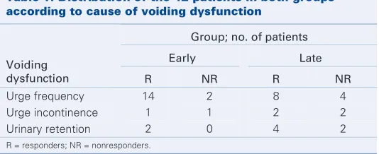
In the early group, 16 patients were diagnosed with UF with suprapubic discomfort or pain, com-pared with 12 patients in the late group. There were 2 patients with urinary retention in the absence of evidence of outlet obstruction in the early group, whereas there were 6 in the late group. Two patients in the early group and 4 in the late group had UI (Table 1).
Screening test (PNE)
There were 12 PNE attempts performed for 12 patients in the early group. For the late group,
Table 1: Distribution of the 42 patients in both groups according to cause of voiding dysfunction
Group; no. of patients
Early Late
Voiding
dysfunction R NR R NR
Urge frequency 14 2 8 4
Urge incontinence 1 1 2 2
Urinary retention 2 0 4 2
R = responders; NR = nonresponders.
Voiding diary
Patients with voiding dysfunction
Percutaneous nerve evaluation
Improvement of > 50% of voiding parameters
Permanent implantation
< 3 weeks > 6 months
Early group Late group
24 PNE attempts were performed for 22 patients. Two patients had their PNE repeated owing to tech-nical difficulties.
Waiting time
In the early group, all 20 patients had their perma-nent implant within 2–3 weeks. In the late group, patients had to wait for 6–24 months, with a mean waiting time of 18 months.
Location of the permanent electrode
All patients had a single electrode (model 3889 Medtronic Inc.) implanted. Of patients, 25 had the implantation of a permanent neurostimulator on the left third sacral nerve root, and 15 patients had it on the right third sacral root. In 2 patients, the permanent electrode was implanted on the right fourth sacral nerve root.
Response during the follow-up
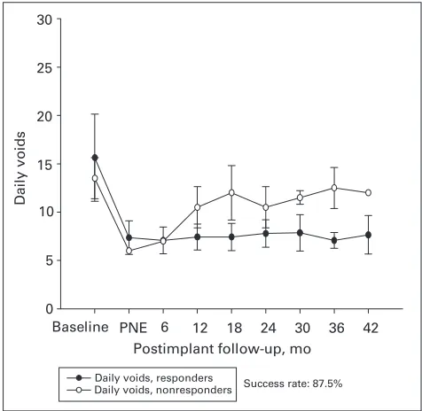
In the early group, 17 of 20 patients (85%) showed good response in terms of their symptoms and their overall satisfaction for a follow-up period of 32.5 (mean) months. Among the subgroup of patients with UF, 14 of 16 patients had a good response (87%) both in the voided volume per void and the number of daily voids (Fig. 2 and Fig. 3). The improvement in the voiding parame-ters was maintained through the follow-up period, as depicted in Figure 2. Three patients had poor response; 1 was due to wound infection, and the other 2 showed poor response 8 and 12 months after the implantation of the permanent stimula-tor, respectively.
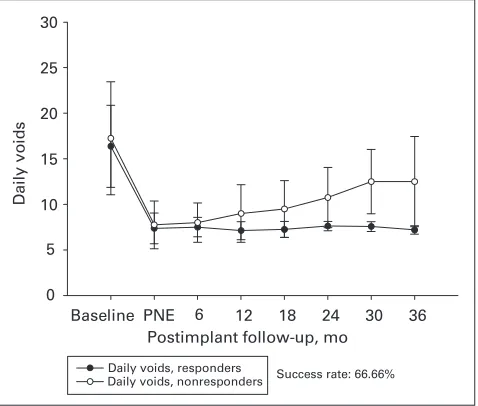
In the late group, 14 of 22 patients (63.6%) showed good response in their symptoms and their overall satisfaction. Six of the 22 patients had worse symptoms than those reported in most post-screening diaries; however, 6 months after the implantation of the permanent stimulator, their symptoms were still better than those reported in the baseline diaries. Figure 4 depicts the responses of patients in the subgroup with UF. Two patients were excluded because of lack of efficiency after 6–12 months (mean 9 mo). The mean time between the implantation and the onset of the poor response was 9 months. The patients in this group who showed an improvement in the voiding parameters maintained their response through the follow-up period. (Fig. 4 and Fig. 5)
Discussion
Pelvic neuromodulation has been an established modality of treatment for voiding dysfunction for the past 10 years. Results of several recent studies have shown level 3 evidence that SNM benefits patients with UI, UF and urinary retention. The discrepancy in the success rates, as pointed out in
Postimplant follow-up, mo Baseline PNE 6 12 18 24 30 36 42 50
100 150 200 250 300 350 400
Success rate: 87.5%
Voi
d
e
d
volume
/
void
Urinary frequency, responders Urinary frequency, nonresponders
Fig. 2.Voided volume per void of patients with urge frequency in the early group (n = 16). PNE = Percutaneous nerve evaluation.
Postimplant follow-up, mo
Baseline PNE 6 12 18 24 30 36 42
D
a
ily vo
id
s
0 5 10 15 20 25 30
Success rate: 87.5% Daily voids, responders
Daily voids, nonresponders
the literature, can be attributed to change in the technique, the disease progression or both, lead-ing to voidlead-ing dysfunction.
The current study addresses an important issue — the time factor between the screening of the patient and the implantation of a permanent device. Over the past 7 years, the Division of Urology at the Toronto Western Hospital has been a centre of referral for all patients with voiding dysfunction throughout Ontario. Those patients are referred because of intractable voiding dysfunction and because they did not show a satisfactory response to conventional therapy. Patients were screened according to the protocol that has been previous-ly described.7In short, patients were asked to fill out a voiding diary to record voiding parameters. The patients were then screened using a PNE for the sacral nerve roots bilaterally to test the integri-ty of the sacral nerve roots. The nerve root that showed a good response (motor, sensory or both) was chosen for stimulation via a temporary electrode. The patients were sent home with a temporar electrode connected to an external pulse generator for 4–7 days. During that time, the patient was asked to complete a second voiding diary. Those patients who showed a 50% improvement in one or more of the voiding parameters, as record-ed in the second voiding diary, were schrecord-edulrecord-ed to have a permanent device implanted. The loca-tion of the temporary electrode was recorded through an x-ray film of the sacrum in the lateral and antero–posterior views. Those films were used as a guide during the implantation of the perma-nent electrode. All patients were followed up every 6 months, using a voiding diary to record their post-operative response to the implanted device.
The discrepancy in the waiting period between the 2 groups of patients is owing to local circum-stances. Due to budget constraints, there has been a ceiling of 12 set on the number of annual implan-tations. Because the Toronto Western Hospital was the only centre in Ontario offering the Intrestim therapy for voiding dysfunction, we had to accept all patients referred from the entire province. Because 1 of 3 patients screened with PNE ended up having a permanent implant, those patients who were qualified for the permanent implant had to wait for up to 24 months, owing to budget restric-tions, before receiving the permanent device. Conversely, patients referred from outside Ontario had the screening test and implantation of the
per-manent stimulator within a few weeks. Owing to logistic arrangements, those patients did not have to wait for the extended period of time, because their respective provincial health insurance plans were paying for the devices.
The discrepancy in the outcome of the Interstim therapy favoured better response in the early group of patients (85% compared with 63.6% in the late group). Once patients responded to the SNM, this response was maintained during follow-up, irre-spective of whether the patient received treatment
Postimplant follow-up, mo
Baseline PNE 6 12 18 24 30 36
D
a
ily vo
id
s
0 5 10 15 20 25 30
Daily voids, responders
Daily voids, nonresponders Success rate: 66.66%
Fig. 5. Number of daily voids of patients with urge frequency in the late group (n = 12). PNE = Percutaneous nerve evaluation.
Postimplant follow-up, mo
Baseline PNE 6 12 18 24 30 36
Voi
d
e
d
volume
/
void
0 100 200 300 400 500
Urinary frequency, responders
Urinary frequency, nonresponders Success rate: 66.66%
early or late after the PNE. Upon comparison, these 2 groups of patients had similar disease duration (mean 34 months in the early group v. 32 months in the late group). The patients’ ages were similar in the 2 groups. The 2 groups also showed simi-larity in comorbidities, such as hypertension dis-ease and diabetes. They were also similar in terms of medications. None of the poor outcomes in either group could be attributed to a technical reason in the hardware since the latter was implanted equal-ly in the patients of both groups. The lack of efficacy of the SNM could be attributed to the emer-gence of such concomitant diseases as diabetes mel-litus or cereberovascular accidents. Those diseases have a negative impact on bladder function even after SNM. The patients in the 2 groups were very similar in terms of diagnosis of voiding dysfunction, age group, comorbidity and medication intake; the only difference was the latent period before the implantation of the permanent stimulator. Moreover, we did not find any difference in the time taken to the onset of poor response in either group (mean 9 mo). In clinical practice, the trend has been to offer the SNM to the patient after the failure of either treatment with pharmacotherapy, pelvic floor reha-bilitation or both to control the voiding dysfunction. A study from The Netherlands showed that patients who present with voiding dysfunction that lasted a short period of time had a higher chance of a suc-cessful outcome after test stimulation, compared with patients who had dysfunction for a relatively longer period of time.8
We strongly believe that the pathological process causing the voiding dysfunction is progressive, because the only difference in the 2 groups in the current study was the patients’ wait time for the permanent implant. If voiding dysfunction remains untreated for a long time, it makes it more diffi-cult for SNM to restore the normal balance between the afferent and the efferent components of the sacral reflex arc. The current study does not address the critical time beyond which SNM therapy may fail to restore or revert the voiding function. Studies have shown that the development of aberrant spinal
reflexes are the basis of neuroplasticity.9,10Spinal neuroplasticity could be responsible for the high failure rate in the patients who had to wait for the permanent implant. Currently, we recommend the rescreening of the patients who had to wait for 6 months to note whether they still show an appro-priate response. Staged implants would be anoth-er altanoth-ernative to introduce before the implanta-tion of the permanent device.
RReeffeerreenncceess
1. Schmidt RA, Jonas U, Oleson KA, et al. Sacral nerve stimulation for treatment of refractory urinary urge incontinence. Sacral Nerve Stimulation Study Group. J Urol 1999;162:352-7.
2. Hassouna MM, Siegel S, Nyeholt AA, et al. Sacral neuromodulation in the treatment of urgency-frequency symptoms: a multicenter study on efficacy and safety. J Urol 2000;163:1849-54.
3. Jonas U, Fowler CJ, Chancellor MB, et al. Efficacy of sacral nerve stimulation for uri-nary retention: results 18 months after implantation. J Urol 2001;165:15-9. 4. Siegel SW, Catanzaro F, Dijkema HE, et al. Long-term results of a multicenter study
on sacral nerve stimulation for treatment of urinary urge incontinence, urgency-frequen-cy, and retention. Urology 2000;56(Suppl 1):87-91.
5. Shaker HS, Hassouna M. Sacral nerve root neuromodulation: effective treatment for refractory urge incontinence. J Urol 1998;159:1516-9.
6. Shaker HS, Hassouna M. Sacral root neuromodulation in idiopathic nonobstructive chron-ic urinary retention. J Urol 1998;159:1476-8.
7. Elabbady AA, Hassouna MM, Elhilali MM. Neural stimulation for chronic voiding dys-functions. J Urol 1994;152:2076.
8. Scheepens WA, Van Koeveringe GA, De Bie RA, et al. Long-term efficacy and safety results of the two-stage implantation technique in sacral neuromodulation. BJU Int 2002;90:840-5.
9. de Groat WC. A neurologic basis for the overactive bladder. Urology. 1997;50 (6A Suppl):36-52; discussion 53-6.
10. de Groat WC. Plasticity of bladder reflex pathways during postnatal development. Physiol Behav 2002;77:689-92.
Division of Urology, Toronto Western Hospital, University Health Network, University of Toronto, Toronto, Ont.
AAcckknnoowwlleeggeemmeennttss:: This work was supported by the Department of Surgery at the University Health Network, University of Toronto, Toronto, Ont.
This article has been peer reviewed.
CCoommppeettiinngg iinntteerreessttss:: None declared.