THE PHYSIOLOGICAL EXPRESSION OF INDUCIBLE NITRIC OXIDE SYNTHASE(iNOS) IN THE HUMAN COLON.
PHILIP JOHN ROBERTS MB BS MRCP
ProQuest Number: U644292
All rights reserved
INFORMATION TO ALL USERS
The quality of this reproduction is dependent upon the quality of the copy submitted.
In the unlikely event that the author did not send a complete manuscript and there are missing pages, these will be noted. Also, if material had to be removed,
a note will indicate the deletion.
uest.
ProQuest U644292
Published by ProQuest LLC(2016). Copyright of the Dissertation is held by the Author.
All rights reserved.
This work is protected against unauthorized copying under Title 17, United States Code. Microform Edition © ProQuest LLC.
ProQuest LLC
789 East Eisenhower Parkway P.O. Box 1346
Statement of authenticity
I hereby confirm that all the experiments described in this thesis were designed and performed by myself in the Department of Gastroenterology laboratories, Addenbrookes hospital, Cambridge, UK
Acknowledgments and dedications
I would like to acknowledge the advice, support and encouragement in writing this thesis of: Dr SJ Middleton, consultant physician. Department of Gastroenterology,
Addenbrookes hospital for his close supervision of the research and writing of the thesis, and for clinical training. Dr JO Hunter, consultant physician. Department of
A bstract Background
Inducible nitric oxide synthase(iNOS) is expressed in the colonic epithelium in both inflammatory bowel disease(IBD) and colorectal cancer.-Nitric oxide(NO ), the product of this enzyme has been implicated in the pathogenesis o f both conditions. There is conflicting data, however, on whether iNOS is expressed in the normal, uninflamed human colon.
Hypothesis
In preliminary work using a colonocyte cell line(HT-29), I was able to show expression of iNOS under bacterial lipopolysaccharide(LPS) stimulation. I postulated therefore, that normal human colonic mucosa might express iNOS in the same manner in the light of its close proximity to such stimulants as LPS.
Patients and methods
RT-PCR, immunoblotting and immunohistochemistry were used to investigate iNOS expression in 17 histologically normal specimens obtained at colectomy performed for colorectal neoplasia. In addition, 14 normal mucosal biopsies were obtained
endoscopically and evaluated. Twelve surgical specimens and 16 endoscopic biopsies from patients with active ulcerative colitis(UC) were used as inflammatory controls. Results
in the majority o f the normal specimens and also to the crypt epithelium and inflammatory cells in the colitic specimens.
Conclusions
Contents Page no.
1. Title page
2. Statement of authenticity, acknowledgments, and dedications 3. Abstract
5. Contents 9. Summary
17. Abbreviations
C hapter 1. Introduction
19. Historical background and discovery o f NO* 19. Identification of EDRP as NO*
20. The biosynthesis o f NO* 21. NO*-Physiological effects 21. Cardiovascular system 21. Neurological system
22. NO* as effector of immune system 22. NO* and the gastrointestinal tract(GI) 22. Motility
23. Maintenance o f mucosal defence 23. Gastric protection
23. Intestinal protection
24. Immunomodulatory effects
24. NO* and inflammatory bowel disease (IBD) 24. 1. Cell culture systems
25. 3.NOS inhibition in models o f colitis 26. 4.Human IBD studies
27. NO and tissue damage in IBD
27. Peroxynitrite and tissue damage in IBD 28. The carcinogenic potential of NO*
28. Possible anti-inflammatoiy effects of NO* in IBD 30. INOS ‘knock-out’murine models of IBD
3 1. The constitutive expression of iNOS 32. Preliminary colonocyte experiments 32. Aims
33. Chapter 2-materials and methods 34. Antibodies and peptides
34. Buffers and stock solutions 37. Cell culture experiments 37. RT-PCR-HT 29 colonocytes 40. INOS immunoblotting
40. Immunohistochemistry HT 29 colonocytes 42. Human tissue and iNOS expression Characteristics o f patient groups
43. Table 1-surgical specimens 44. Table2-endoscopic specimens 45. RT-PCR of colonic tissue
45. Extraction and immunoblotting of iNOS 47. Immunoblotting of colonic tissue
48. Chapter 3. Results
49. Table 3- iNOS immunohistochemistry in colon 50. Fig la-UnstimulatedHT 29 cells-iNOS antibody 51. Fig Ib-Stimulated HT 29-normal rabbit serum 51. Fig Ic-Stimulated HT 29-iNOS antibody
52. Fig 2a-Qualitative iNOS mRNA-surgical tissue/colonocytes 52. Fig 2b-Kinetic iNOS mRNA-surgical tissue
53. Fig 3-iNOS immunoblotting
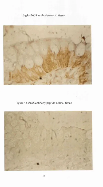
54. Fig 4a-H&E-normal colonic mucosa 54. Fig 4b-normal rabbit serum-normal tissue 55. Fig 4c-iN0S antibody-normal tissue
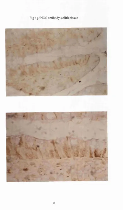
55. Fig 4d-iN0S antibody/peptide-normal tissue 56. Fig 4e-H&E-inflamed colonic mucosa 56. Fig 4f-Normal rabbit serum-colitic tissue 57. Fig 4g-iN0S antibody-colitic tissue 58. Staistical analysis(results)
59. Chapter 4-Discussion
66. The carcinogenic potential of NO in the colon 68. Summary of discussion
69. Chapter 5-COX-2 expression in IBD 69. Abstract
70. Introduction
72. Materials and methods 72. RT-PCR
74. Immunohistochemistry 75. Results
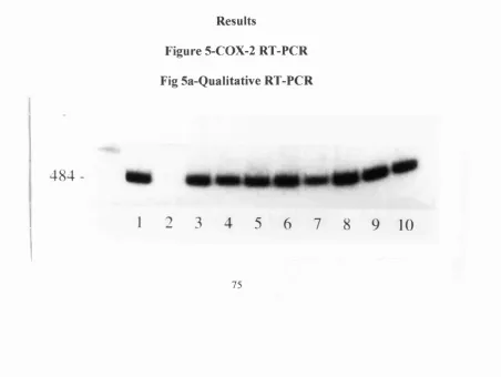
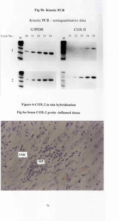
75. Fig 5a-Qualitative COX-2 RT-PCR 76. Fig 5b-Kinetic COX-2 PCR 76. In situhybridisation
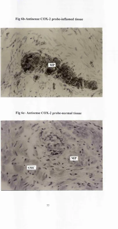
76. Fig 6a-Sense probe-inflamed tissue 77. Fig 6b-antisense probe-inflamed tissue 77. Fig 6c-antisense probe-normal tissue
78. Fig 7a-H&E myenteric plexus-inflamed tissue 78. Fig 7b-normal rabbit serum-inflamed tissue 79. Fig 7c(i,ii)-COX-2 antibody-inflamed tissue 80. Fig 7d-COX-2 antibody-normal tissue 80. Fig 7e-COX-2 antibody-inflamed mucosa 81.Fig 7f-COX-2 antibody-normal mucosa 82. Discussion
89. Chapter 6-Final conclusions 93. Future work
94. Captions to figures and legends 97. Publications from thesis
Summary
At the time of beginning the work contained in this thesis there were several reports su
ggesting that the inducible isoform of nitric oxide synthase i.e. iNOS is expressed in the colonic epithelium in active I B D ( 92-95,99-io4), and that the resultant nitric oxide(NO*) produced at this site was directly toxic to the colonocyte itself(99), thereby incriminating NO* as a pathogenic factor in this condition. Following on from this, it has been noted that the product o f the reaction between NO and superoxide radical, peroxynitrite (119,120) is also expressed by colonocytes in active I B D and it is postulated that it is this reactive product of NO* oxidation that is cytotoxic in IBD and not NO* itself (99,104). Peroxynitrite will nitrosate tyrosine residues in tissues, forming nitrotyrosine, which can be
demonstrated in situ by immunolabelling techniques (126), which have enabled the demonstration o f co-expression of iNOS and nitrotyrosine in the epithelium in active IBD(99,io4), with the suggestion that tissue damage in this condition is proportional to the amount of nitrotyrosine labeling at this site, an indirect marker o f peroxynitrite
production. Therefore reports have suggested that increased expression o f iNOS and peroxynitrite in I B D is fundamental to the pathogenesis o f this condition.
ileum (152). In all these cases there is an absence of tissue inflammation and the authors hypothesize that iNOS is induced by some luminal agent as in each case it is the surface epithelium of these tissues that express iNOS. In the light of these findings 1 formulated a similar hypothesis that the surface colonocytes in the human colon would express iNOS.
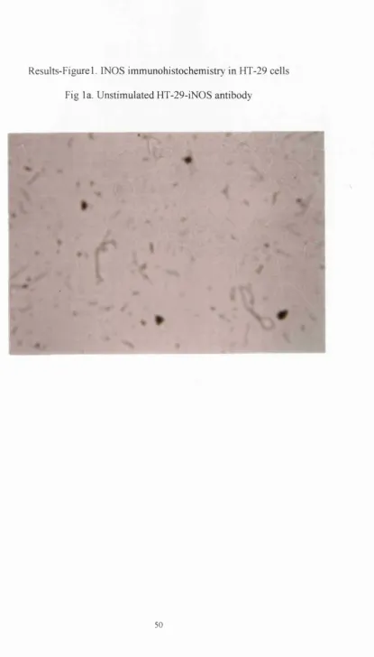
Initial work with the colonocyte cell line ‘HT-29’ showed that if these cells were grown in a germ free environment then they would not express iNOS mRNA or protein. However when these colonocytes were incubated with bacterial lipopolysaccharide, they were shown to express iNOS mRNA and protein (See Figsl&2a).
In the normal, uninflamed human colon, although there may be an absence of
inflammatory cytokines and chemokines, it is plausible that due to continuous contact of the surface epithelium with a variety of luminal agents, then iNOS could be produced in the colonocyte due to these interactions. In the light o f this hypothesis 1 sought to answer the question more thoroughly as to whether iNOS is expressed in the epithelium of the normal human colon.
Using the techniques of RT-PCR, immunohistochemistry and immunoblotting, the expression of iNOS was evaluated in 17 histologically normal surgical specimens
obtained at colectomy for colorectal neoplasia, and in 14 endoscopic biopsies taken from patients who were shown either at colonoscopy or barium enema to have normal colons. As it has been shown that carcinogenic factors can induce iNOS(25-28), tissue was
obtained from a site distant to the tumour in the colectomy specimens to attempt to avoid such stimulants, and of course the endoscopic specimens were obtained from patients without any adenomas or carcinomas, thus controlling for any such factors.
colon. Again the endoscopic biopsies were not subject to such manipulation and 4 full thickness biopsies were taken immediately prior to colectomy, thus avoiding surgical manipulation.
O f the 17 surgical ‘normal’ specimens, 9 were obtained from patients with left-sided lesions and the remaining 8 from patients with right-sided colonic lesions. The histology of the tumours revealed 14 adenocarcinomas and 3 adenomas with high-grade
dysplasia.(see table 1, pg. 43). It is important to note that most o f these patients received antibiotics at induction of anaesthesia, but were on no other medications prior to
colectomy.
As there is a wealth of data showing iNOS expression in active I B D ( 92-95,99-104), 12 colectomy specimens taken from patients with refractory UC, and 16 endoscopic biopsies o f the same condition were analyzed in a similar manner, acting as inflammatory controls. Most of the patients with UC that went to theatre were on
immunosuppressants(table l.pg 43), and were suffering from pancolitis that had failed to settle on medical therapy. None of the patients were deemed to have toxic dilatation o f the colon at the time of surgery.
RT-PCR revealed the presence of iNOS mRNA in all the surgical specimens
evaluated (see fig 2a). Kinetic analysis revealed some 6-8 fold increase in iNOS message in the colitic specimens compared to the normal tissues when standardised for G3PDH mRNA levels (see fig 2b). When looking at iNOS protein production, using
immunoblotting and a primary anti iNOs antibody monospecific for iNOS, some
peptide, confirming the presence of iNOS protein in the specimens as described (see fig
3).
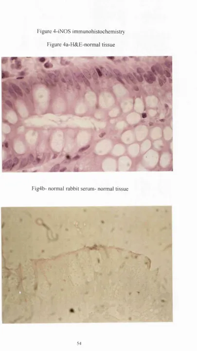
Further to this, immunohistochemistry localised iNOS protein to the surface epithelium in 14/17(82%) of the histologically normal surgical specimens and in
14/16(88%) of the normal endoscopic biopsies, (see fig 4c). INOS was also expressed in inflammatory cells, as well as the epithelium in 11/12(90%) of the surgical and
15/16(93%) of the endoscopic biopsies of patients with UC(see fig 4g).
Therefore I have shown that iNOS is expressed in a ‘constitutive’ manner in the epithelium of the normal human colon. This data compliments and extends that of Moochala et a/(i53) who have shown similar data in a smaller number o f patients, i.e. 12 endoscopic specimens using the technique of immunohistochemistry alone. A further report suggested iNOS enzyme activity in the mucosa of only a few normal specimens but an absence o f iNOS protein expression itself in these specimens (i54). This study also suggested that colonic iNOS expression was related to the stage o f cancer development with the highest levels in precancerous adenomas. Our surgical specimens were obtained as detailed from patients with colorectal cancer or adenomas although tissue was
obtained from a site distant to the lesion. As local carcinogenic factors may play a role in iNOS induction, I also examined colonic tissue taken endoscopically thereby eliminating such effects. All other studies to date, with the exception of the two mentioned above have failed to demonstrate iNOS expression in the normal colon(92-95,99-104). My data has convincingly shown such iNOS production in the colonocytes of the normal human colon in a substantial number of subjects..
in the factors contributing to iNOS expression in the inflamed, and normal colon. Of course I did not examine the effect of iNOS expression on apoptosis in the colonocyte, another mechanism whereby cellular death can be provoked in the absence of
inflammation, with reports of NO* both stimulating and inhibiting apoptosis in a variety of cell systems (I83-188). But on the evidence shown in my experiments, iNOS appears to be produced in the surface colonocyte without causing cellular death or necrosis.
The expression of this enzyme in the normal colon is likely to occur in response to some luminal agent such as bacterial LPS or some dietary component, as yet undefined. It can be seen that such expression of iNOS in the surface epithelium of the normal colon is similar to that already described in the respiratory epithelium in mancisi). As in the lung, colonocyte iNOS production could be construed as part o f the local barrier function of the tissue. The resultant production of NO* from epithelial iNOS activation could provide a means of defence against luminal bacteria, via its ability to provide an
oxidative barrier thereby preventing translocation of potentially pathogenic organisms to the deeper layers of the colon ( i 46).
Further effects of local NO* production could be in regulating micro vascular patency, permeability, tissue perfusion and mucosal barrier function as discussed above
(47,48,64). These effects have principally been shown for the constitutive NOS enzymes but recent work has shown that iNOS induction is critical in maintaining such mucosal homeostasis under conditions o f endotoxaemia (145). Currently there is no data describing such effects in the normal colon, but it seems plausible.
certain cells such as endothelial cells ( i 86, i 87), and human lymphocytes from apoptosis ( i 89). At the cellular level, the chemistry of NO* is ill-defined and complex (i89,203), with the potential of N 0 ‘ to either induce or protect against apoptosis in the normal colonic epithelium, depending upon the luminal and cellular microenvironment.
What about the possible deleterious effects of local NO* production in the normal colon? Apart from the above discussed effects on apoptosis, NO* and its oxides will nitrosate amines (134,135), which in the colonic epithelium would lead to increased levels of luminal nitrosamines with their carcinogenic potential In addition to this, NO* has been shown to deaminate DNA bases thereby having the ability to alter cellular DNA directly
(137,138). Dietary factors are implicated in colorectal carcinoma (200,201), including red meat which increases faecal concentrations of N-nitroso compounds (202). The mechanism by which this increase in nitrosamine concentration occurs is uncertain, but the surmise is that it may be via epithelial iNOS expression, with resultant nitrosation of luminal
amines by NO*, thus creating a luminal microenvironment which is more likely to lead to carcinogenesis (202).
I have shown the expression of iNOS in the mucosa o f normal human colon. The resultant NO* produced from this enzyme may have beneficial effects by both
maintaining epithelial barrier function with prevention o f bacterial translocation, and by stimulating apoptosis. Conversely, epithelial NO* if in the right luminal environment may possibly be carcinogenic to the colonic epithelium. Therefore mucosal iNOS
expression might be a link between diet or some other luminal component and colorectal carcinogenesis.
and interact (222,223). COX-2 is upregulated in IBD (218-220), with primarily epithelial expression in man, as opposed to sub-epithelial expression in an animal model of colitis
(218), In the light of this I sought to establish whether COX-2 is expressed in a similar physiological manner to iNOS in the human colon. Using the same surgical specimens as already described for iNOS, COX-2 expression was evaluated using RT-PCR, in situ hybridisation and immunohistochemistry. I also looked at 6 specimens from patients with colonic Crohns disease.
might suggest that in the human colon there are infact differing stimulants to iNOS and COX-2 expression which are as yet undetermined.
C O X - 2 is upregulated in cancer of the colon (227-229), and increased expression in active IBD may explain the increased risk of colorectal cancer in patients with chronic IBD ( 136,180).
Abbreviations.
APS-ammonium persulphate
cDNA-complimentary deoxyribonucleic acid Ci-Curie(measurement of radioactivity) DEPC-diethylpyrocarbonate
DTT-dithiotreitol
d-NTPs-deoxynucleotides
032P-CTP-DLD-1-human intestinal epithelial cell line
DAB-3'3'diaminobenzidine tetrahydrochloric dihydroate EDT A-ethylenediaminetetra-acetic acid
ECL-enhanced chemiluminescence
G3PDH-glycerol-3 -phosphate dehydrogenase HT-29-colonic epithelial cell line
IBD-inflammatory bowel disease IFNa-interferon alpha
IL-la-interluekin-1 alpha IEC-6-intestinal epithelial cell-6
LPS-lipopolysaccharide MgCLz-magnesium chloride NaCL-sodium chloride
Na2HP04-sodium phosphate (disodium phosphate) NaOH-sodium hydroxide
NOS-nitric oxide synthase: iNOS- inducible NOS eNOS-endothelial NOS nNOS-neuronal NOS NF-xB-transcription factor
NP40(tergitol)-nonylphenoxypolyethoxyethanol NH4CL-ammonium chloride
N-methylaminoacetic;N-methyl glycine-sarcosine
PCR(RT)-reverse transcription polymerase chain reaction PMSF-phenylmethy 1 sulphonyl fluoride
PBS-phosphate buffered saline RNA-ribonucleic acid
RIPA buffer(see immunoblotting) SDS-sodium dodecyl sulphate
TNF-a-tumour necrosis factor alpha
Tris base-(Trizma base)-tris(hydroxymethyl aminomethane) Tris-HCL-tris-hydrochloric acid
TBS-tris buffered saline
T ween-20-polyoxyethylenesorbit2in UC-ulcerative colitis
Chapter 1 Introduction
Historical Background of Nitric Oxide(NO*) The Discovery of NO*
Nitric Oxide(NO-), an atmospheric gas is one of the smallest known products of
mammalian cells and is measured directly as a chemiluminescent product o f its reaction with ozone.
The first suggestion that mammals were capable of producing oxides of nitrogen was as
long ago as 1916(i) when it was shown using dietary studies o f nitrogen balance that more nitrate was excreted than ingested. Further evidence did not follow until it was revealed that rodents produce nitrate in response to endotoxin(bacterial
lipopolysaccaharide-LPS) stimulation and that a nine-fold increase in urinary nitrate excretion was shown in humans with infective diarrhoea(2-5).
Identification of EDRF as NO*
I t w a s s h o w n t h a t v a s o d i l a t a t i o n o f b o v i n e a o r t i c s t r ip s i n d u c e d b y a c e t y l - c h o l i n e w a s d e p e n d e n t u p o n t h e e n d o t h e l i u m , b e i n g m e d i a t e d b y a l a b i l e , h u m o r a l f a c t o r t e r m e d ‘E n d o t h e l i u m D e r i v e d R e l a x i n g F a c t o r ’( E D R F )(8 ) . T h e e f f e c t s o f t h i s E D R F w e r e i n h i b i t e d b y h a e m o g l o b i n a n d m e t h y l e n e b l u e , p o t e n t i a t e d b y s u p e r o x i d e
d i s m u t a s e ( S 0 D ) ( 9 ) , a n d m e d i a t e d b y t h e s t i m u l a t i o n o f c y c l i c G M P , la t e r s h o w n t o b e t h e s e c o n d m e s s e n g e r o f N O * .
F u r c h g o t t s u g g e s t e d a t t h e t i m e o f t h i s s e m i n a l r e s e a r c h t h a t E D R F c o u l d b e N O * a n d f u r t h e r t o t h i s w o r k i n s e p a r a t e l i n e s o f s t u d y f i r s t l y P a l m e r e t a l(io ) a n d t h e n
I g n a r r o e t a l i i ) w e r e a b l e t o s h o w t h a t E D R F w a s i n d e e d N O * . T h i s l e d t o
t h e i d e n t i f i c a t i o n o f L - a r g i n i n e a s t h e s u b s t r a t e f o r e n d o t h e l i a l N O * p r o d u c t i o n ( i 2) a n d t h e c o n f i r m a t i o n o f t h e r o l e o f N O * a s t h e b i o l o g i c a l l y a c t i v e i n t e r m e d i a t e o f
t h e L - a r g i n i n e t o n it r it e / n it r a t e p a t h w a y . T h i s p a t h w a y w a s a l s o s h o w n i n a c t i v a t e d murine macrophages(i3-i5).
The Biosynthesis of NO
NO* i s t h e p r o d u c t o f 5 e l e c t r o n o x i d a t i o n o f L - a r g i n i n e c a t a l y s e d b y t h e e n z y m e n i t r i c
o x i d e s y n t h a s e ( N O S ) , a h a e m o p r o t e i n w h i c h r e s e m b l e s c y t o c h r o m e P450 r e d u c t a s e ( i6 ) a n d r e q u i r e s f l a v i n e a d e n i n e d i n u c l e o t i d e ( F A D ) , f l a v i n e m o n o n u c l e o t i d e ( F M N ) , c a l m o d u l i n , t e t r a h y d r o b i o p t e r i n a n d N A D P H a s c o f a c t o r s ( i 7 ,i8 ) .
N O S e x h i s t s a s t h r e e d i s t i n c t i s o f o r m s , r e p r e s e n t i n g t h r e e d i s t i n c t g e n e
p r o d u c tS (i6 ,i9 -2 i) w h i c h v a r y i n c e l l u l a r s i t e , a m i n o a c i d s e q u e n c e , r e g u l a t i n g m e c h a n i s m s a n d h e n c e f u n c t i o n a l r o l e s . F u n c t i o n a l l y t h e N O S i s o f o r m s a r e s e p a r a t e d i n t o
NOS(nNOS or NOSI), and endothelial NOS(eNOS or NOSIII), and calcium/calmodulin independent inducible NOS(iNOS or NOSH).
NOSIII i s l o c a t e d i n t h e v a s c u l a r e n d o t h e l i u m ( 22) b e i n g m o s t l y m e m b r a n e
b o u n d . NOSI h a s b e e n l o c a l i s e d t o t h e c y t o s o l o f b o t h c e n t r a l a n d p e r i p h e r a l n e u r o n s ( i6 ) , b u t i s a l s o f o u n d i n e x t r a n e u r o n a l s i t e s s u c h a s s k e l e t a l m u s c l e , t h e p a n c r e a s a n d t h e k id n ey(23,24). NOSII f r o m n o w o n r e f e r r e d t o a s iNOS i s i n d u c e d i n a v a r i e t y o f
m a m m a l i a n c e l l s b y LPS, a n d a n a r r a y o f c y t o k i n e s a n d c h e m o k i n e s s u c h a s i n t e r l e u k i n -Ifi, t u m o u r n e c r o s i s f a c t o r - a a n d g a m m a in t e r f e r o n . C e l l s c a p a b l e o f e x p r e s s i n g iNOS
i n c l u d e ; n e u t r o p h i l s , m a c r o p h a g e s , h e p a t o c y t e s , e n d o t h e l i a l c e l l s , c o l o n o c y t e s a n d s m o o t h m u s c l e cells(25-28). As c o m p a r e d t o t h e c o n s t i t u t i v e i s o f o r m s iNOS p r o d u c e s la r g e r a n d m o r e s u s t a i n e d p r o d u c t i o n o f NO* i n b i o l o g i c a l s y s te m s ( 2 9 -3 i) w h i c h e x p l a i n s i t s r o l e i n c e l l m e d i a t e d i m m u n e r e s p o n s e s .
NO*-Physiological effects
It is now known that NO* has pluripotent effects in man, with its three main classical
functions being regulation o f vascular tone(S), neurotransmission(32) and cell mediated immune response(7,i4). It acts by activating guanylate cyclase with increased cyclic GMP which in turn activates protein kinases.
Effects on cardiovascular system
NO* is well established regulator of vascular tone. Its main effect in the vascular system is that of endothelium dependent relaxation of smooth muscle(s, 11,33). Within the
vasculature, NO* is important in the inhibition of both platelet adherence(34) and
Effects on neurological system
NO* is involved in signal transduction in both the central and peripheral nervous
systems(i6,32), but also in the non-adrenergic, non-cholinergic neuronal system(NANC) of the gastrointestinal and urogenital tracts where such nerves are termed ‘nitrergic’(38,39).
NO as an effector of immune function
The first suggestion that the L-arginine/NO* pathway was involved in immune functions in mammalian cells was established in murine macrophages, when it was demonstrated that macrophage cytotoxicity was dependent upon activation o f this pathwayc?, w).
Macrophage NO* produced via stimulation o f iNOS is now recognized as a major effector molecule in host defence in man against a variety of microbes including bacteria, parasites such as malaria and schistosomiasis and a multitude o f viruses(4o-43). This
system also seems to be important in the tumouricidal activity o f macrophages. It is felt that NO* is cytotoxic to tumour cells by a variety of mechanisms. These include its ability to inhibit haem containing enzymes o f the mitochondrial respiratory chain such as aconitase of the Krebs cycle, NADPH ubiquinone oxidoreductase and succinate-
ubiquinone oxidoreductase of the electron transport chain(i5,44), thereby inhibiting cellular respiration. Other enzymes affected are non-haem metalloenzymes, and ribonucleotide reductase with its obvious effects on cellular DNA synthesis(45,46). Therefore in summary NO* is undoubtedly important as an effector molecule in mammalian cells.
NO and the gastrointestinal tract(GI tract) Motility
Gastro-intestinal(GI) tract(38,39) is o f paramount importance in the regulation o f motility throughout the GI tract(4?,48). It is involved in the regulation o f peristaltic contractions o f
the distal portion o f the oesophagus and relaxation o f the lower oesophageal sphincter In pathological specimens o f patients with achalasia and infantile pyloric stenosis, there is
an absence o f nitrergic nerves and consequent failure o f NO* driven relaxation o f the lower oesophageal sphincter and the pylorus respectively(49,50).
NO* also stimulates adaptive gastric dilatation, and regulates relaxation o f other important sphincter systems in the GI tract such as the ileocaecal valve and the anal sphincter(5i). Lastly, the tonic relaxation of the distal colon is mediated by neuronal NO* synthesis(52)
Maintenance of mucosal defence
Many studies have shown NO* to be a critical mediator of mucosal defence in the GI tract as exhibited by the following:
1.Gastric protection
In r a t s , NO* d o n o r s ( c h e m i c a l s u b s t a n c e s w h i c h r e l e a s e NO*) p r o t e c t t h e g a s t r i c m u c o s a
f r o m e t h a n o l i n d u c e d d a m a g e . F u r t h e r m o r e , e n d o t o x i n t r e a t m e n t a f f o r d s p r o t e c t i o n a g a i n s t i n g e s t e d e t h a n o l i n t h e s e r a t s , a n e f f e c t w h i c h i s a b o l i s h e d b y L-NAME, a n o n -s e l e c t i v e i n h i b i t o r o f NO* s y n t h e s i s , d e m o n s t r a t i n g t h e p r o t e c t i v e e f f e c t s o f NO*(53- 57).N0* a p p e a r s t o b e o n e o f t h e m o s t i m p o r t a n t s i g n a l s f o r m u c u s s e c r e t i o n a n d i t s
o b v i o u s p h y s i c a l b a r r ie r p ro p erties(5 8 ,5 9 ). NO* m e d i a t e s t h e h y p e r a e m i c r e s p o n s e t o l o c a l ir r it a n t s i n t h e s t o m a c h a n d p l a y s a k e y r o l e i n m o d u l a t i n g b a s a l g a s t r i c b l o o d f l o w ( 60).. NO a l s o a p p e a r s t o h a v e u l c e r h e a l i n g p o t e n t i a l , v i a e f f e c t s o n e p i t h e l i a l r e stitu tio n (6 i-6 3 ).
Maintenance of intestinal epithelial mucosal barrier function appears to be critically dependent upon NO* (47,48,64), as demonstrated by evidence that NO* maintains epithelial tight junctions and protects against ischaemia-reperfusion damage o f the feline
intestine(65).
Immunomodulatory effects of NO* in the GI tract.
NO* inhibits mediator release from mast cells in vitro(66,67) and mast cell NO* itself appears to have an immunomodulatory effect via inhibition o f other inflammatory mediators such as platelet activating factor(65-68),
NO* also regulates neutrophil recruitment by inhibition o f the expression o f adhesion molecules, including intercellular adhesion molecule I (ICAM-1) in vascular
endothelium(69).
NO* and inflammatory bowel disease(IBD)
There is a growing body of evidence suggesting that overproduction of NO* as a result of iNOS expression could play an important role in the pathogenesis of IBD, a condition characterised by increased local production of many inflammatory mediators(7o-75), which are known to induce iNOS (18,25,27,28,76).
Evidence for a pathogenic role for iNOS and NO* in IBD is suggested from cell culture experiments, animal models of colonic inflammation and the use o f both selective and non-selective NOS inhibitors in these systems, and human IBD studies.
l.C ell culture systems
LPS and the resultant increased NO* produced was shown to alter colonocyte viability(77). The same was also shown o f a human colonic epithelial cell line HT29,
which expresses iNOS mRNA and functional NOS activity as represented by increased nitrite production under cytokine stimulation with TNFa, IFNy, and IL-lo(74).
Further to this, a small intestinal cell line(IEC-6) exhibits increased NOS activity when stimulated by bacterial LPS and other pro-inflammatory cytokines(78). More
recently, it has been shown that iNOS expression is potentiated by cAMP, an
intracellular mediator o f several pro-inflammatory agents in a human intestinal cell line,
DLD-1(79).
Therefore it is apparent that both colonic and small intestinal epithelial cells are able to express iNOS under appropriate inflammatory conditions.
2.Animal models of IBD
In animal models o f colitis there is evidence o f increased luminal nitrite and nitrate levels, increased iNOS protein immunoreactivity, and increased calcium independent iNOS activity as shown by increased enzymatic conversion o f arginine to L-citrulline(the end product o f the biosynthetic pathway),(8o-85).
3.NOS inhibition in animal models of colitis
H ow ever, it has also been show n that the sam e inhibitor in similar or different m odels
had som e beneficial effects(89), w hilst in another set o f experim ents the inflam m ation was
exacerbated by this inhibitor(90).
The effects o f selective iNOS inhibition in similar experim ents has also produced
discordant results(80,82,88). In summary, these variable effects o f NOS inhibitors m ay be due to the use o f differing drug regim es, achieving partial or com plete inhibition o f
constitutive NOS, inducible NOS or both enzym es.
Therefore the benefit o f inhibition o f iNOS in animal models o f colitis has not been categorically demonstrated and o f course to date there are no experiments in human IBD patients using selective NOS inhibitors.
4.Human IBD studies
A ctive IBD is associated w ith increased levels o f various proinflammatory mediators such as IL-1, IL-6, IL-4, platelet activating factor(PAF), leukotienes ,TNFa, IFNy,
prostaglandins and complement(7o-75).
Specifically in ulcerative colitis(UC) there is increased mucosal levels o f IL-1 p, IL-6, TNFa, and IFNy ,all known ‘inducers’ o f iNOS(i8,25-28).
In view o f these findings there should be increased luminal production o f the product o f this enzyme activity,i.e. NO*. Indeed this has been shown using a chemiluminescence method(96), infra red diode laser spectroscopy(97) and by using a selective NO*
microelectrode(98) in patients with IBD.
Direct evidence for over expression o f iNOS protein in active IBD, has been shown
repeatedly being localised to the colonic epithelium and inflammatory cells o f the mucosa (99- 104), and in patients with toxic megacolon(io6). It is evident that many o f the features o f active IBD such as mucosal hyperaemia, vasodilatation, and consequent
‘leaky’ epithelium are consistent with the biological actions o f NO* (65).
NO and tissue damage in IBD
It is postulated by some workers that over production o f iNOS in both Crohns and Ulcerative colitis is critical to the pathogenesis o f this disease and tissue injury(99,ioo,io4), although this has by no means been demonstrated conclusively. Mechanisms whereby NO* can cause cellular injury in IBD include:
1 NO shows great affinity for iron-sulphur clusters which are critical to the function o f various enzymes such as glyceraldehyde-3-phosphate dehydrogenase(io7), aconitase o f the Kreb’s cycle(io8-iio), and a number o f electron transport enzymes(io8,i 11- 113). The result o f NO* binding to these active sites renders the enzyme nonfunctional.
2. NO* induces the release o f intracellular iron which inhibits mitochondrial function and DNA synthesis(ii4-ii9).
Peroxynitrite and tissue damage in IBD
NO* in the presence o f superoxide radical(02”), will produce the highly reactive
molecule peroxynitrite, ONOO"(i2o,i2i), which indiscriminately attacks biomolecules
rats when administered rectally(i25) Peroxynitrite will nitrosate tyrosine residues to form nitrotyrosine(i26) in tissues which can be labeled in situ using specific antibodies. Two
groups have exploited this feature by demonstrating a correlation between the degree o f
tissue injury in IBD and the degree o f nitrotyrosine immunolabelling(99,io4), suggesting a role for peroxynitrite induced tissue damage in active IBD. There is evidence that NO*-and peroxynitrite(ONOO ) have different targets in the mitochondrial chain and hence inhibit separate enzymes(i27,i28).
In summary, although NO* itself is a weak oxidant, peroxynitrite is a highly reactive species and both NO* and ONOO" themselves or via reactive intermediates may induce cytotoxicity via a variety of mechanism including; tyrosine nitration, lipid peroxidation, DNA strand breaks,and the consequent activation of poly-ADP ribose synthase(i22,i23,i29-
132). Furthermore nitrogen centered oxidants also degrade iron-sulphur clusters of mammalian mitochondrial enzymes, rendering them non-functional.
The carcinogenic potential of NO in UC
Apart from the potential for cellular toxicity the other possible biological consequence o f overproduction o f iNOS protein in UC is carcinogenesis. NO*, as discussed, freely reacts with oxygen radicals to yield nitrosating species and the formation o f carcinogenic
nitrosamines(i33-i35) which may be important in explaining the increased incidence o f colorectal cancer in chronic UC, as high levels o f such nitrosamines have been
The possible anti inflammatory effects of NO in IBD
It can be seen that there is compelling evidence implicating NO* and its reactive products as causing tissue damage in active IBD and furthermore NO has the potential to produce
increased permeability, mucosal dilatation and effect motility, all features o f an exacerbation o f this condition.
What about possible beneficial effects o f NO* in IBD? NO* may diminish epithelial cell damage by scavenging oxygen free radicals and reducing their harmful effects(i39). Also due to the ability o f NO* to prevent neutrophil adhesion under inflammatory conditions, it will further reduce oxidant stress(37,i4o,i4i). This effect on neutrophil
adherence is in part mediated by down regulation o f ICAM-1 expression on the endothelial surfacecvo), which may infact be a direct effect o f local NO* as
supplementation o f NO* has been shown to down regulate adhesion molecule expression and thereby leukocyte-endothelial cell interactions (uiy Further evidence for an
inhibitory role o f NO* in inflammation comes from work showing that inhibition o f NO* synthesis and function causes an increase in pro-inflammatory cytokines such as IL-6 and TNFa, two such cytokines that up regulate adhesion molecules(i43).
Further evidence has shown that NO* can inactivate the transcription factor NF-kB in endothelial cells(i44), with resultant down regulation o f a whole host o f pro-inflammatory mediators. Whether such effects occur in human colonocytes is currently unknown.
NO is a critical regulator o f microvascular patency, permeability, tissue perfusion and mucosal barrier function(47,48,64). Although these functions o f NO* are principally
mediated by the constitutive NOS enzymes, it has been shown that the maintenance o f vascular integrity is iNOS dependent in endotoxaemic intestinal damage in the rat(i45).
Specific effects o f iNOS expression in the colonic mucosa with resultant local i.e. luminal production o f NO* might be to provide an oxidative barrier to bacterial
invasion(i46), thereby preventing translocation o f micro-organisms to deeper layers o f the colon. Indeed in a recent study it was shown that parental supplementation o f L-arginine
reduced bacterial translocation through an NO* dependent mechanism(i47).
INOS ‘Knock-out’ murine models of IBD
Advances in recombinant DNA technology have permitted the development of mice that lack the capacity to express the iNOS gene. These animals can produce NO* in a
constitutive manner but are unable to ‘overproduce’ NO* via iNOS induction. In other words, these are iNOS knockout micecus). These mice would seem ideal for examining the effects of iNOS in experimental colitis but again the data is far from conclusive. On the one hand, a functional iNOS gene seemed to exacerbate the inflammation in TNBS induced colitis in these mice with increased lethality(i49). It was shown in these
experiments that the absence of a functional iNOS gene conferred a significant resistance to TNBS induced death and colonic damage, and reduced nitrotyrosine formation,
suggesting that iNOS is critical to the production of peroxynitrite and colonic inflammation in this model. Also there was an absence o f an effect of iNOS on neutrophil endothelial adherence in this model suggesting no role for NO* on this function.
the inflammatory response and that only under very particular situations does iNOS activity contribute to cellular toxicity. A possible explanation for the differing effects o f
iNOS gene ablation in these two models o f colitis could be the agent used to induce the inflammation. Whereas, TNBS produces transmural changes, acetic acid causes damage confined to the colonic epithelium.
Therefore even with genetic manipulation o f iNOS production as seen in these murine models o f colitis there is conflicting data as to whether iNOS expression and consequent
production o f large quantities o f NO* is beneficial or in fact deleterious and causing tissue damage.
The constitutive expression of iNOS
There is extensive data suggesting that iNOS requires inflammatory
stimuli(cytokine/mitogen, LPS etc.) for its expression , such as exist in IBD. Indeed currently iNOS expression in the human colon is believed to be dependent upon such
inflammatory conditions that occur in LBD(92-io6). However recent evidence suggests that iNOS can be ‘constitutively’ expressed in some tissues such as the epithelium o f human lung(i5i), and mouse ileal mucosa(i52), with the plausible hypothesis that luminal antigens are the stimulus for iNOS expression at these sites. Whether such ‘constitutive’
expression occurs in the non-inflamed human colon is uncertain. One previous study demonstrated mucosal iNOS protein expression in normal colon, with an absence o f such
expression in colorectal cancer(i53). Another study showed evidence o f calcium independent NOS activity consistent with iNOS in normal colonic mucosa but were
used(i54). All other studies have failed to demonstrate iNOS expression in the non-inflamed colonic mucosa(92-io6).
Preliminary colonocyte experiments
In preliminary experiments I found that HT-29 colonocytes would express iNOS mRNA and protein under LPS stimulation. I postulated therefore, that as the colonocyte in vivo is in constant contact with luminal agents such as LPS, it would seem likely then, that the normal human colon would exhibit mucosal iNOS expression.
Aims
CHAPTER 2 Materials and methods. The following items were purchased from: Sigma Ltd.
Acetate buffer, acetone, ammonium acetate, ammonium chloride, aprotinin, B- mercaptoethanol, boric acid, BSA, DEPC, DTT, DAB, EDTA, glycerol, glycine, guanidine thiocyanate, hydrogen peroxide, haematoxylin, HCL, isopropanol, LPS, leupeptin, methanol,n-ethylmaleimide, NP40, potasium dihydrogen phosphate, pepstatin, PMSF, paraformaldehyde, phenol/cholroform5:l, sodium citrate, sarcosine, SDS, sodium chloride,sodium phosphate(disodium phosphate), sodium hydroxide, sodium acetate, sodium azide, sodium deoxycholate, tris base(Trizma), TEMED, tris-HCL, tween-20.
BioRAD Ltd.
APS, Bromophenol blue, TEMED.
Amersham Ltd.
ECL, nitrocellulose membranes, rainbow markers.
Bioline Ltd.
Magnesium chloride, lOX PGR buffer, a32PdCTP, dNTPs.
Gibco UK Ltd.
Molecular weight markers(100bp ladder)
Scotlab Ltd.
Acrylamide-bis-acrylamide.
Pierce laborotories Ltd. Immunopure
Antibodies and peptides
Below lists all the antibodies used and their origins. Santa Cruz Biotechnology, inc.USA.
1.NOS2(N-20): affinity purified rabbit polyclonal antibody raised against human iNOS(see immunohistochemistry text).
2.NOS2 blocking peptide. 3. Anti-rabbit IgG-HRP
DAKO Ltd.
4.Cytokeratin -rabbit polyclonal raised against epithelial cell marker ‘cytokeratin’. 5.Biotinylated goat anti-rabbit (2"^ layer of ABC peroxidase immunolabelling)
Buffers and stock solutions
PGR homogenisation buffer PBS(pH 7.4)
DEPC treated sterile water-85mls Dissolve in 10 Ls distilled water
Guanidine thiocyanate-64.5g NaCL-87.9g
Sodium citrate-0.97g KH2?04-2.72g
l3-Mercaptoethanol-90pls Na2HP04-11.35g(anhydrous)
TBE(pH 8) Tris base-55g/L Boric acid-27.5g/L EDTA 0.5M-2mls Adjust to pH with HCL TBS(pH 8)
Tris-HCL-lOmM NaCL-150mM
Adjust to pH with HCL
Sample (SDS reducing) buffer Tris-HCL-62.5mM(pH 6.8) Glycerol-10%(w/v)
SDS-2%(w/v)
Bromophenol blue-0. l%(w/v) DTT-lOOmM
EDTA-lmM
Blotting buffer(pH 8.3) Tris base-5.8lg(50mM) Glycine-2.93 g(40mM) SDS-0.37g(1.3mM) Methanol-200mls
Made up to IL with distilled water.
RIPA buffer
Tris-HCL(pH 6.8)-25mM EDTA-lmM
SDS-0.1%(w/v)
NP40-l%(w/v)SDS-0. l%(w/v) Sodium deoxycholate-1 %(w/v) NaCL-0.9%(w/v)
5X Running(electrode) buffer Tris base-15g
Glycine-72 g SDS-5g
SDS-PAGE gel
Stacking gel(4% gel, 0.125M Tris-HCL, pH 6.8) Distilled water-6.1ml
Tris-HCL 0.5M(pH 6.8)-2.5ml SDS-10%(w/v)-100pls
Acrylamide(30% w/v)/bis-acrylamide( 1.034% w/v)-1.3mls Add just before use:
Ammonium persulphate(10% w/v)-50pls TEMED-lGpls
Total volume of monomer-1 Omis
Separating gel(7.5% gel, 0.375M Tris-HCL,pH8.8) Distilled water-4.85mls
Tris-HCL 1.5M, (pH8.8)-2.5mls SDS-10%(w/v)-100pls
Acrylamide(30% w/v)/bis-acrylamide( 1.034% w/v)-2.5mls Add just before use:
Ammonium persulphate(10% w/v)-50pls TEMED-5pls
Total volume of monomer-1 Omis PAGE GEL-6%
5X TBE-5mls Distilled water-15mls
Acrylamide(30% w/v)/bis-acrylamide 1.034% w/v)-5mls TEMED-15pls
Cell culture experiments.
HT-29 cells, a human tumour colonocyte cell line was obtained from the European Collection of Animal Cell Cultures. Epithelial cell origin was confirmed by the presence of immunolabelling using a mouse monoclonal anticytokeratin antibody(obtained from Dako Ltd.). HT-29 cells were cultured in McCoy 5(obtained from Sigma Ltd), a specific germ-free media supplemented with 10% fetal calf serum, containing penicillin( 10 pg/ml and streptomycin( 1 Ou/ml). Cells were incubated at 37°C in humidified air containing 5%C02. Cells were grown to confluence in 25 ml flasks and passaged up to 5 times with trypsin/ EDTA(0.25%). Viability was assessed using trypan blue exclusion and
experiments continued if 80% or more cellular viability was obtained. Stimulation experiments were performed using E coli. LPS(20ng/ml, Sigma Ltd) for 6 hours when cells were washed and either spun and frozen at -70C or plated on glass slides and fixed in acetone. Aliquots of frozen colonocytes were evaluated for iNOS mRNA expression using RT-PCR, and the acetone fixed cells were evaluated using immunohistochemistry for iNOS protein expression. Atleast 12 sets of colonocyte stimulation versus
unstimulated experiments were performed and analysed as detailed above.
RT-PCR- HT-29 cells. RNA extraction
Total RNA was purified from frozen HT-29 colonocytes according to the method of Chomczynski and Sacchi(is5), involving homogenisation in guanidine
phenol/chloroform(pH 4) and O.Smls of 2M sodium acetate were added, mixed well and left for up to 20 minutes on ice. All centrifugation steps were performed in a bench-top microfuge at 10000 rpm, the initial separation step being performed for 20 minutes. The aqueous supernatant was mixed with an equal volume of acid phenol/chloroform, spun for 20 minutes at 10000 rpm, the aqueous supernatant now being mixed with an equal vloume o f isopropanol. This was left mixing at -20°C for 1 hour. The mixture was further spun at lOOOOrpm for 20 minutes and the supernatant discarded to leave the RNA pellet adherent to the centrifuge tube. This was dissolved in 3 mis o f homogenistation buffer, mixed with 3 mis of isopropanol and as before allowed to mix at -20°C for 1 hour. This was then spun at 10000 rpm for 20 minutes and the resultant RNA pellet was washed in 70% ethanol, then dissolved in 300pl of sterile(DEPC) water in a sterile eppendorf tube, and stored at -70°C until analysed.
Reverse transcription
Aliquots o f colonocyte RNA(5pg) were reverse transcribed to produce single stranded cDNA with a kit(Invitrogen, USA) using random primers and AMV reverse transcriptase as follows:Frozen RNA samples were thawed on ice,and then spun at 13000 rpm for 10 minutes. The pellet was then washed in 70% ethanol, spun again at the same 13000 rpm for a further 10 minutes and redissolved, after discarding the supernatant in 30pl of streile, DEPC treated water. 5 pi aliquots of the RNA solution were taken and to this were added the following( from cDNA cycle kit,Invitrogen, USA):6.5pl of kit water and Ipl of random primer. This was then heated at 65°C for 10 minutes inorder to denature the primer and RNA. After leaving the sample to stand at room temperature, it was spun at
80mM sodium pyrophoshate, and lastly 0.5pl AMV reverse transcriptase. The mixture was briefly spun at 13000 rpm and incubated at 42°C for 1 hour. After this period of reverse transcription the samples were incubated at 95°C for 2 minutes to denature RNA- cDNA hybrids. A further period of reverse transcription was performed after adding more AMV reverse transcriptase and incubating at 42°C for 1 hour. After a further 2 minutes incubation at 95°C, 80pls of DEPC water was added to the samples, which were stored at -20°C pending further evaluation. Control experiments were performed as detailed above except with the omission o f the reverse transcriptase enzyme.
PCR amplification
Samples of cDNA(50ng) were then analysed by PCR amplification using pairs of oligonucleotide primers designed according to the nucleotide sequences encoding human iNOS(i55). They were then synthesized and gel-purified by R&D Systems Europe
Ltd.(Abingdon, UK.). Primer sequences were as follows; 5' gAg AAA gCC CCC TgT gCC 3' iNOS sense, 5' TAC CgC TTC CAC CCT ggC 3' iNOS antisense amplifying a 424bp(base pairs) cDNA fragment, spanning exon 2 to exon 6 o f human iNOS gene. Synthetic primers specific for human G3PDH were used as an internal standard, having been designed from the reported cDNA sequencecis?) These were as follows:5'CAT CAC CAT CTT CCA GGA GC 3' sense, 5'ATG CCA GTG AGC TTC CCG TT 3' antisense, corresponding to codon postions +73 to +231.
Each PCR reaction contained the appropriate volumes of: cDNA sample, lxNH4CL PCR buffer(Promega. WI. U.S.A.), 0.8mM MgCLi ,0 .5 pi each primer,
seconds, extension 72°C 30 seconds, and dénaturation 93°C 30 seconds) and run for up to 45 cycles.
PCR products were analysed by non-denaturing 6% polyacrylamide gel
electrophoresis and film autoradiography using radiolabelled DNA size markers(100bp ladder, labelled with aszP-dCTP), the radioactivity being counted using a scintillation counter, enabling verification of densitometric measurements. Products separated in this way were excised and eluted from the gel into 0.5M ammonium acetate and ethanol precipitated. The identity of the PCR product was confirmed by direct cycle sequencing using a kit(New England Biolabs sequencing kit, Letchworth, UK), and performed by Dr K. Morgan.
Immunoblottmg of iNOS
The monospecificty of the primary iNOS antibody(N20) to be used in immunolocalisation studies was initially assessed by immunoblots. Purified
iNGS(NGSn), eNGS(NGSIII) and nNGS(NGSI) obtained from transfected insect cells were used as standards(gifi from R. Knowles, Glaxo-Wellcome, Stevenage). No cross reactivity was observed with the antibody and eNGS or nNGS but strong labelling was seen with the iNGS standard. Therefore it was apparent that the primary antibody was monospecific for Inos.
Immunohistochemistry HT-29 colonocytes.
initially washed in buffered PBS and then endogenous peroxidase activity was blocked using an endogenous peroxidase suppressor ‘Immunopure’(Pierce Laborotories) applied for 22 minutes. After thorough washing in PBS, 1:20 dilution of normal goat serum was applied to the slides inorder to block non-specific staining. The excess goat serum was blotted from the sections, and not washed with PBS. The primary antibody, a rabbit polyclonal raised against a peptide corresponding to amino acids 3-22 mapping at the amino terminus of human iNOS was applied to the sections at concentrations of
0.5-Ipg/ml, dissolved in PBS containing 0.05%BSA and 0.01%sodium azide, and incubated for 1 hour at room temperature or overnight in a 4°C fridge. Following washing in PBS, the secondary antibody, a goat anti-rabbit biotinylated antibody was applied at a
concentration of 5pg/ml, dissolved in PBS and 0.05%BSA and incubated for 30 minutes. Another set of washes with PBS was performed followed by the addition of AB
vectastain(equal volumes of A, and B Vectastain in PBS with 0.05% BSA ,made up one hour before application) to the sections for 30 minutes. Rinses with PBS were again performed. Sections were then washed in 0. IM acetate buffer for 5 minutes, and
o f the primary antibody with iNOS peptide, obtained from Santa Cruz, US A( Applied to sections starting at a peptide:antibody concentration o f 2:1, dissolved in PBS with 0.05%BSA and 0.01% sodium azide).
Human tissue and INOS expression
colectomy inorder to make any correlation between iNOS expression and tumour type and invasiveness.
Table 1. Characteristics of patient groups Surgical specimens
Histologically normal tissue UC specimens Age(mean years) 55 years(9(?,8$)
(range:40-78 years)
40 years(7(?,5$) (range;33-60 years)
Medication Nil
Azathioprine
(dose:2.5mg/kg n=7) Methylprednisolone (mean dose:500mg/day n=12)
5-ASA*(dose2.4g/day n=10)
Site o f disease Left sided neoplasm(n=9) Right sided neoplasm(n=8)
Pancolitis (n=12)
Histological diagnosis Tubulovillus adenoma(n=3) Adenocarcinoma(n=14)
Active (ulcerative colitis)
*5 aminosalicylic acid. J'-male, $ -female Endoscopic biopsies
have full colonoscopy, who were given Klean preparation as laxative prior to the procedure. It was felt necessary to analyse both of these types of biopsies as they were not influenced by any local carcinogenic factors or surgical stress, and manipulation, factors known to influence iNOS expression. Inflamed mucosal biopsies from patients with active UC(n=16) served as disease controls. All endoscopic biopsies were frozen in liquid nitrogen and post fixed in fresh l%(w/v) paraformaldehyde, pending
immunohistochemical evaluation. The characteristics o f the patients from which the endoscopic biopsies were taken are shown in table2
Characteristics of patient groups-Table 2. Endoscopic specimens
Normal specimens UC specimens
Method of biopsy Rigid sigmoidoscopy Colonoscopy Rigid sigmoidoscopy Colonoscopy Age(mean years) 46 years (range 24-73) 46 years (3(?,39) (range 24-73) 40 years (3(?,3$) (range 22-60) 40 years (6a',4g) (range 22-60)
Medication Nil Nil Prednisolone
(n=4) 5-ASA* (n=6) Azathioprine (n=3) prednisolone (n=5) 5-ASA* (n=8)
Bowel prep Nil(n=8) Klean prep
(n=6)
Nil(n=6) Klean prep (n=10) Histology Normal(n=8) Normal(n=6) Active colitis
No dysplasia
Active colitis No dysplasia *5 aminosalicylic acid
RT-PCR of colonic tissue
The evaluation o f INOS mRNA in the colonic specimens using RT-PCR was as for the HT-29 colonocytes with some minor changes as described below.
RNA extraction
Total RNA was purified from 200mg of colonic tissue according to the method of Chomczynski and Sacchi(i55) described above(page38). Obviously larger volumes of homogenisation buffer(10mls/lg tissue) were required to homogenise the surgical specimens.
Reverse Transcription and PCR amplification
These were as for the colonocytes(see page38-39), but with semi-quantitative PCR analysis being performed on the surgical specimens using kinetic PCR with G3PDH as the ‘house-keeper’ gene. Kinetic PCR amplification was for up to 45 cycles. Levels of iNGS mRNA were compared to the level of G3PDH mRNA by assessment of
transmitted light through the autoradiographic film using a Joyce-Loebl Chromoscan, thereby assessing radiographic band ‘densitometry’ for each sample.
Extraction and immunoblotting of iNGS in colonic tissue
Tissue extraction
Colonic mucosa was homogenised in ice cold RIPA buffer, with the addition of protease inhibitors as follows:PMSF lOOpg/ml; aprotinin containing 5-10 trypsin inhibitor
units(TIU)/ml diluted to SOpl/ml; pepstatin Ipg/ml; leupeptin O.Spg/ml and n- ethylmaleimide 625pg/ml. Homogenates(containing equal amounts of protein-20 pg) were then solubilised in sample buffer by boiling to 95°C to denature tertiary protein structure, and loaded onto premade 4% stacking SDS PAGE gels and 7.5% separating SDS PAGE gels and electrophoresed in running buffer at100- 200Volts for 45 minutes. An aliquot of the iNOS standard was also loaded in a similar fashion. Rainbow coloured molecular weight markers were also loaded in sample buffer in a similar manner to the samples in order to provide a standard for protein molecular weight, in each gel run. The following incubations were all carried out at room temperature unless stated otherwise.
Separated proteins were transferred to nitrocellulose membrane from the separating gel in blotting buffer at a constant current of 420milliAmps for 1 hour. The membrane was then washed in 0.1% Tween-20/PBS and agitated for 10 minutes. Blocking of possible background, non-specific binding of the primary antibody was achieved by placing the membrane in Tween-20/PBS with 3% BSA for 1 hour (or overnight at 4°C). The membrane was then washed in Tween-20/PBS followed by the application of the primary iNOS antibody at the same concentration as for immunostaining(0.5-2pg/ml) for
membranes for a period of 30 seconds to 10 minutes in the dark room. Evident protein bands seen can be compared to the protein standards for molecular weight on the film.
Immunohistochemistry of colonic tissue
The technique of ABC Vectastain immunoperoxidase was used to evaluate iNOS protein expression in colonic tissue and was exactly the same as for HT-29 colonocytes(see page 41). The tissues were however frozen immediately on receipt and then cut into 10pm sections on APES coated slides. These were then fixed in fresh l%(w/v)
paraformaldehyde, washed in PBS and probed for iNOS protein localisation using the method described above.
Statistical analysis
Chapter 3. Results
A total of 12 stimulation and 12 unstimulated experiments were performed
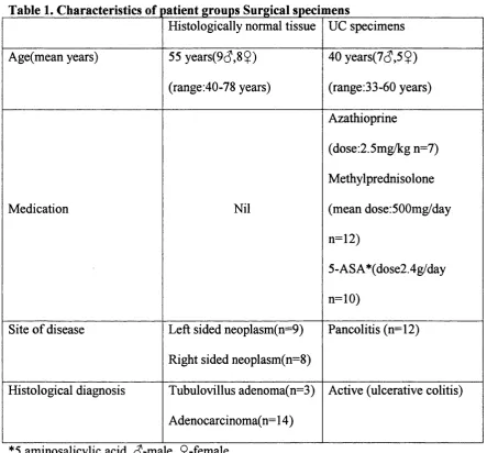
On the colonocytes.Decreasing doses of LPS were used, starting at 10 pg/ml with the concentration of 20ng/ml found to be the lowest concentration o f stimulant at which iNGS expression was seen in these cells. We were able to demonstrate iNGS expression in all viable HT-29 colonocytes, in all stimulation experiments performed after 6 hours post LPS stimulation both at the level of gene expression(see fig 2a), and protein production(see figlc).
When analysing the surgical tissues we found iNGS mRNA in all specimens tested and in comparison to the level of G3PDH on kinetic analysis, there was some 6 to 8 fold increase in iNGS mRNA in the colitic as compared to the normal tissue, on
densitometry(fig 2b)
Immunoblotting of the surgical specimens revealed that some 76%(13/17) of normal specimens and 81(9/11)% of colitic specimens expressed a 130kDa protein that was seen to co-migrate with the iNGS standard. This protein band was inhibited by the
Tables. iNOS immunohistochemistry in human colon
Normal tissue UC tissue
Surgical Endoscopic Surgical Endoscopic
% samples staining positive for iNOS
82(14/17) 88(14/16) 90(10/11) 93(15/16)
Cellular localisation
Surface epithelium
Surface epithelium
Surface and crypt
epithelium Inflammatory cells of LP*
Surface and crypt
Results-Figurel. INOS im munohistochemistry in HT-29 cells
Fig la. U nstim ulated HT-29-iNOS antibody
Fig lb.Stim ulated HT-29 -norm al rabbit serum
Fig Ic.Stim ulated HT-29 cells-iNOS antibody
V
\
0
Figure2-iN 0S RT-PCR
Fig2a-Q ualitative iNOS mRNA expression-surgical tissue/stimulated colonocyte;
MW 1 2 3
4 0 0
-Fig2b-Kinetic iNOS mRNA analysis-surgical tissue
G3PDH iNOS G3PDH INOS
30 34 41 45 30 34 41 45
r
F ig3-iN 0S im munoblotting-normal surgical tissue
iNOS S
K
Figure 4-iNOS im munohistochemistry
Figure 4a-H&E-normal tissue
«t- %
«
%
Fig4b- normal rabbit serum- normal tissue
Fig4c-iN 0S antibody-normal tissue
Figure 4d-iNOS antibody/peptide-normal tissue
4 6"
Figure 4e-H&E-colitic tissue
»
A
n
/
I
Figure 4f-normal rabbit serum-colitic tissue
Fig 4 g-iN 0S antibody-colitic tissue
I
L
Statistical analysis
On comparing the proportion of normal and diseased samples that stained positive for iNOS using immunohistochemistry:
95% confidence interval: -0.4138-0.062(p-value-0.14), were found indicating no significant difference in the proprtion of normal and diseased samples staining positive for iNOS protein.
Chi square analysis also revealed non-significant differences in these proportions. Chi square value-1.127
p-value-0.835
On similar analysis but for western blotting:
C hapter 4. Discussion.
My data demonstrates iNOS mRNA and protein expression in HT-29 colonocytes(Figs lc,&2a) after stimulation with LPS alone, in an otherwise germ free environment. Similar such expression was shown by Kolios et alç^) by using a ‘cocktaiT of
proinflammtory cytokines(TNF-a, IFN-y, and IL-l-a) to induce iNOS expression in HT- 29 cells. Further to this Kolios et al have now identified those cytokines which have a down-regulatory effect on HT-29 iNOS expression, noteably; IL-13,and IL-4(io2).
These prelimenary results and previous reports o f colonocyte ability to express iNOS led me to investigate whether intact colonocytes in the normal, non-inflamed colon express iNOS in a similar manner.
My results on human tissue suggest that iNOS is consistently expressed in the normal colon. The surgical specimens were obtained from patients undergoing colectomy for colorectal neoplasia and as a consequence although tissue was taken from a site distant from the lesion, local carcinogenic factors such as cytokines might have played a role in the iNOS expression. Therefore, I also analysed a group o f patients who were shown to have completely normal colons, using endoscopically obtained tissue to examine iNOS protein expression and found similar data with the same cellular localisation of iNOS protein. Another problem with the surgical tissue and iNOS expression is that of the effect of surgical manipulation itself. Hence, full thickness biopsies were taken prior to any handling of the colon and analysed using immunohistochemistry. Again similar results were obtained. Therefore it appears that iNOS is expressed, on average in up to 85% o f normal colons, with the prime cellular localisation being the surface
could suggest that iNOS is ‘constitutively expressed’ in the normal colon in the same manner as has been demonstrated in the respiratory epithelium in mancisi), the ileal mucosa in the mouse(i52),and in the rat kidneyciss). My data parallels and extends that of Moochhala et akm ) who demonstrated iNOS in the epithelium of normal colon when compared to colonic carcinomas,although the study was limited by small numbers of patients and using immunohistochemistry alone. On the contrary, Ambs et a/(i54) suggested the reverse, i.e. that iNOS expression was most marked in colonic adenomas and carcinomas and concluded that the disparate results obtained were due either to different genetic populations or a lack of specificty of the iNOS antibody used in the initial studies. My immunoblotting data showed that the antibody used in the protein studies was monospecific for iNOS and therefore one would be happy that the staining observed in the normal colon was due to iNOS protein.
My data also confirms many previous reports of increased mucosal iNOS production in active IBD, with similar cellular localisation found in my experiments(92-io6), although all these reports have failed to convincingly demonstrate iNOS expression in normal colon, the reasons for which are not apparent and need further analysis. The functional role of increased NO* production in IBD is unknown. There is evidence that NO* is a molecular ‘double-edged sword’ with both possible protective and damaging roles in this condition.
Tiao(i43) showed that inhibition of NO* synthesis and function causes increased production of IL-6 and TNF-a, two cytokines that up regulate adhesion molecules and thereby perpetuate the inflammatory response. Further evidence o f the anti-inflammatory effects o f NO' is its ability to inactivate NF-kB in endothelial cells(i44) via stimulating the production of the inhibitor of this transcription factor. As a consequence o f this effect on NF-kP there follows down regulation o f a variety o f pro-inflammatory cytokines and consequent salutory effects on the inflammatory cascade. As yet there is no such evidence specifically shown for NO'in human colonocytes but there is obviously the possibility o f such an effect in these cells in the presence of inflammation. NO' may also contribute to host defence in conditions such as active IBD by maintaining blood flow to tissues with increased metabolic needs and by modulating epithelial permeability(65). Horton et akui) demonstrated a benefit of parental supplementation of arginine on bacterial translocation via a NO' dependent mechanism ,all features of NO' that could be construed as beneficial to colonocyte homeostasis under the stress o f inflammation.
Data using NOS inhibitors in animal models of colitis in some cases suggest a deleterious effect. For example Pfeiffer et al (90) showed that the administration of the non-specific NOS inhibitor L-NAME exacerbated TNBS induced colitis in rats, suggesting an anti-inflammatory role for NO' in this setting. However, other studies using NOS inhibitors have given differing results and are discussed later. The above described effects of NO' relate to inflammatory states such as occurs in IBD. Obviously in the non-inflamed colon such actions o f NO' would not be expected to occur. What could be the possible benefit then of epithelial iNOS induction in the normal colon as shown by my studies?
Firstly epithelial iNOS induction and local sustained NO' release could provide an
NO* exerts important anti-microbial effects against a wide range of microbes(i59) such as Escherichia coli and Salmonella spim ) and iNOS deficient mice are highly susceptible to certain bacterial infections mchxàmg Listeria monocytogenes{\6\\ suggesting the paramount importance of iNOS expression as a means of preventing invasive bacterial infection of the colon. More recently. Forte et al (i62) have shown a marked increase in endogenous nitrate synthesis in patients with confirmed infective gastroenteritis. Although not definite it seems plausible that this increase nitrate is coming from
colonocyte iNOS induction, as previously, Kolios et al (los) and Islam et al (i63) showed such epithelial induction in biopsy specimens o f patients with acute Shigella and
Salmonella colitis, with the suggestion that NO* is limiting tissue damage in the presence of infection. Therefore it is plausible that colonocyte NO* production not only helps to kill invasive bacteria but also helps to prevent initial invasion. Furthermore, NO* has been shown to regulate microvascular patency, permeability, tissue perfusion and mucosal barrier function as discussed above(4?,48,64). These effects are principally
mediated by the constitutive NOS but it has been shown by Hutcheson et al (i45) that the inducible isoform is critical in maintaining the above effects under conditions of
endotoxaemia. Therefore it is possible that epithelial iNOS production in the normal human colon may play a role in mucosal homeostasis although there is currently no data to confirm or refute this hypothesis.