O R I G I N A L R E S E A R C H
Long Noncoding RNA RP11-334E6.12 Promotes the
Proliferation, Migration and Invasion of Breast Cancer
Cells Through the EMT Pathway by Activating the
STAT3 Cascade
This article was published in the following Dove Press journal:
Cancer Management and Research
Dongjun Sun1
Hengming Liu2
Tiantian Wang3
1Department of General Surgery, Chiping
District People’s Hospital, Liaocheng, Shandong, People’s Republic of China;
2Department of Anesthesiology, Chiping
District People’s Hospital, Liaocheng, Shandong, People’s Republic of China;
3Department of Thyroid and Breast
Surgery, Shandong Provincial Hospital Affiliated to Shandong First Medical University, Jinan, Shandong, People’s Republic of China
Background: RP11-334E6.12 is a dysregulated long noncoding RNA (lncRNA) that has never been studied in breast cancer. The biological function and potential mechanism of RNA RP11-334E6.12 in tumorigenesis are still unknown.
Methods: We scanned the Cancer Genome Atlas (TCGA) database and identified RP11-334E6.12 as one of the most dysregulated lncRNAs. The level of RP11-RP11-334E6.12 was assessed in breast cancer (BC) tissue samples and BC cell lines. The survival and RP11-334E6.12 expression of patients were analysed. The biological influence of RP11-334E6.12 on BC cell lines was studied using proliferation, Transwell migration, and invasion assays.
Results:RP11-334E6.12 was upregulated in both the TCGA database and our own database. Moreover, survival analyses indicated that RP11-334E6.12 was related to poor overall survival. Moreover, RP11-334E6.12 promoted the proliferation, migration and invasion of BC cells. RP11-334E6.12 promotes the epithelial mesenchymal transition of BC by activating the STAT3 pathway.
Conclusion:Taken together, our results demonstrate that RP11-334E6.12 is associated with
the progression of breast cancer. Our findings indicate that long noncoding RNA
RP11-334E6.12 promotes the proliferation, migration and invasion of breast cancer cells by activating the STAT3 pathway.
Keywords:breast cancer, RP11-334E6.12, survival, proliferation, migration, invasion
Background
Breast cancer (BC) is one of the most common cancers worldwide and is the second
leading cause of cancer-related mortality in women.1 Although advances in the
therapy have been made, many patients still suffer from metastasis and recurrence. Immunotherapeutic strategies, such as CAR T cells and PDL1, have been applied,
but immunotherapy was not suitable for all patients.2 Hence, more therapeutic
targets are urgently needed.
Long noncoding RNAs (lncRNAs) are involved in many physiological and
pathological processes in humans, such as cell division and differentiation.3,4
LncRNAs are reported to be involved in the progression of multiple kinds of
carcinomas.5,6 Many studies have also shown that lncRNAs contribute to the
progression of breast carcinoma through processes such as proliferation, angiogen-esis and tissue invasion.7,8
Correspondence: Tiantian Wang Department of Thyroid and Breast Surgery, Shandong Provincial Hospital Affiliated to Shandong First Medical University, Jinan, Shandong, People’s Republic of China
Email [email protected]
Cancer Management and Research
Dove
press
open access to scientific and medical research
Open Access Full Text Article
Cancer Management and Research downloaded from https://www.dovepress.com/ by 118.70.13.36 on 24-Aug-2020
In the present study, we scanned the TCGA database
and identified RP11-334E6.12 as a significantly
upregu-lated lncRNA in BC. We next detected the expression difference between normal tissue and BC. We next uncov-ered the biological function of RP11-334E6.12 through a series of experiments and found that RP11-334E6.12 promotes the proliferation, migration and invasion of BC by activating STAT3.
Methods
Tissue Samples
All human tissues were obtained from the surgical suite in the Department of Thyroid and Breast Surgery,
Shandong Provincial Hospital affiliated with Shandong
University after confirmation by a pathologist. Tissues
were obtained with the patients’ written consent under
a protocol approved by the institution’s Institutional
Review Board.
Quantitative Real-Time Polymerase Chain
Reaction (qRT-PCR)
Total RNA was extracted using TRIzol reagent
(Invitrogen, NY, USA) according to the manufacturer’s
instructions. qRT-PCR was performed with the SYBR green detection RT-PCR system (Takara, Japan). Actin was used for normalization. All conditions were repeated
in triplicate. The 2−ΔΔCtmethod was used to calculate the
relative RNA expression.
Cell Culture and Cell Transfection Assay
The human mammary cancer cell lines MCF-10A, MDA-453, BT-549, BT-474, MCF-7 and MDA-231 were gifts from Professor Tian of the Department of Thyroid and
Breast Surgery, Shandong Provincial Hospital affiliated
with Shandong University and was approved by institu-tional review board or ethics committee of Shandong
Provincial Hospital affiliated with Shandong University.
Figure 1RP11-334E6.12 was upregulated in BC and was negatively corelated with prognosis.
Notes:(A) The relative level of RP11-480I12.5 in TCGA database (student‘s two tailed paired test *p<0.05). (B) Survive analysis of patients in TCGA database (Log rank test). (C) The relative level of RP11-480I12.5 in our own database (student‘s two tailed paired test ***p<0.001). (D) Survive analysis of patients in our own database (Log rank testp<0.001).
Cancer Management and Research downloaded from https://www.dovepress.com/ by 118.70.13.36 on 24-Aug-2020
Cells were cultured in Dulbecco’s Modified Eagle Medium (DMEM), Minimum Essential Medium (MEM) and RPMI 1640 supplemented with 10% foetal bovine serum (Life Technologies, Grand Island, NY, USA), 1% penicillin G,
and streptomycin in the incubator at 37°C with 5% CO2.
Cell Counting Kit-8 Assay
The Cell Counting Kit-8 (CCK-8, Dojondo, Tabaru, Japan) assay was applied to measure the proliferation ability. Cells (500) were seeded into 96-well plates. The absorp-tion values were measured at 24, 48, and 72 hrs after primary seeding. The experiments were repeated three times, and the data are shown as the mean±standard devia-tion (SD).
Colony Formation Assay
Stable cell lines were seeded in a 6-well plate at a density of 500 cells per well. After being cultured for 2 weeks, cell
colonies were fixed with 4% paraformaldehyde and
stained with 1% crystal violet. The colonies were exam-ined and counted under a microscope.
Transwell Migration and Invasion Assay
A Transwell assay was applied to detect migration and invasion ability. For the invasion assay, Matrigel chambers (BD Biosciences, San Jose, CA, USA) were used to deter-mine the effect of cells on invasion according to the
manufacturer’s instructions. Cells (7.5×10⁴ cells/well)
were resuspended in 250 μL of medium in the upper
chamber (8-μm pore size, Costar, Corning, NY, USA) of
a Transwell system, whereas the lower chamber wasfilled
with 0.5 mL of medium supplemented with 10% FBS. After incubation for 24 hrs at 37°C, the invasive cells
were fixed with 100% methanol and stained with 0.5%
crystal violet before counting under an inverted micro-scope. For the migration assay, the indicated cells were plated on uncoated Matrigel upper chambers. The number of migrated cells was estimated under a microscope
(Nikon, Tokyo, Japan) at 200× magnification.
Lentivirus Production and Stable Cell Line
Construction
Lentiviral vectors expressing shRNA and control were cotransfected with packaging vectors psPAX2 and pMD2G (Addgene) into HEK293FT cells for lentivirus production using Lipofectamine 3000 in accordance with the manufac-turer’s instructions. To establish stable cell lines, cells were
transduced by using the above lentiviruses and polybrene (8 mg/mL, Sigma). After incubating for 72 hrs, cells were selected with 2 mg/mL puromycin for 3 days.
Statistical Analysis
All data analyses were performed with SPSS 20.0
statis-tical software. Theχ2test was used to analyse the
relation-ships between categorical variables. The differences
between groups were compared by Student’s t-test. Cox
regression and Kaplan-Meier methods were used to
ana-lyse OS, and p<0.05 was considered to be a significant
difference from the control.
Results
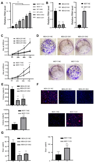
RP11-334E6.12 Is Upregulated in BC
Tissues and Associated with Poor
Prognosis
After TCGA screening, the long noncoding RNA
RP11-334E6.12 was identified as one of the lncRNAs
contribut-ing to the progression of BC (Figure 1A, p<0.05, 1B,
p <0.05). To further verify this hypothesis, we analysed
the expression pattern in 36 paired normal tissues and
tumour tissues. RP11-334E6.12 was significantly
upregu-lated in tumour tissue compared to normal tissue
(p<0.001) (Figure 1C). Moreover, we analysed the
corre-lation between RP11-334E6.12 and the overall survival of patients. Our own database showed that RP11-334E6.12
was negatively associated with prognosis (p <0.001)
(Figure 1D), and patients with a high level of RP11-334E6.12 had a shorter overall survival time than those with a low level. Taken together, RP11-334E6.12 was negatively associated with the tumorigenesis and progres-sion of breast cancer.
RP11-334E6.12 Promotes the
Proliferation of BC
We showed that RP11-334E6.12 is upregulated in BC and negatively associated with prognosis. To further uncover the biological function of RP11-480I12.5, we next detected the expression pattern in BC cell lines. The level of RP11-334E6.12 was higher in BC cell lines than
in the control normal cell line MCF-10A (p <0.001)
(Figure 2A). We next established a stable 334E6.12 knockdown MDA-231 cell line and an RP11-334E6.12-overexpressing MCF-7 cell line. The relative
RNA level is shown inFigure 2B(p<0.001). We applied
a series of experiments to measure the proliferation ability
Cancer Management and Research downloaded from https://www.dovepress.com/ by 118.70.13.36 on 24-Aug-2020
Figure 2RP11-334E6.12 promotes the proliferation in BC.
Notes:(A) The relative level of RP11-334E6.12 in BC cell line (***p<0.001). (B) The relative level of RP11-334E6.12 in stable cell line (***p<0.001). (C) The abs at 450nm of different cell line (**p<0.01, ***p<0.001). (D) The representative image of colony formation assay. (E) The statistical analysis of colony formation assay (***p<0.001). (F) The representative image of Edu assay. (G) The statistical analysis of Edu assay (***p<0.001).
Cancer Management and Research downloaded from https://www.dovepress.com/ by 118.70.13.36 on 24-Aug-2020
of the cell lines described above. Cells with higher RP11-334E6.12 developed an improved proliferation ability in both MDA-231-NC and MCF-7-OV cell lines. Cells with
a higher level of RP11-334E6.12 harboured a higher
absorbance at 450 nm (p <0.01, p <0.001) (Figure 2C)
and a higher colony index (Figure 2D andE,p <0.001).
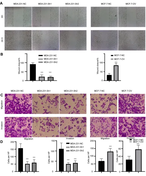
Figure 3RP11-334E6.12 promotes the migration and invasion of BC.
Notes:(A) The representative image of wound healing. (B) The statistical analysis of wound healing (***p<0.001). (C) The representative image of transwell and invasion chamber. (D) The statistical analysis of transwell and invasion chamber (***p<0.001).
Cancer Management and Research downloaded from https://www.dovepress.com/ by 118.70.13.36 on 24-Aug-2020
We next detected the DNA synthesis rate in the cell lines described above through an EdU assay. Both MDA-231-NC and MCF-7-OV cells harboured a higher percentage of
Edu+ cells. These results indicate that RP11-334E6.12
promotes DNA synthesis (Figure 2FandG,p<0.001).
RP11-334E6.12 Promotes the Migration
and Invasion of BC Cells
We have previously demonstrated that RP11-334E6.12 pro-motes proliferation in BC cell lines. Recurrence and metas-tasis are the major reasons for cancer-related death. To uncover the biofunction of RP11-334E6.12 in migration and invasion, we applied wound healing, Transwell and invasion assays. Cells with higher levels of RP11-334E6.12 developed improved migration and invasion abilities in
wound healing (Figure 3Aand3B,p<0.001) and transwell
and invasion chamber assays (Figure 3Cand3Dp<0.001).
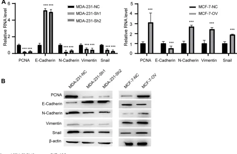
RP11-334E6.12 Promotes the Epithelial
Mesenchymal Transition of BC Cell Lines
We proved that RP11-334E6.12 promotes proliferation, migra-tion and invasion in BC cell lines. Epithelial mesenchymal
transition is one of the most popular pathways reflecting the
status of BC cells. We next examined EMT markers and proliferation markers in the different cell lines. The results showed that mesenchymal markers and proliferation markers decreased in stable knockdown cell lines compared to control cells, while epithelial markers increased at both the RNA level (Figure 4A,p<0.001) and protein level (Figure 4B).
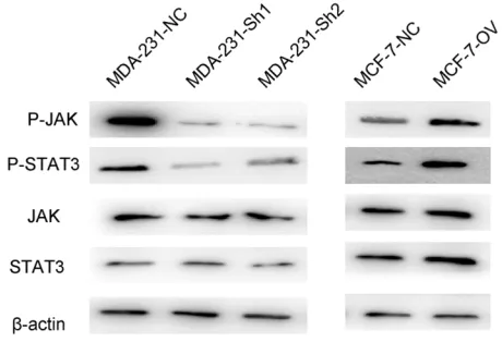
RP11-334E6.12 Activates the STAT3
Pathway in BC
We next tried to determine the potential mechanism of promoting EMT. We examined some critical pathways,
such as theβ-catenin, STAT3 and NOTCH pathways (data
not shown). Finally, we found that the STAT3 pathway was the most dysregulated pathway. P-JAK and P-STAT3
decreased in stable knocking cell lines (Figure 5A). We
hypothesized that RP11-334E6.12 may exert its function through the phosphorylation of the JAK family.
Discussion
Breast carcinoma is the most common malignancy in women worldwide. Although many advancements have
Figure 4RP11-334E6.12 promotes EMT of BC.
Notes:(A) The relative RNA level of EMT markers and PCNA of different cell line (***p<0.001). (B) The Western blot of EMT markers and PCNA of different cell line.
Cancer Management and Research downloaded from https://www.dovepress.com/ by 118.70.13.36 on 24-Aug-2020
been made, many patients still suffer from recurrence and metastasis. However, the mechanism of the tumorigenesis of breast carcinoma is still unknown. Recent studies have shown that many lncRNAs participate in the tumorigenesis and progression of BC through multiple aspects. Dong proved that FBXL19-AS1 promotes cell proliferation and inhibits cell apoptosis via the miR-876-5p/FOXM1 axis in
breast cancer.9 Tang found that LncCCAT1 promotes
breast cancer stem cell function by activating WNT/β
-catenin signalling.10lncRNA GAS5 was recently reported
to promote apoptosis in triple-negative breast cancer by
targeting miR-378a-5p/SUFU signalling.7Rossi found that
lncRNAs interact with proteins to further facilitate breast
progression.11
STAT3 is one of the key pathways sustaining the stem-ness of stem cells, and it has been reported to be
over-activated in multiple kinds of cancers.12 After activation
by inducers, STAT3 is phosphorylated by the JAK family and thus enters the nucleus to exert its function as
a transcription factor.13 STAT3 was reported to be
over-activated in the breast and to promote proliferation and migration. However, STAT3 is one key transcription factor shared in both normal cells and malignant cells. Therapies directly targeting STAT3 are not available.
In the present research, we found that RP11-334E6.12 is commonly overexpressed in BC. RP11-334E6.12 pro-motes proliferation, migration and invasion through the EMT pathway by activating STAT3 pathways.
Conclusion
Taken together, our results indicate that RP11-334E6.12 is associated with tumour progression in breast cancers. Our
findings indicate that the long noncoding RNA
RP11-334E6.12 promotes the proliferation, migration and inva-sion of breast cancer cells through the EMT pathway by activating STAT3 pathways.
Abbreviations
TCGA, The Cancer Genome Atlas; BC, breast cancer; LncRNA, long noncoding RNA.
Ethics and Consent
Tissues were obtained with the patients’ written and
informed consent under a protocol approved by the
insti-tution’s Institutional Review Board in accordance with the
Declaration of Helsinki. Tissues were obtained with the
patients’written consent for publication.
Data Sharing Statement
The data are available in this article.
Author Contributions
All authors contributed to data analysis, drafting and
revis-ing the article, gave final approval of the version to be
published, and agree to be accountable for all aspects of the work.
Disclosure
The authors report no conflicts of interest in this work.
References
1. Bray F, Ferlay J, Soerjomataram I, Siegel RL, Torre LA, Jemal A. Global cancer statistics 2018: GLOBOCAN estimates of incidence and mortality worldwide for 36 cancers in 185 countries. CA Cancer J Clin.2018;68(6):394–424. doi:10.3322/caac.v68.6
2. Keren L, Bosse M, Marquez D, et al. A structured tumor-immune microenvironment in triple negative breast cancer revealed by multi-plexed ion beam imaging.Cell.2018;174(6):1373–1387. doi:10.1016/ j.cell.2018.08.039
3. Luo ZH, Walid AA, Xie Y, et al. Construction and analysis of a dysregulated lncRNA-associated ceRNA network in a rat model of temporal lobe epilepsy. Seizure. 2019;69:105–114. doi:10.1016/j. seizure.2019.04.010
4. Huang T, Wang J, Zhou Y, Zhao Y, Hang D, Cao Y. LncRNA CASC2 is upregulated in osteoarthritis and participates in the regulation of IL-17 expression and chondrocyte proliferation and apoptosis.Biosci Rep.2019;39. doi:10.1042/BSR20182454
5. Taheri M, Ghafouri-Fard S. Long non-coding RNA signature in cervi-cal cancer.Klin Onkol.2018;31(6):403–408.
6. Xu YD, Shang J, Li M, Zhang YY. LncRNA DANCR accelerates the development of multidrug resistance of gastric cancer.Eur Rev Med Pharmacol Sci. 2019;23(7):2794–2802. doi:10.26355/eurrev_20190 4_17554
7. Zheng S, Li M, Miao K, Xu H. lncRNA GAS5-promoted apoptosis in triple-negative breast cancer by targeting miR-378a-5p/SUFU signaling.J Cell Biochem.2019.
Figure 5RP11-334E6.12 promotes the phosphorylation of STAT3 pathway.
Cancer Management and Research downloaded from https://www.dovepress.com/ by 118.70.13.36 on 24-Aug-2020
8. Wang N, Hou M, Zhan Y, Sheng X. LncRNA PTCSC3 inhibits triple-negative breast cancer cell proliferation by downregulating lncRNA H19.J Cell Biochem.2019;120(9):15083–15088.
9. Dong G, Pan T, Zhou D, Li C, Liu J, Zhang J. FBXL19-AS1 promotes cell proliferation and inhibits cell apoptosis via miR-876-5p/FOXM1 axis in breast cancer. Acta Biochim Biophys Sin (Shanghai).2019. doi:10.1093/abbs/gmz110
10. Tang T, Guo C, Xia T, et al. LncCCAT1 promotes breast cancer stem cell function through activating WNT/beta-catenin signaling.Theranostics.
2019;9(24):7384–7402. doi:10.7150/thno.37892
11. Rossi T, Pistoni M, Sancisi V, et al. RAIN is a novel enhancer-associated lncRNA that controls RUNX2 expression and promotes breast and thyroid cancer. Mol Cancer Res. 2019;18 (1):140–152.
12. Palmitoylation alters STAT3 activity and tumor response to high-fat diet.Cancer Discov.2019.
13. Kang S, Tanaka T, Narazaki M, Kishimoto T. Targeting interleukin-6 signaling in clinic.Immunity.2019;50(4):1007–1023. doi:10.1016/j. immuni.2019.03.026
Cancer Management and Research
Dove
press
Publish your work in this journal
Cancer Management and Research is an international, peer-reviewed open access journal focusing on cancer research and the optimal use of preventative and integrated treatment interventions to achieve improved outcomes, enhanced survival and quality of life for the cancer patient.
The manuscript management system is completely online and includes a very quick and fair peer-review system, which is all easy to use. Visit http://www.dovepress.com/testimonials.php to read real quotes from published authors.
Submit your manuscript here:https://www.dovepress.com/cancer-management-and-research-journal
Cancer Management and Research downloaded from https://www.dovepress.com/ by 118.70.13.36 on 24-Aug-2020