FPGA Implementation of EEG Feature
Extraction and Seizure Detection
Sreethu Raj 1, Anuja George 2
PG scholar, Department of Electronics and Communication, MG University, St.Joseph’s College of Engineering and
Technology, Kerala, India1
Assistant Professor, Department of Electronics and Communication, St.Joseph’s College of Engineering and
Technology, Kerala, India2
ABSTRACT: Seizure is a chronic neurological disorder. Seizures are the result of transient and unexpected electrical disturbances in the brain. Seizure is more likely to occur in young children or people over the age of 65 years; however, it can occur at any age. The detection of seizure is possible by analyzing EEG (electroencephalogram) signals. The electroencephalogram (EEG) signal is very important in the diagnosis of seizure. Long-term EEG recordings of a seizure patient contain a huge amount of EEG data. The detection of seizure activity is, therefore, a very demanding process that requires a detailed analysis of the entire EEG data, usually performed by an expert. This paper describes an automated classification of EEG signals for the detection of seizures using wavelet transform and support vector machines. The decision making process consists of three main stages: (a) filtering operation by FIR filter, (b) feature extraction based on discrete wavelet transform (DWT) and (c) classification by support vector machines (SVM) classifiers. The proposed methodology is applied on EEG data sets that belong to two subject groups: a) healthy subjects and b) seizure subjects. Based on the data sets the boundaries of all the features are identified for the proper detection of the test signal. After processing all the data sets, the test signal is given to the system. Decision is made by
comparing the features of the test signal with the maximum and minimum values of all the features of data sets
.
TheEEG feature extraction and seizure detection system is verified by Verilog in ISim simulator and implemented on Xilinx Spartan6.
KEYWORDS:EEG, DWT, Support Vector Machines.
I. INTRODUCTION
Personalized healthcare depends crucially on large volumes of data about both individuals and populations. In the near future it is common to wear a number of biosensors that continuously monitor various aspects of our physiological state. There are two aspects of this enterprise, i.e., gathering the data and efficiently doing something useful with it. In a clinical setting, electroencephalography (EEG) combined with video monitoring is the de facto gold standard for the detection and diagnosis of various neurological conditions, including seizures. Although detecting seizures that occur in daily life is important for the safety and well-being of those with seizures and those around them, clinical systems are far too resource intensive for ambulatory settings. One of the many challenges in the automated detection of seizures is to distinguish between seizure activity and non-seizure activity. To accomplish this task, identification of the EEG features and there extraction plays a key role. EEG measures the electrical activity of the brain and it represents summation of post-synaptic potentials.
accurately and at the same time be robust. The classification accuracy can be improved by proper identification of maximum and minimum limits of all the extracted features, and classifiers that can accurately classify the two groups of EEG signals based on the selected and reduced feature space.
II. RELATED WORK
Adam Page et al.,[1] presents a low power, flexible, and multichannel electroencephalography (EEG) feature extractor and classifier for the purpose of personalized seizure detection. Various features and classifiers were explored with the goal of maximizing detection accuracy while minimizing power, area, and latency. Additionally, algorithmic and hardware optimizations were identified to further improve performance. The classifiers studied include k-nearest neighbor, support vector machines, naive Bayes and logistic regression (LR).This work presented a low-power, flexible, and multichannel architecture designed to perform personalized seizure detection. Siddharth Biswal et al.,[2] have developed a supervised learning to automatically detect with high accuracy EEG reports that describe seizures and epileptiform discharges. They manually labeled 3,277 documents as describing one or more seizures vs no seizures, and as describing epileptiform discharges vs no epileptiform discharges. Then used Naive Bayes to develop a system able to automatically classify EEG reports into this categories. In this paper they described an extremely effective and effcient automated method for classifying free text EEG reports. Osman Salim et al,.[3] propose a new approach for the early detection of epileptic seizure in EEG. The proposed approach is based on Discrete Wavelet Transform (DWT) and Ant Colony (AC) Classifier. The results prove the effectiveness of this approach, which can achieve a high detection ratio with low false classification ratio.
Carlos Guerrero et al.,[4] describes a new approach in features extraction using time-frequency distributions (TFDs) for detecting epileptic seizures to identify abnormalities in electroencephalogram (EEG). Particularly, the method extracts features using the Smoothed Pseudo Wigner-Ville distribution combined with the McAulay-Quatieri sinusoidal model and identifies abnormal neural discharges. Rajeev Yadav et al .,[5] present a novel, simple, computationally efficient seizure detection system to enhance the sensitivity with a low FDR by proposing a dual-stage classifier. An overall improvement has been observed in terms of sensitivity, specificity and FDR. Naveen Verma,[6] presents a low-power SoC that performs EEG acquisition and feature extraction required for continuous detection of seizure onset in epilepsy patients. The SoC corresponds to one EEG channel, and, depending on the patient, up to 18 channels may be worn to detect seizures as part of a chronic treatment system. R. Yadav et al.,[7] presents a novel model-based patient specific method for automatic detection of seizures in the intracranial EEG recordings. The proposed method overcomes the complexities in the practical implementation of the patient-specific approach of seizure detection.
III. RESEARCH ELABORATIONS
A. Seizure
Seizure is a sudden surge of electrical activity occurring in brain. A seizure usually affects how a person appears or acts for a short period of time. Whatever the brain and body can do normally can also occur during a seizure. During seizure the following changes are occur in brain:
The electrical activity is caused by complex chemical changes that occur in nerve cells [1]
Brain cells either excite or inhibit (stop) other brain cells from sending messages. When a seizure occurs, this
balance collapses. This imbalance can cause chemical changes and can lead to surges of electrical activity that cause seizures.
Idiopathic seizure is a type of seizure whose cause cannot be found and are usually seen in children and young adults, but it can occur at any age. There may be a family history of epilepsy or seizures. If seizures occurring repeatedly even after the problem is treated, the condition is called epilepsy. Most seizures stop by themselves. During seizure, the person can be hurt or injured, so the main goal is to protect the person from injury. Try to prevent a sudden fall, clear the area of furniture or other sharp objects, cushion the person's head, loosen tight clothing, stay with the person until he or she recovers, call for medical help.
B. Seizure Detection
Seizure detection uses an automated algorithm to detect whether a seizure is occurring through the analysis of recorded biological signals of the patient. The main goal of seizure detection is to receive and analyse a set of biological signals and convert the set of information signal that is, the input signal into an output signal or indicator of whether the patient is having seizure or not. The main objectives of this are to make the detection as quickly as possible with minimum time and accurately. For most cases, real-time seizure detection is needed, requiring online analysis of signal as soon as they are available, with detection decisions being made without delay and with only using information that is available at the time the detection is made.
A seizure detection system must be capable of determining the presence or absence of ongoing seizures and provide clinicians with a detailed seizure data useful for the management of epilepsy. Even prior to clinical onset of a seizure, various algorithms of different biometric signals can detect seizure. Two main steps are involved in every seizure detection system. First, appropriate features (EEG features) extracted from the information signal. Second, to determine a threshold value to determine the presence or absence of a seizure. This second step is called classification. Sometimes it might be as simple as setting a threshold value or might require models derived from supervised machine learning algorithms. Features for the seizure detection purpose is chosen by a compromise between the need for speed and the need for detection accuracy. The seizure detection system is having two phases namely, training phase and test phase. Derivation of a model from machine learning algorithms is done during a training phase [3] and involves three sub steps and are filtering, feature computation, and feature extraction.
C. EEG
Electroencephalography (EEG) is [4] electrophysiological monitoring method to record electrical activity of human brain, it is typically non-invasive [4], with the electrodes are placed along the scalp of the patient. EEG measures voltage fluctuations resulting [5] from ionic current within the neurons of the brain. The electrical activity of brain during a period of time is collected by placing electrodes at different cerebral locations placed on the scalp. For all the diagnostic purposes the main focus is on the spectral content of the EEG.
EEG is most often used for the diagnosis of epilepsy, which causes abnormalities in EEG signal readings. EEG continues to be a valuable tool for research and diagnosis. EEG has several advantages over the other methods:
Temporal resolution is higher.
Directly measures the electrical activity of the brain.
EEG has become a very useful and popular clinical tool, especially in the field of epileptology, but also in other areas of neurology and psychiatry [6]. Even though EEG is an important clinical tool there are distinct difficulties associated with EEG analysis and interpretation. Traditional method of EEG analysis is based on visually analysing the EEG activity using strip charts and is very laborious and time consuming task which requires skilled interpreters. The manual analysis often fails to detect the uncover features in the input signal. So here comes the need of an automated seizure detection system.
D. Proposed system
The object of wavelet analysis is to decompose signals into several frequency bands. The third stage is support vector machines. It is an automated machine learning algorithm and based on the concept of decision planes that define decision boundaries. Features of EEG signals are extracted and the boundaries of all the features are determined using support vector machines. The block diagram of the proposed system is shown in Fig.1.
Fig.1 : Block diagram
For this system 20 databases are used. Each database contains 2560 values. It uses 10 samples from patients having seizure and 10 samples from normal people. All these samples were filtered and given to DWT. Since the input signal contains large amount of information, the processing of this sample is very time consuming. Due to this the data is down sampled by two in DWT. Here four stage DWT is incorporated to reduce the amount of useful information. After passing through the four stages of DWT, the data is reduced to 160 values from 2560 values. Mean, variance, standard deviation, frequency, maximum amplitude, minimum amplitude and energy are the features used in this system. These seven features are extracted from all the 20 samples. The EEG signals obtained from seizure patients are different from that of normal people. Therefore the values of all these features will also be different. Then the maximum and minimum limits of all these features are identified and classified as two groups using support vector machines. After processing all the 20 databases, the test signal is given to the system. The system will extract all these features of the test signal and then check whether it belongs to seizure category or not. And it will indicate the person is having seizure or not.
1. Discrete Wavelet Transform (DWT)
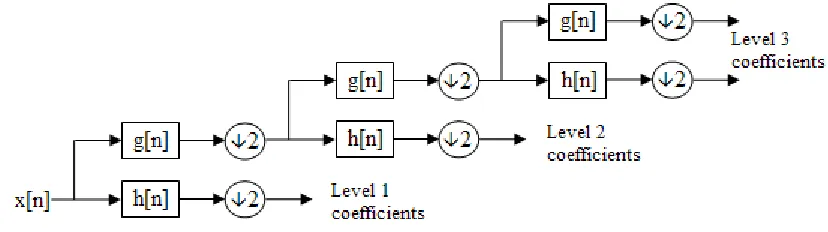
Wavelet transform is the most promising method for extracting features from EEG signals [10] and is used to extract features from EEG signals. The wavelet transform, as a linear time-frequency transform, represents an efficient analytical tool in signal processing, pattern recognition and classification. Wavelet transform is widely used in biomedical signal processing since it is capable of localizing the information in both time and frequency. DWT is a sampled version of continuous wavelet transform and is capable of providing the sufficient information to meet the analysis and synthesis of the original signal in a reduced computational time. The block diagram of DWT is shown in Fig.2.
Consider an EEG signal x(n) of length n, starting from x(n), in the first step it produces two sets of coefficients: approximation coefficients and detail coefficients. These vectors are obtained by convolving the input signal x(n) with the low-pass filter for approximation and with the high-pass filter for detail, followed by downsampling by two.
2. Support Vector Machines
Support vector machines (SVMs) are a set [11] of supervised learning methods used for classification. It is a classification algorithm and is used to find the boundaries of various features of EEG signals. In this algorithm, each data item is plotted as a point in n-dimensional space (where n is number of features) with the value of each feature being the value of a particular coordinate, then perform classification by finding the hyper-plane that differentiate the two classes very well.
IV. RESULTS AND DISCUSSIONS
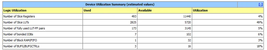
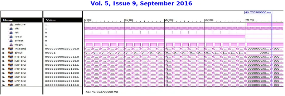
The Xilinx ISE design suite 14.2 is used for the coding of the EEG feature extraction and seizure detection system. The proposed design has been synthesized using Xilinx synthesizer and place & route tools. The functionality of the design has been verified extensively by using Xilinx ISim Simulator verification tool. The coding is done in Verilog language. The EEG feature extraction and seizure detection system is implemented on the Spartan 6 tyro kit designed by Pantech Solutions.The proposed system consists of Finite Impulse Response (FIR) filter, Discrete Wavelet Transform (DWT) and Support Vector Machines (SVM).
Fig. 3 : Resource ulitization summary of proposed system
Fig. 4 : Output waveform of proposed system
V. CONCLUSION
An EEG feature extraction and seizure detection system have been implemented on Spartan 6 tyro kit designed by Pantech Solutions. Traditional method of analysis of the EEG is based on visually analyzing the EEG activity using strip charts. This is very time consuming task which requires skilled interpreters, who by the nature of the task are prone to subjective judgment and error. The proposed system identifies the presence or absence of seizure in an easier and faster manner.
Verilog is used as Hardware Description Language (HDL) to program all the components of the EEG feature extraction and seizure detection. Verification of functionality of all components has done by giving different input and output. Proposed system is synthesized using Xilinx ISE design suite 14.2 xc6slx9-3tqg144 device and simulated in ISim.
REFERENCES
[1] Adam Page, Chris Sagedy, Emily Smith, Nasrin Attaran, Tim Oates, Tinoosh Mohsenin, “A Flexible Multichannel EEG Feature Extractor and Classifier for Seizure Detection”, IEEE Transactions On Circuits And Systems II: Express Briefs, Vol. 62, no. 2, February 2015.
[2] Siddharth Biswal, Zarina Nip, Valdery Moura Junior, Matt T. Bianchi, Eric S Rosenthal, M Brandon Westover , ”Automated Information Extraction from Free-Text EEG Reports”, IEEE Transactions On Biomedical Engineering, Vol. 59, no. 5, 2015
[3] Osman Salem, Amal Naseem , Ahmed Mehaoua, “Epileptic Seizure Detection From EEG Signal Using Discrete Wavelet Transform and Ant Colony Classifier “, IEEE ICC Selected Areas In Communications Symposium, 2014.
[4] Carlos Guerrero-Mosquera , Angel Navia Vazquez, “ New approach in features extraction for EEG signal detection “, 31st Annual International Conference of the IEEE EMBS Minneapolis, Minnesota, USA, September 2-6, 2009
[5] Rajeev Yadav, Rajeev Agarwal, M.N.S. Swamy, “A Novel Dual-Stage Classifier for Automatic Detection of Epileptic Seizures “, 30th Annual International IEEE EMBS Conference Vancouver, British Columbia, Canada, August 20-24, 2008.
[6] Naveen Verma, Ali Shoeb, Jose Bohorquez, Joel Dawson, John Guttag Anantha P. Chandrakasan, “A Micro-Power EEG Acquisition SoC With Integrated Feature Extraction Processor for a Chronic Seizure Detection System “, IEEE Journal Of Solid-State Circuits, Vol. 45, no. 4, April 2010.
[7] R. Yadav, M. N. S. Swamy, R. Agarwal, “Model-Based Seizure Detection for Intracranial EEG Recordings "”, IEEE Transactions On Biomedical Engineering, Vol.59, no. 5, May 2012
[8] Kaveh Samiee, Peter Kovacs, Moncef Gabbouj, “Epileptic Seizure Classification of EEG Time-Series Using Rational Discrete Short-Time Fourier Transform "”, IEEE Transactions On Biomedical Engineering, Vol. 62, no. 2, February 2015
[9] Chen Zhang, Muhammad Awais Bin Altaf , Jerald Yoo, “ Design and Implementation of an On-Chip Patient-Speci_c Closed-Loop Seizure Onset and Termination Detection System “, IEEE Journal Of Biomedical And Health Informatics, Vol. 20, no. 4, July 2016
[10] LI Nian-qiang, NIE Yun-jie, ZHU Wei, “The Application of FPGA-based Discrete Wavelet Transform System in EEG Analysis”, International Conference on Intelligent Systems Designs and Engineering Applications, 2012.
[11] Inan Guler, Elif Derya Ubeyli , “Multiclass Support Vector Machines for EEG-Signals Classification”, IEEE Transactions on Information Technology in Biomedicine ,Volume: 11, Issue: 2, March 2007.