ABSTRACT
BALBIN, MICHELLE MACUGAY. Surveillance and Development of Detection Platform for Antimicrobial Resistance and Pathogenic Bacteria. (Under the direction of Dr. Siddhartha Thakur).
The study aimed to identify the antimicrobial resistance (AMR) determinants and virulence factors in Salmonella spp. and Escherichia coli from different anthropogenic areas in North Carolina. Soil samples were collected from different anthropogenic areas: urban and natural. Minimum inhibitory concentration (MIC) was determined by using the broth
microdilution method. Whole genome sequencing and analysis were done to identify the AMR determinants and virulence factors among the isolates. The urban environment was observed to have a higher prevalence of Salmonella spp. and E. coli. The Salmonella spp. isolates showed resistance to Sulfisoxazole and Streptomycin, while E. coli was resistant to Sulfisoxazole, Cefoxitin, and Ampicillin. The WGS analysis identified Salmonella serotypes Schwarzengrund and Mississippi. Aminoglycoside resistance genes and IncFIB and IncFIC(FII) plasmids were detected among Salmonella spp. In general, E. coli was predominated by isolates from
phylogroup B1, B2, and D. Multidrug transporter mdfA gene was detected in most of the E. coli from the natural and in all isolates from the urban environment. FosA7 gene was detected in an isolate from a residential yard. The pCoo and pB171 plasmids were detected in urban, while col 156 and pHN7A8 plasmids were detected in natural environments. The detection of AMR
determinants and virulence factors in these bacteria is significant in understanding the occurrence and even the development of AMR. The presence of these determinants in different
There are several laboratory techniques used in the research and surveillance of bacterial pathogens such as Salmonella spp. and antimicrobial resistance (AMR). However, these
techniques require expensive laboratory facilities and highly trained individuals, creating a gap in surveillance and research especially in developing counties. Therefore, there is a need to develop a simple, rapid, specific, sensitive, and low-cost detection platform that can be used in field testing or laboratories with lesser capabilities.
Gold nanoparticles (AuNPs) are widely used in nanosensors due to their tunability, biocompatibility, and optical properties, particularly surface plasmon resonance. We designed and performed a simple, rapid, sensitive, specific, and cost-effective detection platform using functionalized AuNPs based on colorimetric assay.
Surveillance and Development of Detection Platform for Antimicrobial Resistance and Pathogenic Bacteria
by
Michelle Macugay Balbin
A thesis submitted to the Graduate Faculty of North Carolina State University
in partial fulfillment of the requirements for the degree of
Master of Science
Comparative Biomedical Sciences
Raleigh, North Carolina 2019
APPROVED BY:
_______________________________ _______________________________ Dr. Siddhartha Thakur Dr. Megan Jacob
Committee Chair
ii DEDICATION
iii BIOGRAPHY
Michelle Macugay Balbin was born on February 18, 1989 in Tumauini, Isabela, a province in the northern part of Luzon island in the Philippines. She attended the Regional Science High School (Region II) where she was initially exposed to research. In 2005, she attended the College of Veterinary Science and Medicine in Central Luzon State University (CLSU-CVM), Nueva Ecija where she graduated with Bachelor of Science in Animal Husbandry (2009) and Doctor of Veterinary Medicine (2011). After passing the Veterinary Licensure Exam in 2011, she joined a research group in CLSU-CVM and studied coronavirus in swine. Being interested in infectious diseases in livestock, in 2013, she transferred to the Animal Health Unit of the Philippine Carabao Center (PCC) National Headquarters and Gene pool, a livestock research agency under the Department of Agriculture. In 2014, she became a recipient of Thailand’s National Center for Genetic Engineering and Biotechnology (BIOTEC) Advanced Training program where she was stationed at King Mongkut’s University of Technology in
iv ACKNOWLEDGMENTS
My sincerest thanks to my advisor, Dr. Siddhartha Thakur for his guidance and support during the entire course of study. I will always be grateful for his encouragements especially during the lowest time of my life. I would also like to thank the committee members, Dr. Megan Jacob and Dr. Thomas LaBean for their valuable suggestions and advice.
To everyone in the Thakur Laboratory Ayanna, Chloe, Nigatu, Ms. Joy, Anggie, Hailey, Annie, Luke, Kim, and Suvendu and all the undergraduate students, I’m grateful to have a
chance meet and learn from all of you! To Shivaramu and Shivasharan, I am grateful for sharing your knowledge and I wish all the best on your future careers. To P’Joe and P’Chaon thank you
very much for sharing your knowledge and research expertise. To Ms. Lyndy, Ms. Erin, and Mitsu, I’m so grateful for all your help in the lab. To Anubha and Wayne, I am forever grateful
for your encouragements. Sa aking mga kasamahan sa 2017 Philippines Fulbright Classic Graduate Program: Dr. Elfren, Sir Tim, Atty. Ryan, Lt. Bryner, Sir Jim, Judge Cyrus, Ms. Roselyn, and Ms. Hannah, nawa’y samahan tayo ng mga bituin sa ating paglalakbay. To all my
friends in the Philippines, NCSU Fulbright scholars, and to everyone that I met here in the US, thank you for all the messages of encouragement.
To my family, especially to my parents, my siblings, niece and nephews for their continuous love and support. To my pets Popeye, Mort, and Miggy for the joy and good memories.
v TABLE OF CONTENTS
LIST OF TABLES ... vii
LIST OF FIGURES ... viii
CHAPTER 1: Literature Review ... 1
1.1 Introduction ... 1
1.2 Antimicrobial Resistance in the Environment ... 2
1.3 Antimicrobial Resistance: dissemination and impact ... 3
1.4 Bacterial pathogens in AMR studies... 6
1.5 Laboratory techniques used in AMR studies ... 7
1.6 Nanosensors ... 9
1.7 Gold nanoparticle and colorimetric-based sensors ... 10
1.8 References ... 14
CHAPTER 2: Antimicrobial resistance and virulence factors profile of Salmonella spp. and Escherichia coli isolated from different environments exposed to anthropogenic activities ... 23
2.1 Abstract ... 23
2.2 Introduction ... 24
2.3 Materials and methods ... 25
2.3.1 Sample collection and bacteria isolation... 25
2.3.2 Resistance determination ... 26
2.3.3 Analysis of antimicrobial resistance genes (ARGs) ... 26
2.3.4 Whole genome sequencing ... 26
2.3.5 Data assembly and analysis ... 27
2.4 Results ... 28
2.4.1 Prevalence of Salmonella spp. and E. coli ... 28
2.4.2 AMR in Salmonella spp. and E. coli... 28
2.4 3 AMR determinants and virulence factor analysis ... 28
2.5 Discussion ... 29
2.6 Accession numbers ... 33
2.7 Abbreviations ... 33
2.8 Acknowledgment ... 33
vi CHAPTER 3: Enzyme-free detection of bacterial pathogen through use of functionalized
gold nanosensors ... 41
3.1 Abstract ... 41
3.2 Introduction ... 42
3.3 Materials and Methods ... 44
3.3.1 Reagents ... 44
3.3.2 Thiol modified oligonucleotide and extracted genomic DNA ... 44
3.3.3 Synthesis of citrate-stabilized gold nanoparticles ... 45
3.3.4 pH-assisted functionalization of AuNPs with thiol-modified oligonucleotide ... 45
3.3.5 Optimization of enzyme-free direct detection conditions ... 46
3.3.6 Detection of Salmonella spp. ... 47
3.4 Results and Discussion ... 47
3.5 Acknowledgment ... 50
3.6 Ethical approval ... …51
3.7 Competing Interest ... 51
3.8 References ... 52
APPENDICES ... 60
Appendix A. Resistance and MIC distribution (squashtogram) of Salmonella spp. isolates from urban (n=15) and natural (n=5) environment ... 61
vii LIST OF TABLES
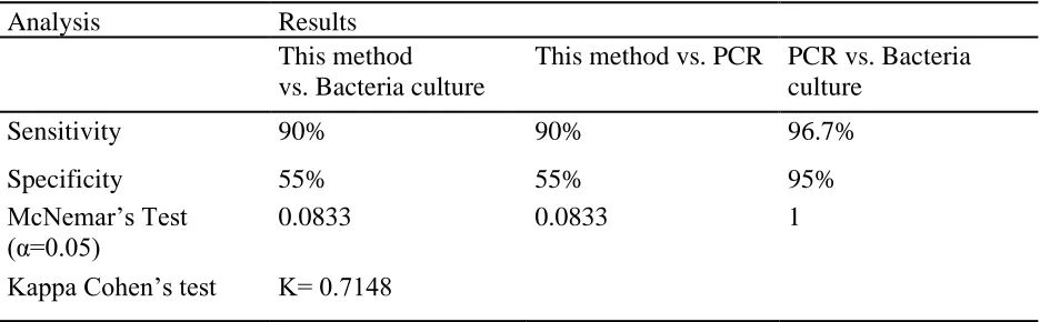
Table 2.1 Resistance and MIC distribution (squashtogram) of E. coli isolates from urban (n=41) and natural (n=25) environment ... 39 Table 3.1 Analysis of the performance of this method in comparison to bacterial culture and
viii LIST OF FIGURES
Figure 1.1 Phylogenetic tree and genotypic characteristics of E. coli isolated in the urban and
natural environment………37
Figure 1.2 Phylogenetic relationship of fosfomycin resistance genes……….38
Figure 2.1 Working principle of the described method……….……..56
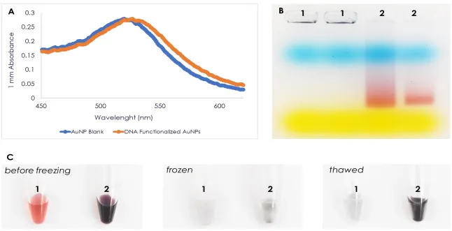
Figure 3.2 Characterization of AuNP...57
1 CHAPTER 1: Literature Review
1.1 Introduction
The increasing problem of antimicrobial resistance (AMR) is causing an alarm
worldwide, posing a risk both in humans, animal health, and the environment [1-3]. According to an eminent economist Jim O’Neil, AMR is a more certain threat than climate change [4]. With
limited choices of antimicrobials, it is expected that the cost of health care will increase due to longer duration of illness, additional medical tests, and the use of more expensive drugs [5]. Currently, the estimated annual deaths due to AMR is 700,000 and by the year 2050, it is projected that this number would reach up to 10 million along with $100 trillion economic loss [6]. Recognizing this urgent problem, the World Health Organization (WHO) issued a Global Action Plan on AMR to ensure the continuity of successful treatment and prevention of infectious diseases through the responsible use of quality and effectively safe medicines. To achieve this goal, surveillance programs and research has been launched to further understand AMR.
Different research and surveillance programs are being implemented in many countries, providing more comprehensive data on diversity as well as the mechanisms of AMR [1,7]. The majority of these studies have focused on AMR in clinical and agricultural environments such as hospitals and livestock farms. This left a knowledge gap regarding AMR determinants and virulence factors [8-10] found in the natural environment as well as in areas subjected to anthropogenic impacts.
2 developing countries where most of the emerging AMR determinants are reported. One of the WHO’s strategic objectives on the Global Action Plan on AMR is the development of diagnostic
tools (Objective 5) to help medical practitioners better identify and treat the pathogens causing infections [11]. Therefore, a need to develop for a simple, rapid, specific, sensitive and low-cost detection platform with an efficiency comparable to the existing methods that can be used in laboratories with lesser capabilities. Recent advances in research and technology have facilitated the development of platforms that focuses on eliminating the need for sample transportation and further processing. The early and accurate detection of these pathogens can reduce the financial cost by up to 20-35%, increase food production and food safety, and improve global economy [12]. More importantly, these diagnostic platforms can improve the quality of data and pace of reporting [13,14] which is important in tackling the issues related to AMR [15].
1.2 Antimicrobial Resistance in the Environment
The environment plays a significant role in the emergence and transmission of resistant bacteria. Soil is a vast source of AMR determinants as it has very diverse microbial composition, varying with the biochemical and geographical gradient [16] as a result of constant selective pressure. The majority of the antimicrobials being used today were initially isolated from the natural environment, particularly from the soil Actinomyces [6,17,18]. Normally, the
3 as genetic mutations in the microbe’s chromosome, horizontal transfer of AMR genes, and
plasmids allowing the organism to survive and thrive in the environment [4] in the environment to humans and animals.
The presence of human activities in an environment increases the chance of
contamination [6,8] such as spillage of antimicrobials, heavy metals, biocides and even resistant bacteria [8-10,22]. Such contamination creates a selective pressure or environmental hotspot for the development and dissemination of AMR [8,22]. In a heavy metal polluted environment, this plasmid containing resistance genes is maintained and so the AMR determinants, even in the absence of antimicrobials explaining the persistence of AMR in the environment [10]. Aside from the presence of antimicrobial producing microorganisms in the natural environment, the role of wildlife was also cited in the dissemination of resistance and pathogenic bacteria [9]. Several reports of AMR contamination in the natural environments have been associated with wildlife such as migratory birds and foxes [9,10,23].
1.3 Antimicrobial Resistance: dissemination and impact
The unwise and prudent use of antimicrobial drugs, both in animals and humans has even contributed to the rise of bacterial resistance [21,24]. Accordingly, the AMR is commonly seen in environments where microbes routinely come into contact with antimicrobials such as in hospitals and livestock farms [9,19]. It is believed that livestock animals are the most important source of AMR since they are constantly exposed to different antimicrobials as a treatment to diseases, prophylaxis and growth promotant. This has improved the food animal production and stabilized the meat supply and demand however, this approach greatly contributed to the current problem of AMR [24,25]. In the US alone, it is estimated that 80% of its antimicrobial
4 has been difficulty in establishing a direct causality between the prevalence of AMR in humans, animals, and the environment [24]. However, recent studies revealed a strong correlation
between AMR patterns found in the bacteria isolated from animal, human and its surrounding environment [26-30] and the antimicrobial drugs being used in animal production or farm system [26].
The emergence of the Extended-Spectrum Beta-Lactamases (ESBLs) produced by E. coli mainly contributed to the increase in antimicrobial resistance [21]. The ESBLs are enzymes inhibited by clavulanic acid and other inhibitors of the class A B-Lactamases, making the bacteria resistant to penicillin, cephalosporins, and monobactams. ESBL bacteria are also reported to be resistant to other antibiotics such as aminoglycosides, chloramphenicol,
fluoroquinolone, tetracycline and trimethoprim-sulfamethoxazole [21]. This resistance to a wide spectrum of antibiotics created reliance on reserved antibiotics such as carbapenems [21,31]. However, there has been an increasing report of carbapenem resistance in the US, and some countries in the western part of Asia and Europe [32]. In 2010, a highly resistant
Enterobacteriaceae isolated from a patient in Sweden was reported. This Enterobacteriaceae was found to carry New Delhi Metallo-B-Lactamase-1 (NDM-1) carbapenemases, making it highly resistant to many classes of antibiotics including Beta-lactams, fluoroquinolones, and
5 plasmid-colistin mediated resistance (MCR-1) in animals and humans. The emergence of MCR-1 resistant bacteria is deeply concerning and calls for an urgent re-evaluation of the use of
polymyxins, especially in animal production. High prevalence was reported both in animals and humans [34] and it is believed that this type of resistance is already extensive, considering that China is one of the largest producers and exporter of swine and poultry products.
Indeed, antimicrobial resistance has rapidly spread worldwide, and humans are the most affected by this scenario. Different interrelated factors among humans, animals, and the
environment play an important role in the development and persistence of antibiotic resistance. According to an eminent economist Jim O’Neil, antimicrobial resistance is a more certain threat
than climate change [4]. Without effective antimicrobials, treatment of common diseases as well as the management of medical procedures (such as organ transplantation) can be prolonged or can result in death. Resistance to common antimicrobials also increases the cost of health care due to longer duration of illness, additional medical tests and the use of more expensive drugs [5,6,11]. By the year 2050, the estimated economic losses due to AMR is $100 trillion. It is estimated that 700,000 people die annually due to AMR and by 2050, it is estimated that this number would reach up to 10 million [6]. The same can happen in livestock and food production, especially in areas where intensive farming is practiced. It is estimated that there will be a 67% increase of antimicrobial use by 2030 and that one-third of this will be used in livestock
production [24]. Therefore, it is only necessary to conduct continuous surveillance and
6 1.4 Bacterial pathogens in AMR studies
The Salmonella spp. and E. coli are the two of the most commonly reported bacterial pathogens affecting millions of humans annually [5,11,35]. These are gram-negative bacteria that are commensals and commonly found in the gastrointestinal tract of humans, livestock, birds, and even insects. While some serotypes or strains of these bacteria are host specific, others can be transmitted from animals to humans and the environment.
Salmonella is a complex group of medically important pathogens in human and
veterinary medicine. This bacterium, specifically the non-typhoidal Salmonella (NTS) serovars are normally isolated from the intestinal tract of humans, livestock, and bird and even insects. Globally, there are an estimated 78,707,591 [5,11,35] cases annually due to NTS. In the United States, NTS is estimated to cause 1.2 million illnesses and 450 deaths annually [5,11,36].
Similarly, E. coli is a commensal bacterium that thrives in the intestine of the human and animal host. Some E. coli strains such as Shiga-toxin E. coli (STEC) have acquired virulence factors and properties that allowed it to survive in different hosts and environments. Globally, the estimated number of illnesses due to STEC is 1,176,854 [5,11,35] including the 265,000 reported cases in the US [36]. Disturbingly, the highest percentage of deaths due to infection from these pathogens was reported among children under 5 years of age [5,11,35].
7 These bacteria are known to acquire and preserve AMR determinants and virulence factors from other microorganisms allowing them to thrive in the environment. Often, the reintroduction of these resistant and pathogenic bacteria into their primary host is a health concern due to their new properties that could cause serious diseases [36]. Surveillance reports have also shown international travel and trade as major risk factors in the dissemination of exotic bacterial strains and AMR determinants [8,38]. This was highlighted when a multidrug-resistant S. Schwarzengrund, a predominant cause of Salmonellosis in South East Asia was reported among isolates from persons and chickens in Thailand and food imported from Thailand to Denmark and the US [38]. For these reasons, the Salmonella spp. and E. coli are often used as indicator bacteria in AMR surveillance programs and other public health-related studies. 1.5 Laboratory techniques used in AMR studies
Different laboratory techniques are being used in the surveillance of AMR and resistant bacteria. Bacterial culture is an indispensable technique used in disease investigation as well as in AMR studies. The use of selective and differential media can identify a certain bacterium [39] up to the genus level. Differentiation of species is more important in the clinical setting since this can affect the treatment regimen, medication approach, and management [25]. With regards to antimicrobial resistance, the conventional technique based on the Clinical and Laboratory Standards Institute (CLSI) is very informative, providing the antimicrobial concentration that will inhibit the growth of bacteria. Moreover, a bacterial culture is needed to obtain pure isolates for further molecular analysis. The molecular technique has always been a reliable tool in investigating diseases or antimicrobial resistance overcoming the difficulty in identification, differentiation and quantification pathogens [40]. This can provide more comprehensive
8 AMR mechanism [39]. It can also provide information on the origin and compare relatedness between pathogens or isolates. This is very important in investigating diseases that are related to travel or acquired during a visit to countries such as the case of NDM-1 bacterial infection from a patient in the UK. The Polymerase Chain Reaction (PCR) can identify and amplify DNA markers specific pathogen or plasmid encoding antimicrobial resistance. Multiplex PCR is being widely used because it can detect multiple genes at the same time. Isothermal molecular
techniques such as Rolling Cycle Amplification (RCA) and Loop-Mediated Isothermal
Amplification (LAMP) are rapid and equally sensitive and specific as PCR, but multiplexing is difficult using these methods [4]. Array-based detection of antibiotic resistance genes can identify multiple genes encoding AMR determinants as well as virulence factors in a bacteria genome [41]. Selection pressure can easily change the bacterial genome leading to diversity between strains or isolates. Pulse Field Gel Electrophoresis (PFGE) can identify relatedness of isolates from recent outbreaks, but not well-suited to long-term global epidemiology. Methods such as multilocus enzyme electrophoresis (MLEE) and multilocus sequence typing (MLST) are reported to be highly discriminatory. MLST can distinguish between isolates or clones by
targeting fragments of the housekeeping genes, where genetic variation accumulates slowly. This can also provide information on evolutionary history, predict genotype and reveal the pattern of evolutionary descent of isolates [42,43]. With the continuous decrease in the cost of whole-genome sequencing (WGS), more bacterial whole-genome sequences are being reported in the
GenBank. Like MLEE and MLST, WGS provides information on the evolution of the isolates or origin based on its relatedness to other sequences. Moreover, WGS data has allowed researchers to understand the host’s physiological responses to different organisms and prompted studies on
9 Overall, both conventional and molecular techniques are important in AMR and bacterial pathogen studies [45]. It is important to emphasize that not all laboratories are capable of these techniques, especially in developing counties [46]. These techniques require expensive facilities and highly trained individuals hence, a gap in the epidemiological surveillance and research. The availability of rapid diagnostic tests that can specifically diagnose a bacterial pathogen would not only improve the clinical management of the diseases but would also help in the control of AMR by prescription of correct antimicrobials [30,39]. Therefore, a need to develop for a simple, rapid, specific, sensitive and low-cost detection platform with an efficiency comparable to the existing methods that can be used in laboratories with lesser capabilities.
1.6 Nanosensors
Nanotechnology is a science that deals mainly with the study of matter with a dimension between 1 to 100 nm and its manipulation at an atomic and molecular scale [45].
Nanotechnology has been widely explored and studies have been conducted to evaluate its applicability in different fields especially in medicine [46,47]. One of the major interests in nanotechnology today is the nanosensors or nanobiosensors and their application in point-of-care disease diagnosis [45]. There has been continuous research on the development of simple and rapid diagnostic platforms that would allow on-site diagnosis. Moreover, nanosensors are reported to increase the specificity and sensitivity of a technique. This favors a better diagnosis of a bacterial pathogen that would not only improve the clinical management of the diseases but could also help in the control of AMR by prescription of correct antimicrobials [30].
10 antibody-pathogen interaction or sequence-specific DNA hybridization into a signal which can be electric (voltage, resistance, or current change) or optical (colorimetric, fluorescent,
luminescent, turbidity) [49]. The electrochemical sensor is based on the detection of changes in ions in a solution or a mixture during the reaction process. The change in ions is recognized and recorded as a change in current and potential. Electrochemical biosensors are extremely sensitive and specific. The DNA-based electrochemical sensors commonly rely on the immobilization of single-strand DNA (capture probe) on a solid surface (glass slide or electrode) and the detection of a complementary strand upon hybridization. Electrochemical based detection of DNA through measurement of oxidized gold nanoparticles is widely studied and modification of this technique can further increase the sensitivity of the method. A study conducted by [50]
determined the efficiency of latex particles when gold nanoparticles are deposited on its surface. The modification of the technique allowed the detection of DNA at 0.1 femtomolar (fM), which is lower compared to previous reports. The same technique was used in the detection of
Aphanomyces invadans, a fungus that causes devastating disease in fish and reported a detection limit of 0.5 fM in synthesized linear target and 1 fM using PCR product [51]. Optical rely on the surface plasmon resonance property of nanoparticles. Nanoparticles exhibit intense and distinct colors [52] and changes in its surroundings (such as changes in ion concentrations) can be
observed even thru the naked eye. This property is very suitable especially in the development of colorimetric-based detection platforms [45,53].
1.7 Gold nanoparticle and colorimetric-based sensors
11 drug vehicles, imaging agents in therapy, biomarkers in the pharmaceutical field, and diagnostic tools [47,57,58]. These properties have attracted researchers, especially in the medical field to further explore AuNP especially in the development of different disease detection/screening platforms [56,59-63]. In 1996, Mirkin and his research group first shown the colorimetric assay for DNA detection using oligonucleotide functionalized AuNPs. In this experiment, the
researchers described a method for assembling colloidal gold nanoparticles rationally and reversibly into macroscopic aggregates. The method described attaching two batches of thiol modified oligonucleotides into the surface of 13 nm AuNPs. A solution containing
12 driven by the London-van der Waals forces between the AuNPs, wherein in this method, the repulsive interaction is greatly reduced [60].
A well-dispersed AuNP can be seen as red to pink in color with absorbance spectra of 520 nm. The decrease in the distance of AuNPs in a solution would cause a shift of plasmon resonance which is observed as a change in color from red to blue. Changes in the environment around the AuNPs can lead to aggregation which could be observed directly due to the color change of the solution from red to blue [55,60-66]. A salt solution such as Magnesium Chloride can destabilize the well-dispersed AuNPs because of the loss of electrostatic charges on the surface of the particle [67]. This causes a decrease in the distance between AuNPs as a result of particle coupling, followed by aggregation. Recent studies reported that the hybridization of oligonucleotide functionalized AuNPs on a target DNA could stabilize the AuNPs even after addition of salt solution [54,62,66,68,69,70].
During the last decade, there has been an increase in the utilization of this method as an indicator after PCR amplification eliminating the agarose gel electrophoresis step [65]. Other researchers reported the same method in the detection of pathogens amplified through loop-mediated isothermal amplification (LAMP) [68,69-71]. Molecular methods generate a large amount of product, which is negatively charged due to the phosphate backbone of the DNA. Specific hybridization of the oligonucleotide-functionalized gold nanoparticle to its
13 electrostatic repulsion between the AuNP in the solution is disrupted therefore causes
aggregation [62,66,68-70]. Colorimetric-based detection of DNA is an attractive method because it is rapid and cost-effective. It enables visual or direct detection of DNA without the use of sophisticated equipment.
14 1.8 References
1. Johnson AP. Surveillance of antibiotic resistance. Philos Trans R Soc Lond B Biol Sci 2015;370:20140080.
2. Mo SS, Urdahl AM, Madslien K, Sunde M, Nesse LL, Slettemeas JS, et al., What does the fox say? Monitoring antimicrobial resistance in the environment using wild red foxes as an indicator. PLoS ONE 2018;13:e0198019.
3. Oloso NO, Fagbo S, Garbati M, Olonitola SO, Awosanya EJ, Aworh K, et al. Antimicrobial resistance in food animals and the environment in Nigeria: A review. Int J Environ Res Public Health 2018;15:1284.
4. Anjum MF. Screening methods for the detection of antimicrobial resistance genes present in bacterial isolates and the microbiota. Future Microbiol 2015;10:317-320.
5. World Health Organization (WHO). Global action plan on antimicrobial resistance. 2015. http://www.who.int/iris/handle/10665/193736
6. O'Neill J. Antimicrobial Resistance: Tackling a crisis for the health and wealth of nations. London: Review on Antimicrobial Resistance, London, United Kingdom. 2014.
7. Richardson LC, Bacazo MC, Parker CC, Dewey-Mattia D, Golden N, Jones K, et al. An updated scheme for categorizing foods implicated in foodborne diseases outbreaks: A tri-agency collaboration. Foodborne Pathog Dis 2017;14:1-10.
8. Berendonk TU, Manaia CM, Merlin C, Fatta-Kassinos D, Cytryn E, Walsh F, et al. Tackling antibiotic resistance: the environmental framework. Nat Rev Microbiol 2015;13:310-317. 9. Bengtsson-Palme J, Kristiansson E, Larsson DGJ. Environmental factors in influencing the
15 10. Martinez JL. Antibiotics and antibiotic resistance genes in natural environments. Science
2008;321:365:367.
11. World Health Organization (WHO). WHO estimates of the global burden of foodborne diseases. 2015. http://www.who.int/foodsafety/areas_work/foodborne-diseases/ferg/en/ 12. Neethirajan S, Ragavan KV, Weng X. Agro-defense: Biosensors for food from healthy crops
and animals. Trends Food Sci Technol 2018;75:25-44.
13. Carriço JA, Sabat AJ, Friedrich AW, Ramirez M, on behalf of the ESCMID Study Group for Epidemiological Markers (ESGEM). Bioinformatics in bacterial molecular epidemiology and public health: databases, tools and the next-generation sequencing revolution. Euro Surveill 2013;18:pii=20382.
14. Karp BE, Tate H, Plumbee JR, Dessai U, Whichards JM, Thacker EL, et al. National Antimicrobial Resistance Monitoring System: Two Decades of Advancing Public Health through Integrated Surveillance of Antimicrobial Resistance. Foodborne Pathog Dis 2017;14:545-557.
15. Liu JM, Hu Y, Yang YK, Liu H, Fang GZ, Lu X, et al. Emerging functional nanomaterials for the detection of food contaminants. Trends Food Sci Technol 2018;71:94-106.
16. Gyles C, Boerlin P. Horizontally transferred genetic elements and their role in pathogenesis of bacterial disease. Vet Pathol 2014;51:328-340.
17. Aminov RI. A brief history of the antibiotic era: lessons learned and challenges for the future. Front Microbiol 2010;1:1-7.
16 19. Pal C, Bengtsson-Palme J, Kristiansson E, Larsson DGJ. The structure and diversity of
human, animal and environmental resistomes. Microbiome 2016;4. doi:10.1186/s40168-016-0199-5.
20. Clermont O, Christenson JK, Denamur E, Gordo DM. The Clermont Escherichia coli phylo-typing method revisited: improvements of specificity and detection of new phylo-groups. Environ Microbiol Rep 2013;5:58-65.
21. Krizman M, Avgustinn JA, Zdovc I, Golob M, Trkov, M, Ciglenecki UJ, et al. Antimicrobial resistance and molecular characterization of Extended-Spectrum B-Lactamases and other Escherichia coli isolated from food of animal origin and human intestinal isolates. J Food Prot 2017;80:113-120.
22. Berkner S, Kondradi S, Schonfeld J. Antibiotic resistance and the environment-there and back again. EMBO Rep 2014;15:740-744.
23. Huijbers PMC, Blaak H, de Jong MCM, Graat EAM, Vandenbroucke-Grauls CMJE, Husman AM. Role of the environment in the transmission of antimicrobial resistance to humans: A review. Environ Sci Technol 2015;49:11993-12004.
24. Van Boeckel TP, Brower C, Gilbert M, Grenfell BT, Levin SA, Robinson TP, et al. Global trends in antimicrobial usage in food animals. PNAS USA 2015;112:5649-5654.
25. Kaakoush NO, Castano-Rodriguez N, Mitchell HM, Man SM. Global epidemiology of Campylobacter infection. Clin Microbiol Rev 2015;28:687-720.
26. Keelara S, Scott HM, Morow WM, Hartley CS, Griffin DL, Gebreyes WA, et al.
Comparative phenotypic and genotypic characterization of temporally related non-typhoidal Salmonella isolated from human clinical cases, pigs, and the environment in North Carolina.
17 27. Quintana-Hayashi MP, Thakur S. Longitudinal study of the persistence of
antimicrobial-resistant Campylobacter strains in distinct swine production systems on farms, at slaughter, and in the environment. Appl Environ Microbiol 2012;78:2698-2705.
28. Pornsukarom S, Thakur S. Assessing the impact of manure application in commercial swine farms on the transmission of antimicrobial resistant Salmonella in the environment. PLoS ONE 2016;11:e0164621.
29. Pornsukarom S, Thakur S. Horizontal Dissemination of Antimicrobial Resistance
Determinants in Multiple Salmonella Serotypes following Isolation from the Commercial Swine Operation Environment after Manure Application. Appl Environ Microbiol
2017;83:e1503-e1517.
30. Zellweger RM, Carrique-Mas J, Limmathurotsakul D, Day NPJ, Thwaites GE, Baker S. A current perspective on antimicrobial resistance in Southeast Asia. J Antimicrob Chemother 2017;72:2963-2972.
31. Kumarasamy KK, Toleman MA, Walsh TR, Bagaria J, Butt F, Balakrishnan R, et al. Emergence of new antibiotic resistance mechanism in India, Pakistan and the UK: a molecular, biological, and epidemiological study. Lancet Infect Dis 2010;10:597-602. 32. Holt KE, Wertheim H, Zadoks RN, Baker S, Whitehouse CA, Dance D, et al. Genomic
analysis of diversity, population structure, virulence and antimicrobial resistance in Klebsiella pneumoniae, an urgent threat to public health. PNAS USA 2015;112:e3574-E3581.
18 carbapenemase-producing Klebsiella pneumoniae in London. Sci Rep 2017;7.
doi:10.1038/s41598-017-12637-4.
34. Liu YY, Wang Y, Walsh TR, Yi LX, Zhang R, Spencer J, et al. Emergence of plasmid-mediated colistin resistance mechanism MCR-1 in animals and human beings in China: a microbiological and molecular biological study. Lancet Infect Dis 2016;16:161-168. 35. Havelaar AH, Kirk MD, Torgerson PR, Gibb HJ, Hald T, Lake RJ, et al. World Health
Organization global estimates and regional comparisons of the burden of foodborne disease in 2010. PLoS Med 2015. https://doi.org/10.1371/journal.pmed.1001923.
36. Center for Disease Control and Prevention (CDC). National Salmonella Surveillance Overview. Atlanta, Georgia: US Department of Health and Human Services, CDC. 2011. 37. Center for Disease Control and Prevention (CDC). National Shiga toxin-producing
Escherichia coli (STEC) Surveillance Overview. Atlanta, Georgia: US Department of Health and Human Services, CDC. 2012.
38. Aarestrup FM, Hendriksen RS, Lockett J, Gay K, Teates K, McDermott PF, et al.
International spread of multidrug-resistant Salmonella Schwarzengrund in food products. Emerg Infect Dis 2007;13:726-731.
39. Bauer KA, Perez KK, Forrest GN, Goff DA. Review of rapid diagnostic test used in antimicrobial stewardship programs. Clin Infect Dis 2014;59:S134-S145.
40. Frasao BS, Marin VA, Conte-Junior CA. Molecular detection, typing, and quantification of Campylobacter spp. in foods of animal origin. Compr Rev Food Sci Food Saf
19 41. McNeece G, Naughton V, Woodward MJ, Dooley JSG, Naughton PJ. Array-based detection
of antibiotic resistance genes in Gram negative bacteria isolate from poultry meat in the UK and Ireland. Int J Food Microbiol 2014;179:24-32.
42. Rodriguez-Noriega E, Seas C, Guzman-Blanco M, Mejia C, Alvarez C, Bavestrello L, et al. Evolution of methicillin-resistant Staphylococcus aureus clones in Latin America. Int J Infect Dis 2010;14:e560-e566.
43. Yamashita A, Sekizuka T, Kuroda M. Characterization of antimicrobial resistance dissemination across plasmid communities classified by network analysis. Pathogens 2014;3:356-376.
44. Taveres LS, Silva CSF, de Souza VC, da Silva VL, Diniz CG, Santos MO. Strategies and molecular tools to fight antimicrobial resistance: resistome, transcriptome and antimicrobial peptides. Front Microbiol 2013;4:1-11.
45. Prado M, Espina B, Ferenandez-Arguelles MT, Dieguez L, Fucinos P, Vial S, et al. Detection of foodborne pathogens using nanoparticles: Advantages and Trends. Antimicrobial Food Packaging 2016;183-201. http://dx.doi.org/10.1016/B978-0-12-800723-5.00014-0.
46. Teengam P, Siangproh W, Tuantranont A, Vilaivan T, Chailapakul O, Henry CO. Multiplex paper-based colorimetric DNA sensor using pyrrolidinyl peptide nucleic acid-induced AgNPs aggregation for detecting MERS-CoV, MTB, and HPV oligonucleotide. Anal Chem 2017;89:5428-5435.
20 48. Mocan T, Matea CT, Pop T, Mosteanu O, Buzoianu AD, Puia C, et al. Development of
nanoparticle-based optical sensors for pathogenic bacterial detection. J Nanobiotechnol 2017;15:1-14.
49. Ghosh I, Stains CI, Ooi AT, Segal DJ. Direct detection of double stranded DNA: molecular methods and applications for DNA diagnostics. Mol BioSyst 2006;2:551-560.
50. Pinijsuwan S, Rijiravanich P, Somasundrum M, Surareungchai W. Sub-femtomolar electrochemical detection of DNA hybridization based on latex/gold nanoparticle-assisted signal amplification. Anal Chem 2008;80:6779-6784.
51. Kuan GC, Cheng LP, Rijiravanich P, Marimuthu K, Ravichandran M, Yin LS, et al. Gold-nanoparticle based electrochemical DNA sensor for the detection of fish pathogen
Aphanomyces invadans. Talanta 2013;117:312-317
52. Huschka R, Neumann O, Barhoumi A, Halas NJ. Visualizing light-triggered release of molecules inside living cells. Nanoletters 2010;10:4117-4122.
53. Li F, Li F, Yang G, Aguilar ZP, Lai W, Xu H. Asymmetric polymerase chain assay combined with propidium monoazide treatment and unmodified gold nanoparticles for colorimetric detection of viable emetic Bacillus cereus is milk. 2018;255:1455-1461. 54. Fang WF, Chen WJ, Yang JT. Colorimetric determination of DNA concentration and mismatches using hybridization-mediated growth of gold nanoparticle probes. Sens Actuators B Chem 2014;192:77–82.
55. Jung YL, Jung C, Parab H, Cho D-Y, Park HG. Colorimetric SNP genotyping based on allele-specific PCR by using a thiol-labeled primer. Chembiochem 2011;12:1387–1390. 56. Khan A, Rashid R, Murtaza G, Zahra A. Gold nanoparticles: Synthesis and applications in
21 57. Daniel MC, Astruc D. Gold nanoparticles: assembly, supramolecular chemistry, quantum
size-related properties, and applications toward biology, catalysis, and nanotechnology. Chem Rev 2004;104:293-346.
58. Huo S, Jin S, Ma X, Xue X, Yang K, Kumar A, et al. Ultrasmall gold nanoparticles as carriers for nucleus-based gene therapy due to size-dependent nuclear entry. ACS Nano 2014;8:5852-5862.
59. Mirkin CA, Letsinger RL, Mucic RC, Storhoff JJ. A DNA-based method for rationally assembling nanoparticles into macroscopic materials. Nature 1996;382:607–609.
60. Sato K, Hosokawa K, Maeda M. Rapid aggregation of gold nanoparticles induced by non-cross-linking DNA hybridization. J Am Chem Soc 2003;125:8102-8103.
61. Li HX, Rothberg L. Colorimetric detection of DNA sequences based on electrostatic interactions with unmodified gold nanoparticles. PNAS USA 2004;101:14036-14039. 62. Ahmadpour-Yazdi H, Hormozi-Nezhad MR, Abadi AR, Sanati MH, Kazemi B.
Colourimetric-based method for the diagnosis of spinal muscular atrophy using gold nanoprobes. IET Nanobiotechnol 2015;9:5-10.
63. Castañeda MT, Alegret S, Merkoçi A. Electrochemical sensing of DNA using gold nanoparticles. Electroanalysis 2007;19:743–753.
64. Bakthavathsalam P, Ranjendran VK, Mohammed JAB. A direct detection of Escherichia coli genomic DNA using gold nanoprobes. J Nanobiotechnology 2012;10:
https://doi.org/10.1186/1477-3155-10-8.
65. Jung YL, Jung C, Parab H, Li T, Park HG. Direct colorimetric diagnosis of pathogen
22 66. Xia F, Zuo X, Yang R, Xiao Y, Kang D, Vallée-Bélisle A, et al. Colorimetric detection of
DNA, small molecules, proteins, and ions using unmodified gold nanoparticles and conjugated polyelectrolytes. PNAS USA 2010;107:10837–10841.
67. Levy R, Thanh NTK, Doty RC, Hussain I, Nichols RJ, Schiffrin DJ, et al. Rational and combinatorial design of peptide capping ligands for gold nanoparticle. J A Chem Soc 2004;126:10076-10084.
68. Arunrut N, Kampeera J, Suebsing R, Kiatpathomchai W. Rapid and sensitive detection of shrimp infectious myonecrosis virus using a reverse transcription loop-mediated isothermal amplification and visual colorogenic nanogold hybridization probe assay. J Virol Methods 2013;193:542–547.
69. Jaroenram W, Arunrut N, Kiatpathomchai W. Rapid and sensitive detection of shrimp yellow head virus using loop-mediated isothermal amplification and a colorogenic nanogold
hybridization probe. J Virol Methods 2012;186:36–42.
70. Seetang-Nun Y, Jaroenram W, Sriurairatana S, Suebsing R, Kiatpathomchai W. Visual detection of white spot syndrome virus using DNA-functionalized gold nanoparticles as probes combined with loop-mediated isothermal amplification. Mol Cell Probes 2013;27:71– 79.
23 CHAPTER 2: Antimicrobial resistance and virulence factors profile of Salmonella spp. and
Escherichia coli isolated from different environments exposed to anthropogenic activities
Presented here is the manuscript titled: “Antimicrobial resistance and virulence factors profile of
Salmonella spp. and Escherichia coli isolated from different environments exposed to anthropogenic activities” submitted in the Journal of Global Antimicrobial Resistance.
Additional data are presented in the Appendix. 2.1 Abstract
The study aimed to identify the antimicrobial resistance (AMR) determinants and
virulence factors in Salmonella spp. and Escherichia coli recovered from different anthropogenic areas in North Carolina. Soil samples were collected from different anthropogenic areas: urban and natural. Minimum inhibitory concentration (MIC) was determined by using the broth microdilution method. Whole genome sequencing and analysis were done to identify the AMR determinants and virulence factors.
A higher prevalence of Salmonella spp. and E. coli was detected in the urban
24 The detection of AMR determinants and virulence factors in these bacteria is significant in understanding the occurrence and even the development of AMR. The presence of these determinants in different anthropogenic areas suggests the need to conduct longitudinal studies for comparing the profile of pathogens across different environments.
2.2 Introduction
The emergence, persistence, and continuous spread of antimicrobial resistance (AMR) is considered as one of the greatest threats to humans (1,2,3). Currently, the annual deaths due to AMR are estimated to be 700,000 (4) and by 2050, it is projected that this number could reach up to 10 million (4) and an economic loss amounting to $100 trillion. Recognizing this urgent problem, the World Health Organization (WHO) issued a Global Action Plan on AMR to ensure the continuity of successful treatment and prevention of infectious diseases through the
responsible use of quality and effectively safe medicines (4).
The environment plays a significant role in the emergence and transmission of AMR determinants and pathogenic bacteria (2). The constant interactions of humans, animals, and the environment can give rise to selection pressures leading to changes that would help an organism to survive such as mutations, horizontal transfer of AMR genes, plasmids, and virulence factors (1,3,5). The frequent exposure of the environment to antimicrobials have created hotspots for the selection, proliferation, and spread of AMR determinants (1,5,6). As for pathogenic bacteria such as Escherichia coli O157:H7, the increased virulence happens when there is gene loss or silencing, insertion, and rearrangement which occurs under the same mechanisms associated with the acquisition of AMR determinants in the environment (5). Accordingly, AMR is
25 have focused on clinical and agricultural environments. This left a knowledge gap regarding AMR determinants and virulence factors (1,3,6,7) found in the natural environment as well as in areas subjected to anthropogenic impacts. Salmonella spp. and E. coli are the two of the most commonly reported bacterial pathogens affecting millions of humans annually (2). While some serotypes or strains of these bacteria are host specific, there are others that can be transmitted from animals to humans and to the environment. These bacteria are known to acquire and
disseminate AMR determinants and virulence factors allowing them to thrive in the environment (1). Often, the reintroduction of these resistant and pathogenic bacteria into their primary host is a health concern due to their new properties that could cause serious diseases (3). The E. coli O157:H7 for example, is a pathogenic strain that is well adapted in the environment (5,8). For these reasons, the Salmonella spp. and E. coli are often used as indicator bacteria in AMR surveillance programs and other public health-related studies (1).
In our study, we isolated Salmonella spp. and E. coli from different anthropogenic areas in North Carolina and identified the AMR determinants as well as virulence factors through antimicrobial susceptibility testing (AST) and whole genome sequencing (WGS). The information gathered from this study is important in understanding the occurrence and persistence of resistance determinants and pathogenic bacteria in the environment. 2.3 Materials and methods
2.3.1 Sample collection and bacteria isolation
26 includes Forest A (n=14), Forest B (n=8), Forest C (n=5), Forest D (n=5), and potting soil mix (n=9).
Prior to bacterial isolation, each of the soil samples was sieved and mixed. The Salmonella spp. was isolated using Xylose-Lysine-Tergitol (XLT-4) selective media. Subsequently, 5 colonies from each positive plate were subjected to biochemical tests using Triple Sugar Iron agar slant (TSI agar), Lysine Iron Agar (LIA) and Urea agar slant (9,10). For E. coli, each sample was streaked into a McConkey agar (10,11). From each plate, multiple (n=3) colonies were picked for further isolation until a pure culture was obtained. Confirmation of Salmonella spp. isolates were performed through amplification of the invA gene (9) while the 16s rRNA gene was used to identify E. coli (12).
2.3.2 Resistance determination
The AST was performed by the broth microdilution method in a 96-well gram-negative sensititre plate containing a panel of 14 antimicrobials (CMV3AGNF Gram Negative NARMS plate, Trek Diagnostic System, Cleveland, OH, USA). The Minimum Inhibitory Concentration (MIC) was interpreted based on the Clinical and Laboratory Standards Institute standards (CLSI) [9,10].
2.3.3 Analysis of antimicrobial resistance genes (ARGs)
The presence of ARGs among the isolates was analyzed through the amplification of ARGs by polymerase chain reaction (PCR). The presence of integron was also determined by the amplification of the integron gene-1 (intI 1) (9).
2.3.4 Whole genome sequencing
27 Qiagen DNeasy Blood and Tissue Kit (Qiagen, Germany) following the manufacturer’s
instructions. The quality and concentration of the extracted DNA were determined using the NanoDropTM 2000/2000c Spectrophotometer (Thermo Scientific, USA) and Qubit3.0
Fluorometer (Thermo Fisher Scientific, USA). DNA libraries were prepared using the Nextera XT DNA Library Preparation Kit (Illumina, USA) following the manufacturer's instructions. The resulting DNA libraries were purified using AMPure XP beads (Beckman Coulter, USA) and re-quantified using the Qubit3.0 Fluorometer (Thermo Fisher Scientific, USA). Sequencing was performed on the MiSeq System using v2 sequencing reagent kits (Illumina, USA) (9). 2.3.5 Data assembly and analysis
Raw sequences were assembled using the CLC Genomic Workbench (QIAGEN
Bioinformatics, USA). Draft genomes were annotated using Rapid Annotation using Subsystem Technology (RAST) (http://rast.theseed.org/FIG/rast.cgi). Serotype and phylotype of the isolates were determined through pubMLST (https://pubmlst.org/bigsdb), SeqSero
(http://denglab.info/SeqSero), and EnteroBase (https://enterobase.warwick.ac.uk/). The presence of plasmid and virulence genes were determined using PlasmidFinder
(https://cge.cbs.dtu.dk/services/PlasmidFinder/), VirulenceFinder
(https://cge.cbs.dtu.dk/services/VirulenceFinder/), and Virulence Finder Database
(http://www.mgc.ac.cn/VFs/main.htm). Resistance genes were determined through ResFinder (https://cge.cbs.dtu.dk/services/ResFinder/) (9) and Comprehensive Antibiotic Resistance Database (CARD) (https://card.mcmaster.ca). The phylogenetic tree was constructed using GrapeTree (https://enterobase.warwick.ac.uk/) and visualized through iTOL
28 2.4 Results
2.4.1 Prevalence of Salmonella spp. and E. coli
The prevalence of Salmonella spp. in soil samples from the urban and natural
environments was 10.34% and 2.44% respectively. In E. coli, the prevalence was observed to be 62.1% in urban and 26.8% in the natural environment. From these positive soil samples, a total of 20 Salmonella spp. (urban, n=15; natural, n=5) and 66 E. coli (urban, n=41; natural, n=25) isolates were obtained and analyzed.
2.4.2 AMR in Salmonella spp. and E. coli
The Salmonella spp. from the urban environment were resistant to Streptomycin
(66.67%) and Sulfisoxazole (46.47%) while those from the natural environment were resistant to Sulfisoxazole (100%) (Appendix 1). Similarly, E. coli isolated from the urban environments showed the highest resistance to Sulfisoxazole (78.05%), followed by Ampicillin (7.32%), and Cefoxitin (4.88%). Isolates from the natural environment only showed resistance to
Sulfisoxazole (80%) (Table 2.1).
2.4.3 AMR determinants and virulence factor analysis
The Salmonella spp. from the urban environment carries fox (80%), strA (66.67%), and strB (66.67%) genes while all the isolates from the natural environment encoded for fox (100%)
gene. With E. coli, the ARGs sul1 (4.89%) and blaCMY-2 (4%) were detected from the urban and
natural environments, respectively.
29 aaa(6’)-Iaa gene while S. Schwarzengrund has aac(6”)-Iaa, aph(6)-Id, and aph(3’)-Ib. All S.
Schwarzengrund isolates were detected to carry IncFIB and IncFIC(FII) plasmids (Appendix 2). Similarly, the WGS analysis of E. coli isolates from the urban environment
predominantly belonged to phylogroups A (26.67%), B1 (21.67%), D (20%), and B2 (13.33%). The E. coli from the natural environment (n=13) were determined to be from phylogroups B1 (38.46%), B2 (38.46%), D (7.69%), and E (7.69%). There were 23 virulence genes identified among the isolates, while only 9 were detected at a frequency >10%. These genes include gad (glutamate decarboxylase), lpfA (long polar fimbriae), iss (increased survival serum), air
(enteroaggregative immunoglobulin repeat protein), and eilA (Salmonella HilA homolog) among others. Additionally, virulence factors such as iroN (Salmochelin), espA (secreted proteins), and tsh (temperature-sensitive hemagglutination) were identified. The mdfA gene was detected in 100% and 84.5% of E. coli isolates from the urban and natural environment, respectively (Figure 2.1). A new gene FosA7 conferring resistance to Fosfomycin was detected in an isolate from a residential yard. The activity of the FosA7 gene was assessed by amplifying and cloning the segment into E. coli (TOP10). Using E-test (Liofilchem, USA), the donor isolate and the transformed E. coli showed resistance to Fosfomycin (MIC, >256 ug/mL). Its relatedness to other Fosfomycin resistance genes was analyzed using MAFFT
(https://mafft.cbrc.jp/alignment/software) (Figure 2.2). The pCoo and pB171 plasmids were detected in isolates from a residential yard and garden landscape, respectively. While col(156) and pHN7A8 plasmids were detected from the natural environments, Forest A and D.
2.5 Discussion
30 determinants are expected to be high (13). This study identified AMR determinants and virulence factors in Salmonella spp. and E. coli from different anthropogenic areas in North Carolina using WGS.
S. Schwarzengrund was detected in our isolates from the urban environment. S.
Schwarzengrund was identified as one of the frequently detected serotypes in human, non-clinical sources in the US (14). There have been reports of disease outbreak, food contamination, (14) and multidrug resistance from this serovar (15). Moreover, we detected IncFIB and IncFII plasmids, which were reported to be significantly associated with S. Schwarzengrund from humans, animals, and manure treated environment (15).
The E. coli isolates predominantly belonged to phylogroups B1, B2, and D, which are known to thrive in various ecological niches, including soil (8). Several strains from these phylogroups are commensals and pathogenic and often has large genomes that codes for AMR determinants and virulence factors, allowing them to adapt and survive in different environments (5,6). The plasmids pCoo and pB171 were detected in the urban environment while col(156) and pHN7A8 were identified in the natural environment. Both pCoo and pB171 are virulence
31 and rmtB resistance genes and has the capability to acquire markers involved in plasmid
replication or stability (16).
E. coli has intrinsic and acquired genes that degrade and resist toxic compounds such as
biocides and metals (3,4,18), allowing their survival in the environment. The mdfA is a multidrug efflux protein, and its overexpression results in resistance to several antimicrobials and organic cations (3). The FosA7 gene is a new ARG that was detected from S. Heidelberg from broiler chickens in Canada, was discovered in our E. coli isolate from a residential yard. FosA7 confers resistance to Fosfomycin, a broad-spectrum antibiotic that is used to treat uncomplicated UTI and extensively drug-resistant (XDR) gram- bacteria. Fosfomycin resistance has been reported in bacterial isolates from human and animal origin in China, Japan, France, and recently the US and Canada (19), but was not reported in isolates from the environment.
The majority of the antimicrobials being used today were initially isolated from the natural environment, particularly from the soil Actinomyces (6,7,20). The antimicrobial producing organisms have determinants that would help to resist the action of the
antimicrobial(s) they produce along with the other microbes found in the same environment (3,6,20), and so it is not surprising to detect AMR determinants in the environment (3,18). However, the proximity of humans and its activities in the environment greatly influences the dynamics in the microbial community, genetic variation, resistance selection, and possible emergence of novel mechanisms of resistance (6).
Soil is a huge source of AMR determinants considering its diverse microbial composition that varies depending on the geographical and biochemical gradient (20) as a result of constant selective pressures exerted in the environment. A large amount of antimicrobials being
32 not only alter the microbial community but also hasten and sustain the development of AMR and virulent pathogens (1,5,6). Urban environments are continuously being exposed to different anthropogenic activities making it more vulnerable to changes that may shape the composition of the bacterial community. The presence of human activities in an environment increases the chance of contamination (1,8) such as spillage of antimicrobials, heavy metals, biocides, and even resistant bacteria (1,3,6,18). Such contamination creates a selective pressure or
environmental hotspot for the development and dissemination of AMR (1,5,18). Heavy metals and biocides, for example, was identified to play an essential role in the maintenance and spread of AMR determinants. Specific plasmids can contain genes that confer resistance to several compounds such as antimicrobials, heavy metals, and biocides. In a heavy metal polluted environment, this plasmid containing resistance genes is maintained and so the AMR
determinants, even in the absence of antimicrobials explaining the persistence of AMR in the environment (6).
Salmonella spp. and E. coli are commensal bacteria in humans and animals, but these bacteria are also well adapted in the natural environment. Aside from the presence of
33 2.6 Accession numbers
The paired-end reads used in this study were deposited in the National Center for
Biotechnology Information (NCBI) under the Bioproject accession numbers PRJNA293224 and PRJNA293225 for Salmonella spp. and E. coli isolates, respectively.
2.7 Abbreviations
MIC, Minimum Inhibitory Concentration; ETEC, Enterotoxigenic E. coli; EPEC,
enteropathogenic E. coli; ExPEC, Extraintestinal pathogenic E. coli; UTI, urinary tract infection; MDR, multidrug-resistant; XDR, extensively drug-resistant; MAFFT, Multiple alignment using fast Fourier Transform; AMP, Ampicillin; AUG2, Amoxicillin/Clavulanic acid; AXO,
Ceftriaxone; AZI, Azithromycin; CHL, Chloramphenicol; CIP, Ciprofloxacin; FIS,
Sulfisoxazole; FOX, Cefoxitin; GEN, Gentamycin; NAL, Nalidixic acid; STR, Streptomycin; SXT, Trimethoprim/Sulfamethoxazole; XNL, Ceftiofur; TET, Tetracycline; RY, residential yard; IPP, indoor potted plant; GL, garden landscape; FA, Forest A; FB, Forest B; FC, Forest C; FD, Forest D.
2.8 Acknowledgment
The authors acknowledge the support and assistance of the research staff and students in Thakur Lab and Rob Dunn Lab, NCSU. We are grateful to the Duke Triangle Center for
34 2.9 References
1. Berendonk TU, Manaia CM, Merlin C, Fatta-Kassinos D, Cytryn E, Walsh F., et al. Tackling antibiotic resistance: the environmental framework. Nat Rev Microbiol 2015;13:310-317. 2. Huijbers PMC, Blaak H, de Jong MCM, Graat EAM, Vandenbroucke-Grauls CMJE,
Husman AM. Role of the environment in the transmission of antimicrobial resistance to humans: A review. Environ Sci Technol 2015;49:11993-12004.
3. Bengtsson-Palme J, Kristiansson E, Larsson DGJ. Environmental factors in influencing the development and spread of antibiotic resistance. FEMS Microbiol Rev 2018;fux53:68-80. 4. O'Neill J. Antimicrobial Resistance: Tackling a crisis for the health and wealth of nations.
London: Review on Antimicrobial Resistance, London, United Kingdom. 2014.
5. Gyles C, Boerlin P. Horizontally transferred genetic elements and their role in pathogenesis of bacterial disease. Vet Pathol 2014;51:328-340.
6. Martinez JL. Environmental pollution by antibiotic resistance determinants. Environ Pollut 2009;157:2893-2902.
7. Larsson DGJ, Andremont A, Bengtson-Palme J, Brandt KK, Husman AM, Fegerstedt P, et al. Critical knowledge gaps and research needs related to the environmental dimensions of antibiotic resistance. Environ Int 2018;117:132-138.
8. Elsas JD, Semenov AV, Costa R, Trevors JT. Survival of Escherichia coli in the environment: fundamentals and public health aspects. ISME J 2011;5:173-183.
35 10. Food and Drug Administration. The National Antimicrobial Resistance Monitoring System
(NARMS) Manual of laboratory methods. Rockville, MD: U.S. Department of Health and Human Service, FDA, 2016.
11. Gutierez-Rodriguez E, Gundersen A, Sbodio AO, Suslow TV. Variable agronomic practices, cultivar, strain source, and initial contamination dose differentially affect survival of
Escherichia coli on spinach. J Appl Microbiol 2011;112:109-118.
12. Fratamico PM, DebRoy C, Liu Y. The DNA sequence of the Escherichia coli O22-antigen gene cluster and detection of pathogenic strains belonging to E. coli serogroups O22 and O91by multiplex PCR assays targeting virulence genes and genes in the respective O-antigen gene clusters. Food Anal Methods 2009;2:169-179.
13. Mafiz AI, Perera LN, He Y, Zhang W, Xiao S, Hao W, et al. Case study on the soil antibiotic resistome in an urban community garden. Int J Antimicrob Agents 2018;52:241-250.
14. Akiyama T, Khan AA. Molecular characterization of strains of fluoroquinolone-resistant Salmonella enterica serovar Schwarzengrund carrying multidrug resistance isolated from imported foods. J Antimicrob Chemother 2012;67:10-110.
15. Pornsukarom S, van Vliet AHM, Thakur S. Whole genome sequencing analysis of multiple Salmonella serovars provides insights into phylogenetic relatedness, antimicrobial resistance,
and virulence markers across humans, food animals, and agriculture environmental sources. BMC Genomics 2018;19:801.
16. Johnson TJ, Nolan LK. Pathogenomics of the Virulence Plasmids of Escherichia coli. Microbiol Mol Biol Rev 2009;73:750-774.
36 coli and Klebsiella pneumonia isolated from animals, foods products and humans in China. mSphere 2018;3:e00137-18.
18. Berkner S, Konradi S, Schonfeld J. Antibiotic resistance and the environment- there and back again. EMBO reports 2014;15. DOI.10.15252/embr.201438978.
19. Rehman MA, Yin X, Persaud-Lachmann MG, Diara MS. First detection of Fosfomycin resistance gene, FosA7, in Salmonella enterica serovar Heidelberg isolated from broiler chickens. Antimicrob Agents Chemother 2017;61:e00410-17.
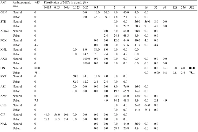
39 Table 2.1 Resistance and MIC distribution (squashtogram)a of E. coli isolates form urban (n=41) and natural (n=25) environment
AMb Anthropogenic
area
%Rc Distribution of MICs in µg/mL (%)
0.015 0.03 0.06 0.125 0.25 0.5 1 2 4 8 16 32 64 128 256 512
GEN Natural 0 0.0 16.0 36.0 4.0 40.0 4.0 0.0
Urban 0 0.0 46.3 39.0 4.8 2.4 7.3 0.0
STR Natural 0 0.0 0.0 56.0 36.0 8.0 0.0
Urban 0 0.0 29.2 58.5 7.3 4.8 0.0
AUG2 Natural 0 0.0 8.0 64.0 28.0 0.0 0.0
Urban 0 2.4 24.4 68.3 4.9 0.0 0.0
FOX Natural 0 0.0 0.0 12.0 44.0 40.0 4.0 0.0
Urban 4.9 0.0 0.0 0.0 53.6 41.5 0.0 4.9
XNL Natural 0 0.0 8.0 84.0 8.0 0.0 0.0 0.0
Urban 0 0.0 14.6 78.1 2.4 0.0 4.9 0.0
AXO Natural 0 100.0 0.0 0.0 0.0 0.0 0.0 0.0 0.0 0.0
Urban 0 100.0 0.0 0.0 0.0 0.0 0.0 0.0 0.0 0.0
FIS Natural 80.0 0.0 0.0 16.0 0.0 4.0 80.0
Urban 78.1 0.0 0.00 9.8 9.8 2.4 78.1
SXT Narural 0 60.0 24.0 12.0 4.0 0.0 0.0
Urban 0 82.9 12.2 2.4 2.4 0.0 0.0
AZI Natural 0 0.0 0.0 0.0 0.0 8.0 76.0 16.0 0.0
Urban 0 0.0 0.0 0.0 0.0 19.5 65.9 14.6 0.0
AMP Natural 0 0.0 24.0 64.0 12.0 0.0 0.0
Urban 7.3 4.9 34.2 48.8 4.9 0.0 2.4 4.9
CHL Natural 0 0.0 4.0 24.0 64.0 8.0
Urban 0 0.0 0.0 14.6 85.4 0.0
CIP Natural 0 44.0 56.0 0.0 0.0 0.0 0.0 0.0 0.0 0.0
Urban 0 78.1 19.5 2.4 0.0 0.0 0.0 0.0 0.0 0.0
NAL Natural 0 0.0 0.0 0.0 44.0 56.0 0.0 0.0
40 Table 2.1 (continued)
TET Natural 0 100.0 0.0 0.0 0.0
Urban 0 100.0 0.0 0.0 0.0
a Areas with white background and numbers (%) indicate the range of dilutions tested for each antimicrobial. Areas with solid-white
background fall outside the range of tested concentrations. Numbers (%) in bold font indicate the percentages of isolates with resistance measured on the broth microdilution plates.
bAntimicrobial: AMP, Ampicillin (1-32 ug/mL); AUG2, Amoxicillin/Clavulanic acid (1/0.5-32/16 ug/mL); AXO, Ceftriaxone
(0.25-64 ug/mL); AZI, Azithromycin (0.12-16 ug/mL); CHL, Chloramphenicol (2-32 ug/mL); CIP, Ciprofloxacin (0.015-4 ug/mL); FIS, Sulfisoxazole (16-256 ug/mL); FOX, Cefoxitin (0.5-32 ug/mL); GEN, Gentamycin (0.25-16 ug/mL); NAL, Nalidixic acid (0.5-32 ug/mL); STR, Streptomycin (32-64 ug/mL); SXT, Trimethoprim/Sulfamethoxazole (0.12/2.38-4/76 ug/mL); XNL, Ceftiofur (0.12-8 ug/mL); TET, Tetracycline (4-32 ug/mL)
41 CHAPTER 3: Enzyme-free detection of bacterial pathogen through use of functionalized
gold nanosensors
Presented here is the manuscript titled: “Enzyme-free detection of bacterial pathogen through use of functionalized gold nanosensors” will be submitted (the present year 2019) to Biochemical
and Biophysical Research Communications for peer review and publication. 3.1 Abstract
There are several laboratory techniques used in the research and surveillance of bacterial pathogens such as Salmonella spp. and antimicrobial resistance (AMR). However, these
techniques require expensive laboratory facilities and highly trained individuals, creating a gap in surveillance and research especially in developing counties. Therefore, there is a need to develop a simple, rapid, specific, sensitive, and low-cost detection platform that can be used in field testing or laboratories with lesser capabilities.
Gold nanoparticles (AuNPs) are widely used in nanosensors due to their tunability, biocompatibility, and optical properties, particularly surface plasmon resonance. We designed and performed a simple, rapid, sensitive, specific, and cost-effective detection platform using functionalized AuNPs based on colorimetric assay.
42 3.2 Introduction
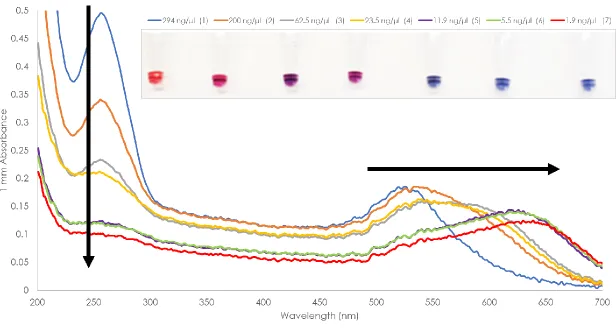
Bacterial pathogens, particularly the non-typhoidal Salmonella (NTS) is one of the most commonly reported bacterial pathogens affecting millions of humans annually [1-3]. Globally, there are an estimated 78,707,591 cases and 59,153 deaths annually due to NTS. In the United States, NTS is estimated to cause 1,027,561 illness and 450 deaths annually [1,2,4]. Disturbingly, the highest percentage of deaths were reported among children under 5 years old [1-3]. Apart from consumption of NTS contaminated foods, the environment, water, and hygiene play an important role in infection, transmission, and dissemination of this pathogen. Moreover, Salmonella spp. can acquire and disseminate antimicrobial resistance (AMR) determinants and virulence factors which complicates the scenario. Globally, it is estimated that 700,000 people die annually due to AMR and by the year 2050, it is projected that this number would reach up to 10 million with an estimated economic loss of $100 trillion [5].
Research and surveillance programs to monitor bacterial pathogens and AMR were established and are being implemented in many countries. Laboratory techniques such as the standard bacterial culture and polymerase chain reaction (PCR) are widely used to provide information on the occurrence of bacterial pathogens and AMR. Bacterial culture is an indispensable technique in the bacterial identification and determination of resistance.
Moreover, a bacterial culture is needed in obtaining pure isolates for further molecular analysis such as pulse-field gel electrophoresis (PFGE) which is time consuming and not suited to long-term global epidemiology [6]. The PCR has been a reliable tool in overcoming the difficulty in identification, differentiation, quantification bacterial pathogens [7], and have provided a more comprehensive information in AMR mechanisms. Currently, the cost of whole-genome
43 sequences in the GenBank. However, not all laboratories are capable of these techniques,
especially in developing counties. These techniques require expensive facilities and highly trained individuals, creating a gap in bacterial pathogens and AMR surveillance and research. The World Health Organization (WHO) has launched its Global Action Plan on AMR to ensure the continuity of successful treatment and prevention of infectious diseases. One of the WHO’s strategic objective on the Global Action Plan on AMR is to encourage development and
production of affordable diagnostic tools (Objective 5) [2,3,5].
One of the major interests in the biomedical sciences nowadays is the nanosensor or nano biosensor and their application in point-of-care (POC) diagnosis [8] of pathogenic organisms, genetic defects, and even harmful chemicals [9-11]. The gold nanoparticles (AuNPs) have been widely explored in the field of nanosensors due to its unique properties such as biocompatibility to different molecules [11,14,15,18-21], tunability, and surface plasmon resonance (SPR) in the visible range depending on size and shape [7,8,14,17,22-24]. The AuNPs are known to appear ruby red in color due to SPR, which is a distant-dependent effect observed based on the interaction of light and electrons surrounding the particle [7]. The changes in the AuNPs surrounding environment such as pH, salt concentration or high ionic concentration results to decrease in the interparticle distance and/or aggregation thus exhibiting colorimetric changes [8,20,24,27-29] that can be directly observed as color shift from red-pink to blue-gray due to plasmonic coupling [18,19,24,30,31]. As a result of aggregation, the position of the AuNPs SPR shifts from 520 nm to new band at 640-650 nm [7,18,19,24,26]. The AuNPs can be stabilized in