Copyright 0 1996 by the Genetics Society of America
Identification
of
Selected Gamma-Ray Induced Deficiencies
in
Zebrafish
Using
Multiplex Polymerase Chain Reaction
Andreas
Fritz,
Marion Rozowski, Charline Walker and Monte Westerfield
Institute of Neuroscience, University of Oregon, Eugene, Oregon 97403 Manuscript received June 11, 1996
Accepted for publication September 16, 1996
ABSTRACT
The ease with which mutations can be generated in zebrafish makes this vertebrate an important resource for developmental genetics and genome studies. We have developed a PCR-based screening method that allows the efficient identification of gamma-ray induced deficiencies targeted to selected sequences. We describe three mutants characteristic of our findings and show that these mutations include deletions and translocations that can affect as much as 1% of the genome. These deficiencies provide a basis for analyzing the functions of cloned zebrafish genes using noncomplementation screens for point mutations induced by high-efficiency chemical mutagenesis.
w
describe here a reverse genetic approach to identify gamma-ray induced mutations affecting any cloned and sequenced zebrafish gene. A genetic approach has been successfully applied to elucidate de- velopmental processes in the invertebrate organismsDrosophila melanogaster and Cmorhabditis elegans. Re- cently, large scale screens to identify and analyze muta- tions that disrupt developmental events have been car- ried out using the zebrafish (Danio rerio) (KIMMEL 1989; MULLINS and NUSSLEIN-VOLHARD 1993; DRIEVER et al. 1994; MULLINS et al. 1994; NUSSLEIN-VOLHARD 1994;
SOLNICA-KREZEL et al. 1994; RILEY and GRUNWALD
1995). These screens employ a classical genetic strategy to isolate mutant phenotypes first before determining the identity of the affected genes. Despite significant progress in establishing a genetic map of the zebrafish genome (JOHNSON et al. 1996; POSTLETHWAIT et al.
1994), identifying the molecular nature of these muta- tions is still a challenge. In zebrafish, as in other verte- brates, many genes have been cloned on the basis of sequence similarity to genes from other organisms. In only a few fortuitous cases has an existing mutant been found to correspond to a mutation in a cloned gene
(SCHULTE-MERKER et al. 1994; TALBOT et al. 1995; SEPICH and WESTERFIELD 1993). A method to generate and identify mutations that affect a chosen gene is an im- portant complementary strategy. We have developed a polymerase chain reaction (PCR)-based method that allows fast and efficient screening for mutations on the basis of their molecular rather than morphological de- fects, providing a basis for analyzing the functions of
these genes.
Gamma-rays are an efficient mutagen in zebrafish (CHAKRABARTI et al. 1983; WALKER and STREISINGER
University of Oregon, Eugene, OR 974031254,
E-mail: [email protected]
Corresponding author: Andreas Fritz, Institute of Neuroscience, 1254
Genetirs 144: 173.51745 (December, 1996)
1983), producing a very high specific locus mutation rate of -1:lOO. Gamma-ray mutagenesis has mainly been used in screens for morphological defects to iden- tify potentially interesting mutants that affect zebrafish embryogenesis (KIMMEL 1989). Several gamma-ray in-
duced mutations have been described in the literature (GRUNWALD et al. 1988; FELSENFELD et al. 1990; WEST-
ERFIELD et al. 1990; HATTA et al. 1991; HALPERN et al.
1993; TALBOT et al. 1995), however, with the exception of the ntlb16" (SCHULTE-MERKER et al. 1994),f2h"327 (TAL
BOT et al. 1995), and n i ~ l " ' ~ ~ (SEPICH and WESTERFIELD
1993) mutations, very little is known about the molecu- lar nature of these events. Ionizing radiation, such as
gamma-rays, can induce a variety of chromosomal changes, that are probably best documented for X-ray induced mutations in Drosophila (CHIA et al. 1985; KEL- LEY et al. 1985; PASTINK et al. 1987, 1988; BATZER et al. 1988; EEKEN et al. 1989, 1994; MAHMOUD et al. 1991;
LINDSLEY and ZIMM 1992). These mutations include point mutations, deletions ranging in size from a few basepairs to megabases, inversions, and translocations. Ionizing radiation induced mutations have also been extensively studied and used in mouse (VAN BUUL et al.
1990; C A ~ A N A C H et al. 1993; GOSSEN et al. 1995; TEASE and FISHER 1996a,b) and medaka (KUBOTA et al. 1992, 1995; S H I M and SHIMADA 1994).
The spectrum of mutations induced by ionizing radi- ation has also been investigated in various human and hamster cell lines, primarily by Southern blot analysis of the hypoxanthine-guanine phosphoribosyltransfer- ase (hprt) locus (TACHIBANA et al. 1990; NELSON et al.
1994, 1995; PUCK and HARVEY 1995). Recently, several groups have developed a multiplex PCR method to ana- lyze these radiation induced mutations at the h p locus (ROSSITER et al. 1991; FUSCOE et al. 1992; YU et al. 1992;
ZU et al. 1993; NELSON et al. 1994; SCHWARTZ et al. 1994;
1736 A. Fritz et al.
showed that 350% of gamma-ray induced alterations were large deficiencies, readily detectable by PCR screening.
We adapted the multiplex PCR method to identify mutations that affect cloned genes in zebrafish by ana- lyzing the genomic DNA (gDNA) of parthenogenetic haploid offspring of females carrying gamma-ray in- duced mutations. We characterized these mutations us- ing the genetic linkage map of the zebrafish genome (POSTLETHWAIT et al. 1994; JOHNSON et al. 1996). Our results demonstrate that gamma rays induce transloca- tions and deletions at high frequency in the zebrafish genome, making the identification and recovery of mu- tations that include selected loci efficient. Some chro- mosomal regions show a significantly higher mutation rate than average, suggesting the existence of "hot spots" for gamma-ray induced mutations. We discuss the type of chromosomal lesions and phenotypes of three mutations isolated in the PCR screen that are representative of the range of molecular and morpho- logical defects observed.
MATERIALS AND METHODS
Fish strains: A substrain of the Oregon AB line (WEST-
ERFIELD 1995) was used for all mutagenesis experiments. This AB substrain was derived from gynogenetic offspring of 21
AB founder females which were free of visible embryonic mutations. The AB strain was also used in crosses with mutant fish. T(msxB)b2'" mutant fish were crossed to leopard" (leo)
fish, originally obtained from the NUSSLEIN-VOLHARD lab at Tubingen and kindly provided by S. L. JOHNSON.
Staging: Embryos were developmentally staged according to standard criteria (KIMMEL et al. 1995), expressed as hours (hr) or days (day) post fertilization at 28.5".
Irradiation: Sperm were collected and irradiated in a Gam- matory-M irradiator with a cesium-137 source as described (WESTERFIELD 1995).
Haploid embryos: Eggs were obtained by gently squeezing the abdomens of anesthetized females. The eggs were then activated by addition of W-treated sperm, which do not make a genetic contribution to the offspring (STREISINGER et al.
1981).
Genomic DNA preparation: Because haploid embryos from each female were analyzed individually, we needed to conduct a large number of genomic DNA (gDNA) extractions and PCRs with maximal reproducibility and minimal handling. Most im- portantly, using a strain of fish carrying a stably integrated transgene (STUART et al. 1988) as a target for PCR amplifica- tion, we learned that it is necessary to remove the chorion from the embryo prior to gDNA extraction to avoid contamina- tion by wild-type sperm gDNA, presumably attached to the chorion (data not shown). For primary screening, 10 haploid embryos from each GO female were randomly collected either at 10 or 24 hr and were manually removed from their chorions. Single embryos were placed into 0.2-ml PCR tubes (Robbins Scientific, #104450-0) and excess liquid removed. Either 20 PI
(10 hr) or 25 p1 (24 hr) of digest buffer was added to each tube. Digest buffer is
lox
ThermoPol Reaction Buffer for Ther- mophilic DNA Polymerases (New England Biolabs, #007-TDP), diluted in sterile deionized water to 1X final concentration, and 100 yg/ml final concentration of Proteinase K The em- bryos were digested for 1-3 hr at 55", followed by 5 min at loo", in a Programmable Thermal Controller with a Hot Bonnet lid(PTC-100-96V; MJ Research, Inc.), and the tubes were stored at -20" until use. Phenotypically sorted embryos either for recovery screening of F1 females or RAPD mapping PCR were processed similarly, except that haploid embryos were digested in 30 p1 digest buffer and diploid embryos in 50 pl.
Oligonucleotide primers: Primers (Table 1) were chosen with the help of the Primer Select Module of the Lasergene Program (DNASTAR, Inc.). Reverse Phase purified (RP-I) primers were obtained from DNA EXPRESS (Macromolecular Resources, Colorado State University). The lyophilized prim- ers were resuspended in 1 ml of sterile, deionized water. Working stocks were prepared by diluting each primer to 10 pM and mixing the forward and reverse primers for each gene 1:l. Compatible primer pair combinations amplifying generally 8-11 different products were established by com- bining these working stocks as described in RESULTS. Aliquots of primer mixes were stored at -20" for convenience and accuracy.
Primer sequences: Listed in Table 1.
Multiplex PCR conditions: All reactions were carried out in a 20 pl volume in a PCR machine. First, 8 y1 of gDNA were combined with 2 p1 of primer pair mix in a 0.2 ml PCR tube and denatured for 5 min at 95" in the PTC machine and then cooled to 4". Then 10 pl of master mix was added to each tube at 4". Master mix contains per reaction: 1.2 pl lox ThermoPol
reaction buffer, 2 p1 dNTP mix (2.5 mM each), 0.4 yl BSA (purified BSA lOOx, New England Biolabs #007-BSA), 0.2 pl AmpliTaq enzyme (1 unit, Perkin-Elmer/Cetus), and 6.2 pl sterile deionized water. The tubes were then set on ice and the PCR program was started. The reaction tubes were re- turned to the heat block after it reached a temperature of
280". The PCR amplification program was: 1.94", 30 s, initial cycle only; 2. 40 cycles of 92", 30 s; 59", 30 s; 75", 2 min; 3. 75", 10 min; 4. 4", indefinite hold. Ten to 15 pl of PCR prod- ucts were analyzed by electrophoresis on 2% ethidium bro- mide containing agarose gels in 0.5X TBE buffer.
To identify embryos by PCR analysis for recovery from the F, generation, generally only three primer pairs were used, the pair that failed to amplify in the embryos from the GO female and two control pairs. For these reactions, the PCR program was reduced to 30 cycles and the 75" extension step shortened to 1.5 min.
RAPD P C R Primers and their sequences defining random
amplified polymorphic DNAs (RAPDs) were obtained from Operon Technologies (Alameda, C A )
.
PCR using RAF'D prim- ers was carried out as described in JOHNSON et al. (1994), except that gDNA from embryos was extracted as described above, and the NEB 1OX ThermoPol reaction buffer was used.Nomenclature: General guidelines for naming zebrafish genes and mutants are published in Genetic Nomenclature Guide (Trends in Genetics) and in The ZebrdfiSh Book
(WESTERFIELD 1995). The chromosomal aberrations de- scribed here have been given a name based on the gene by which they were identified, with a prefix indicating the likely type of chromosomal aberration. The prefixes are based on Drosophila guidelines. Of indicates a simple deletion; T indi- cates a translocation. The 6# is a unique allele designation, where bindicates the Eugene, Oregon, origin of the mutation. Gamma-ray mutagenized fish can carry multiple independent mutations. Thus, each F, line carrying a different mutation inherited from a single GO fish is given a unique b#, which designates an independently segregating mutation originally identified in a GO fish.
RESULTS
PCR-Selected Mutations in Fish 1737
TABLE 1
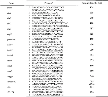
Forward and reverse primers for multiplex PCR of selected zebrafish genes
Gene Primers" Product length (bp)
axial
d1x2
dlx3
elrC
eng2
goosecoid
hoxb4
hoxb5
hoxb6
msxA
msxB
m s d
noggLn
tenascinC
shh
ZF-114
f
r
f
r
f
r
f
r f r f r
f
r
f
r f r
f r
f r
f r f r f
r
f r f
f r r
GACATACGAGCAAGTGATGCA GCGAAAAAAGTCCAAATAACA CCACCTCACCCCTCACC AGCGGCCGAGTCAAAATA ATCTGGTTCCAGAACCGGAG GCTGTAAATGTGATCCCTGC ACGCCACATTACCTTTTCTTTCAC ATGCCGTCAACCGTTTTATCTGTA ACCGAAAGCGACGATAAGCG CAATCGAATAGCGGCTTTGG GTCCCAGGTCTCGTAAACCA TCTCTGCCTCAGTGACTGTC ACGTGGGCACCTTGTCC ACCTTTCGTGCCAGTTGTT TGAGCCCGAACTATTCAGGG CCCTGTTTCTAATGTGCAGG CCTCCACTACCTCGGCGACG CCTTTTGCCGTCTGGTCCAG GGACCTTCGGTAACGCTGGT TAGCTTCGTTCCACATGGAC GTCCCACAATATGCCCTCTC CCAATAGGTGCAAAAGGCAG GCACCTTACGCAAGCACAAG TGGTCTTTCAAGGCACTTCC GAAAGTCCCTGTCCTCTCCG GACGGAGCTAAAATGTTCCG GTAAAAGCGGAACGAGACG GGTGTAGGACCAGAGCCAC CGCCAGTAACATTCAAACAG ATGGCCTGCACTTTTACTGT TCGACAACTCAACGGAAGA TAGGTAACGGTCGGTCAAA CAGTTGTACGGAATAGCAGC TGGCACCGGAGAATCTGTTG
454
229
250
110
987
325
172
430
720
494
373
806
569
546
192'
(295) 931
295
f, forward; r, reverse. The size of each amplification product is indicated. Sources of sequences are as follows: axial, accession No, (acc#) 222762 (STRAHLE et al. 1993); dlx2, acc# U03875, dlx3, acc# X65060 (AKIMENKO et al. (1994); el&, acc# U17601 (GOOD 1995); engl, acc# X68445, eng2, acc# X68446 (EKKER et al. 1992); goosecoid, acc# LO3395 (STACHEL et al. 1993); hoxb4, acc# M24085 (NJOLSTAD et al. 1988a); hoxb5, hoxb6, acc# X68248 (MOLVEN et al. 1993); m s d , acc# U16310, msxB, acc# U16311 (AKIMENKO et al. 1995), 1995; mxD, acc# X65062
(EKKER et al. 1992); noggzn, A. FRITZ, unpublished; tenascinC, acc# U14940 (QUO et al. 1995); sonic hedgehog, acc# 235669 (shh, m u s s et al. 1993); ZF-114, acc# X60095 (MOLVEN et al. 1992).
"Primers are 5'-3'.
*
Expected size of amplification product, actual size indicated in parentheses below.zebrafish genes, based on published sequences or se- quences made available to us prior to publication.
17
pairs of these primer sequences are shown in Table 1 (MATERIALS AND METHODS). Most primers were chosen so that their annealing temperatures were between 56" and 62". Additionally, primer pairs for all the genes were selected to minimize possible primer dimer formation and to amplify a product of unique size for each genewithin a range of 100-1000 bp. Because most primer predictions were based on cDNA sequences, we first tested primer pairs individually on zebrafish gDNA to verify that they yielded a robust amplification product of the expected size. Fourteen of 20 primer pairs ampli-
fied the expected product, one pair produced no ampli- fication product, and five pairs amplified products larger than predicted from the cDNA sequences, presumably because introns were located between the priming sites. Two of these latter five primer pairs were subsequently used for the PCR screen, without further confirmation of the authenticity of the amplified product; in the other cases, new primer pairs were selected.
1738 A. Fritz pt nl.
F K X W I.-Mdtiplex P(:R analysis o f gDNA f r o m single haploid embryos detects gamma-ray induced mutations. Xnal-
ysis o f offspring from three females (female 1, female '2, fe-
male 3) is shown. The target genes are shown on the left side. Embryos 1 and 10 from female 1 lack the msxB specific prodwt.
tions until all products in the mixture were amplified to approximately equal amounts. M'e were able to establish primer pair mixtures that reliably amplified I 11 differ- ent products in a single reaction.
Gamma-ray mutagenesis and PCR screen: We in- duced gamma-ray mutations by irradiating mature sperm obtained from male AB fish. The sperm were exposed to a dose of -250-300 rads, delivered over 35-40 sec (WESTERFIELD 1995). The irradiated sperm was then used to fertilize eggs from AB females. This dose was chosen because it was previously shown by complementation analysis to induce mutations at the
goldpn locus at a high frequency (-1 in 100, see also
Figure 2 in CHAKMRARTI d nl. 1983) and because it leads to a reasonable survival of the fertilized eggs; -25% of the embryos survive to adulthood and consti- tute the GO generation. M'e screened the haploid off- spring from the female fish of this GO generation using the multiplex PCR assay. Haploid embryos were oh-
tained by fertilizing the eggs of the GO females with UV irradiated sperm, which activate development without contributing paternal genetic material (STREISINGER P/
nl. 1981; WESTERFIELD 1995). We routinely assayed 10 embryos, randomly collected from each female, at ei- ther 10 o r 24 hr. Figure 1 shows an example of a multi- plex PCR assay using nine primer pairs per reaction. T h e gDNA from two of 10 embryos from o n e female
(female 1) failed to amplify the msxB (HOLLAND 1991;
AKIMEXKO d nl. 1995) product, suggesting that there was n o msxB genomic template for amplification.
To recover this potential msxR mutation, we crossed the GO female (female 1) to a wild-type male. We raised the FI generation to adulthood and analyzed the h a p loid off'spring from FI females similarly to the embryos of the GO female. We sorted the embryos from the FI
females into morphologically distinct classes, i x . , mu- tant and wild-type phenotypes, and analyzed embryos from each of these classes by multiplex PCR. T h e a b
sence of the mxB PCR product correlated with o n e of
the mutant phenotypic classes, suggesting that the loss of the PCR product was the result of a heritable muta- tion linked to a visible mutant phenotype. Subse-
quently, we isolated male FI fish carrying the msxBmuta- tion by crossing them to their msxB mutant female siblings and confirmed their genotypes by PCR.
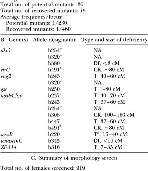
We have similarly identified 30 potential mutations by the absence of a particular PCR product after screen- ing a total of 509 GO females (Table 2A). Due to limita- tions of time and tank space, and to avoid potential PCR artifacts, we attempted to recover only 15 potential mutants for which a given PCR product was missing in a t least two of the 10 embryos. We successfully reidenti- fied all 15 of these mutations in the FI generation (Ta- ble 2B). Thus, considering all the potential mutants, the mutation rate was
-
1 in 230 (0.0043). A more con- servative estimate is 1 in 460 (0.0022), if we include only recovered mutants (Table 2A).In parallel with the PCR screen, we examined the haploid embryos from 919 GO females, of which the 509 of the PCR screen were a subset, for morphologi- cally visible mutations at 24 and 48 h r (KIMMEI. 1989). M'e scored a female as carrying a potential mutation if she produced several embryos displaying the same consistent characteristic phenotype. Phenocopies, i.p., embryos with abnormal phenotypes not due to herita- ble mutations, usually display variable phenotypes. In this screen we found 324 (35%) females that produced offspring with phenotypes that might be due to muta- tions (Table 2C). This is probably an underestimate of the number of mutations present in these 919 females, because a significant fraction carried multiple muta- tions (see also Table 2B, h254 and h320). In addition, we would have missed mutations that are either too subtle to detect morphologically or that produce phe- notypes that are not visible before 3 day, after which haploid embryos die. However, conservatively, the over-
all frequency of morphologically detectable mutations is -0.35 (324/919). Thus the frequency of a mutation detected by the PCR screen is
-
1 % of the overall muta- tion frequency. This estimate agrees with the previously established specific locus frequency for gamma-ray mu- tagenesis of-
1 in 100 (CHAKRZRARTI P/ 01. 1983).The frequency of mutations generated by gamma- rays varied considerably among different chromosomal regions. T h e PCR screens of all 509 females included chromosomal regions, marked by two loci ( d x 2, a g I ) , in which we detected no potential mutations. In con- trast, we recovered six independent alleles that delete the hoxf14, 65, and 116 genes, out of a total of 398 females screened, resulting in a mutation rate of 1 in 66.3 for the chromosomal region containing these loci.
Examples of recovered mutants: T h e mutations we identified with the multiplex PCR screen can be broadly classified into three categories by segregation analysis: class I: reciprocal translocations in which the hvo part- ners segregate and can be propagated independently.
Class 11: balanced reciprocal translocations, in which
PCR-Selected Mutations in Fish 1739
TABLE 2
S u m m a r y of PCR screen
A. Summary of PCR screen
Total no. of females screened: 509 Average no. of loci/screen: 13.6 Total no. of potential mutants: 30
Total no. of recovered mutants: 15 Average frequency/locus:
Potential mutants: 1/230 Recovered mutants: 1/460
B. Gene(s) Allele designation Type and size of deficiency
d1x3
e1rC eng2
gsc hoxb4,5,5
msxB
tenascinC
W-114
b254" b320" b380 b49 1 I,
b243 b320" b250 b237' b245 b254" b308 b447 b491b b220 b345 b316
NA NA Df, <8 cM CR, -80 cM
NA T, -80 cM
T, 37-60 cM NA
T, 37-60 cM CR, -80 cM T", 15-40 cM Df, <10 cM T, 7-35 cM T, 40-60 CM
T, 40-70 CM
CR, 100-160 CM
C. Summary of morphology screen
Total no. of females screened: 919
Total no. of females with potential mutants: 324 Estimated mutation frequency: 1/28
A shows the statistical evaluation of PCR screen. In B, the left column indicates the gene(s) for which mutations were isolated in the PCR screen. The middle column shows allele designations; multiple independent alleles have been found for three different loci. The right column shows the type and size of each deficiency. CR, complex rearrangement; NA, not available; T, translocation; Df, deletion. C provides a summary of the screen of haploid offspring from GO females for mor- phological defects. The 509 GO females used in the PCR screen are a subset of the females used in the morphology screen.
"The 2 GO females from which the F1 lines 6254 and 6320
were generated each carried two independent mutations (and would have been given two 6#s each according to the nomen- clature conventions), leading to a total of 15 mutations identi- fied and recovered; however, the b254 and b320 lines have been terminated.
" b491 delets the hoxb and elre genes; the hoxb cluster and
elrC map to LG3. The other hoxb deficiencies do not affect
elrC.
'The 6237 line has been terminated.
" The 6220 translocation has resolved (see RESULTS).
Class 111: mutations that are inherited as simple defi- ciencies. We describe here one example of each.
Class I, the T ( m s ~ B j ~ ~ ~ " mutation: We found the T(msxB)"22" mutation in a GO female in which two out of 10 embryos were lacking the msxB (AKIMENKO et al.
1995) specific amplification product (see Figure 1). In
the F1 generation, seven of 15 females produced clutches of haploid embryos in which a fraction lacked the msxB specific amplification product and showed a distinct mutant phenotype ( m l , see also Figure 4B). In the clutches of these seven females, the msxB deficient embryos constituted -23% (61/274) of the total hap- loid offspring, while an additional 18% (49/274) of the embryos showed a second, consistent mutant pheno- type (m2). The m2 phenotype includes a shortened, necrotic body axis and a distinct bulb-like tail at 24 hr
(data not shown). The m2 mutant embryos die at 3 day as diploids. The remaining embryos appeared morpho- logically wild type, there were no apparent double mu- tants visible, and both the m2 and wild-type phenotypic classes amplified all 17 PCR products. We subsequently identified 4 male F1 carriers by intercrosses. In all four crosses, the offspring consisted of morphologically wild- type embryos (654/731), the m l (33/731) and m2 (44/ 731) mutant classes of embryos, and no phenotypically obvious double mutants. As in the haploid embryos, all of the diploid embryos displaying the ml phenotype were also missing the msxB amplification product. Thus, the segregation ratios observed among the haploid and diploid offspring suggest that the ?'(m~xB)''~"' mutation was caused by a reciprocal translocation event. How- ever, we have not examined the msxB deficient chromo- some to learn whether it carries sequences from an- other chromosome.
In a reciprocal translocation mutation, both mutant phenotypes are visible in 1/16 embryos of a clutch, while a cross between a translocation mutation and a
noncomplementing simple deletion should produce only one phenotype in 1/8 of the offspring. Consistent with this formal expectation, a subsequent cross of one of the T(msxB)"2" carrier females to a male sibling pro- duced only wild-type (140/157, 89%) and msxB defi- cient mutant embryos (17/157, 11%); no embryos of the m2 phenotype were observed. This suggested that the male F1 fish was heterozygous for the msxB defi- ciency, that the two translocation partners can segre- gate independently, and that fish inheriting only the msxB deficient translocation partner are viable. We con- firmed in subsequent crosses, including the non-com- plementation cross to the b o pigment mutant (see be- low), that the reciprocal translocation partner (m2) of the T(msxB)h22" mutation is not obligate. The T(msxB)"2" designation heretofore refers to the mutation carrying the msxB deficient chromosome in the absence of its original translocation partner.
Because the map position of the msxB gene was un- known at the time, we determined the position of the
T(msxB)b220 mutation o n the zebrafish genetic map
(POSTLETWAIT et a[. 1994; JOHNSON d at. 1996) by sys- tematically comparing RAPD markers between gDNA from T(msxBjh220 mutant embryos and their wild-type siblings. We found three RAPD markers, 17X.640,
A. Fritz et al. 1740
LQ1
bpg
LQ3 b245
1
lIAE.800 t m s t o
-
110.1070 lu7.m 1 M F m fLa 5
t34s
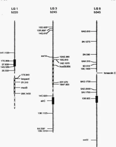
FIGURE 2.-Gamma-rays produce a ran e of chromosomal
defects. Mapping analysis of the T(mxBT" (LGl), T(h~xb)*'~~
(LG3) and Dften)6345 (LG5) mutations. The cross hatching indicates 2cM intervals. Markers and genes on the right side of each LG are missing, markers shown on the left side have been tested and are present. The broken lines indicate re-
gions of uncertainty in and T(ho~b)"~'. The black
boxes indicate the positions of the centromeres (JOHNSON et al. 1996).
tently missing in gDNA extracted from haploid
T(msxB)b220 mutant embryos but present in gDNA from
their haploid wild-type siblings, suggesting that the
T(msxB)b220 mutation affects the region of linkage group
1 (LG1) containing these markers. The two available markers distal to 20K.1450 (Figure 2, LG1) were unin- formative, i e . , the expected PCR products were not seen in gDNA from either wild-type or mutant embryos, probably due to polymorphisms within the
AB
strain. The 33.550 marker and all other tested markers near the centromere or on the other chromosome arm were present in mutant gDNA, indicating that the T ( ~ s x B ) ~ ' ' ~ mutation is a deficiency of 2 1 5 cM and possibly greater than 40 cM,if
the entire distal end of LG1 is missing. Subsequently, we mapped the msxB gene, using a poly- morphism within the gene between theAB
and DAR strains, to LG1 between markers 2V.310 and 20K.1450 (POSTLETHWAIT et al. 1994), in agreement with the map position of the mutation.The extent of the T(G-zsxB)~~~~ deficiency in LG1 pre- dicted that the le0 locus, which is just distal to the
17X.
640 marker, should also be missing. The bot' allele causes a recessive, homozygous viable pigment muta- tion (Figure 3) that can be easily recognized in fish afterFIGURE 3.-The T(~~sxB)~"* mutation also affects Zeo. Off-
spring of a b220/+ X Zeo"/Zeo*' cross at 2 mon of age. (Top)
Zeo"/+ genotype. (Bottom) T(VLSXB)*'~~/Z~O*~ g e n o v ; the Zeo
phenotype is indistinguishable from that of the Zeo /Zeo*' par- ent (not shown). The scale bar indicates 1 cm.
the age of 4 weeks (KIRSCHBAUM 1975). To examine whether le0 was affected by the T ( T x s x B ) ~ ~ ~ ~ mutation,
we raised to adulthood 25 offspring of a cross between a homozygous hot' male and one of the identified F1 females carrying the original translocation mutation. Five individuals from this F2 generation displayed a le0
phenotype that was indistinguishable from the leo" mu- tant phenotype (Figure 3; JOHNSON et al. 1995). The outcome of this cross also confirmed our expectation that the original mutation was a translocation and that, at least, the msxB deficient partner of the original T ( T ~ . s x B ) ~ ~ ~ ~ mutation can be maintained independently. If the b220 mutation had been a simple deletion in
LG1, we would have expected half of the progeny to show the le0 phenotype; if the large deficiency in LG1
had shown haploinsufficiency and could only be main- tained in the presence of its translocation partner, we would not have expected to see offspring displaying the
le0 phenotype. To confirm the predicted genotype of
these fish, we analyzed embryos from intercrosses of putative
lea"/
T ( ~ S X B ) ~ ~ ~ O individuals. As expected, the phenotype associated with the msxB deficiency consti- tuted -25% (42/199) of the embryos.The T ( ~ S X B ) ~ ' ~ ~ mutant embryos, from 16 to 36 hr, suffer from cell death in the dorsal CNS at most axial
levels. Figure 4B shows the phenotype of a live 32
hr
T(msxB)b220 diploid mutant embryo in comparison to a
wild-type embryo of the same age (Figure 4A). Subse- quently, the dying cells are cleared from the spinal cord. Eye formation is severely disturbed, as are the median and pectoral fins. The mutant embryos fail to form blood, and die at -4 day.
Class 11, the T ( h ~ x b ) ~ ' ~ ~ mutation: We identified the
T(hoxbjbZ4' mutation in a GO female, from which three
out of 10 haploid offspring lacked the PCR products of
the hoxb4 (NJOLSTAD et aL 1988a), hoxb5, and hoxb6
genes (MOLVEN et aL 1993). Female and male F1
PCR-Selected Mutations in Fish 1741
F r G W 4.-PCR mutants have morphological defects. Live diploid embryos. Haploid phenotypes appear essentially the same. All views are lateral, anterior is to the left and dorsal is
up. (A) Wild type (34 hr). (B) T(msxB)b””o mutant embryo (34 hr). (C) Left: 32 hr T(ho~b)’~” mutant embTo; right: 36 hr T ( h o ~ b ) ’ ~ ‘ ~ associated phenotype. (D) Df(tm) 345 mutant em-
bryo (34 hr). See text for description of phenotypes. The scale bar in the lower right indicates 250 pm.
among haploid offspring was 24% (266/1107), and among diploid offspring the mutant phenotype consti- tuted 3.8% (84/2194). These frequencies have been maintained through several generations of crosses to wild-type fish. Furthermore, we observed a second mu- tant phenotype among haploid and diploid offspring from T ( h ~ x b ) ~ ’ ~ ~ parents, that consistently cosegregated with the hoxb deficient mutant phenotype (see below and Figure 4C). Together, these observations suggest that the T(h~xb)~’~’ mutation is a reciprocal transloca- tion, in which the two translocation partners can not be maintained separately. We confirmed the translocation nature of the T ( h o ~ b ) ~ ’ ~ ~ mutation directly using an al- lelic polymorphism within the hoxb cluster in the Ore- gon
AB
line; 25% of the haploid embryos from aT(h0~bf’’~’ carrier female inherit both allelic variants of
the hoxb locus (data not shown), one variant from the
wild-type locus on LG3 and the second variant from the translocated copy.
Mapping analysis revealed that an internal piece of LG3 is missing in gDNA from T ( h ~ x b ) ” ” ~ deficient em- bryos (Figure 2, LG3). Additionally, we found that the ssrl4 marker, the first marker distal to the region de-
leted in T ( h ~ x b ) ~ “ ~ embryos, still segregates with the centromere of LG3 in gDNA from mutant embryos, suggesting that the deficiency in LG3 is a large internal deletion. Based on the currently available markers for LG3, the predicted size of the deficiency is minimally 37 cM and may be as large as 60 cM. Because of the size of this deficiency, it is likely that the entire hoxb cluster was translocated, although we have direct evi- dence from PCR for the hoxb2, b4, b5, and b6 genes only.
Development in T ( h o ~ b ) ~ ’ ~ ~ mutant embryos (Figure 4C) proceeds fairly normally through gastrulation and into the early segmentation period. At about the four somite stage (11.5 hr), development begins to slow and essentially arrests after formation of the 12th somite (15 hr)
.
Cell death in the brain begins at the four somite stage, mainly in a medial stripe of cells in the hindbrain. At this stage, cells lateral to the CNS in the trunk also die and further tail development fails; cells instead accu- mulate in the tail bud and eventually die. The embryos die-2
day. Figure 4C also shows a 36hr diploid embryo displaying the second phenotype that consistently co- segregates with the T ( h ~ x b ) ~ ’ ~ ~ mutant phenotype. Cell death in the midbrain and hindbrain becomes apparent -15 hr, and there are no obvious defects in the trunk and tail. The embryos appear developmentally retarded and die -4 day.Class
111,
the Df(ten)””‘ mutation: We identified a mu- tation deleting the zebrafish tenascin C (tenc) (QIAO etal. 1995; TONGIORGI et al. 1995) specific amplification product in six of 10 haploid embryos from a GO female. We inspected the remaining live haploid embryos of this GO female morphologically and found that about half of the embryos (11/21) displayed a visible mutant phenotype by 30 hr and died by 48 hr. The absence of
the tenC product correlated with this observed mutant
phenotype; gDNA from phenotypically mutant embryos failed to amplify the tcnC PCR product. We subse- quently intercrossed F1 progeny of the founder female to analyze the segregation of the Dfl(ten)b345 mutation in diploid embryos. As expected from the segregation frequency of the haploid offspring in the GO female, -25% (84/380) of the embryos displayed the Dflte72)b345 mutant phenotype, and consistently failed to amplify
the tenC specific PCR product.
The Df(ten)b345 mutation has affected <10 cM of the genomic region around the tenC gene; the nearest in- formative markers are all unaffected in the D f l t e 7 ~ ) ~ ~ ~ ’ mutation (Figure 2, LG5). To analyze the mutation fur- ther, we cloned -19 kb of gDNA containing most of
the tenC gene and 3’ flanking sequence and isolated
restriction fragments from this genomic clone to gener- ate probes for hybridization. Southern blots of gDNA
1742 A. Fritz et al.
and the F1 generation show that the Df(ten)*"" mutation is a deletion.
Df(ten)b345 embryos appear normal at 24 hr, although many embryonic tissues express the tenC gene begin- ning at -16 hr (TONGIORGI et al. 1995). Analysis of mutant embryos with a variety of RNA in situ and anti- body probes has thus far not revealed any recognizable patterning or differentiation defects at 24 hr. Morpho- logcally, Df(ten)*345 mutant embryos can be recognized starting at -28 hr. In wild-type embryos at this stage, the head lifts off the yolk and straightens, and the circu- latory system and blood have formed and are visible
(KIMMEL et al. 1995). Both of these processes are im-
peded in mutant embryos (shown in Figure 4C at 32 hr). The head remains in close apposition to the yolk; red blood cells fail to circulate properly and aggregate in clumps, most prominently where the blood island forms. Between 40 and 50 hr, rapid deterioration and cell death throughout the embryo sets in and the mu- tant embryos die and disintegrate.
DISCUSSION
We have shown that a multiplex PCR method pro- vides an efficient strategy for screening gamma-ray mu- tagenized fish to identify mutations affecting specific zebrafish genes. Mutations affecting genomic regions in which genes of interest are located can be detected with frequencies approaching those previously reported using complementation analysis (CHAKRABARTI et aZ. 1983; WALKER and STREISINGER 1983).
The feasibility of this mutant screen largely depends upon the ability to analyze parthenogenetically derived haploid offspring. We can detect mutations in the F1 generation directly, and thus much greater numbers of mutagenized fish can be analyzed than would be possi- ble in a F2 screen. More importantly, mutations caused by deletions are represented in one half of haploid embryos, and translocation mutants are detectable with our screen in one quarter of a haploid clutch. Thus, by screening 10 haploid embryos, randomly selected before morphological phenotypes might be visible, the probability that either type of mutation will be repre- sented in these 10 embryos is >go%. In diploid off- spring, translocation mutations are present in only 1/ 16 of the clutch, thus requiring greater numbers of embryos, or a morphological prescreen to select for potential mutants. A prescreen would almost certainly introduce a bias for or against certain types of mutants. Our current screen can be carried out without any knowledge or assumptions about mutant phenotypes, and it allows the identification of mutations affecting genes that have no visible embryonic phenotype, or whose phenotype is masked by a mutation in a linked gene.
We observed an average locus specific mutation fre- quency of 0.0022 (Table 2A), which is lower than the
previously published value. Complementation analysis at three independent pigment loci (golden, brass, albino;
CHAKRABARTI et al. 1983) had previously shown that the locus specific mutation rate in sperm irradiated with gamma-rays is approximately 0.01 at the dose used in our experiments (see also Table 1 and Figure 2 in CHAK-
MARTI et al. 1983). Furthermore, the ratio of recessive
lethal to specific locus frequencies suggested that most deficiencies are large events, presumably deleting not only the gene analyzed, but also neighboring loci, which should be easily detectable by PCR. However, it is possi- ble that a sizable fraction of mutations that are uncov- ered by complementation analysis escape detection by PCR. Analysis of the mutation spectrum induced by ionizing radiation in various mammalian cell lines sug- gested that -50% of mutations exhibit large deletions affecting more than a single gene (MORRIS et al. 1993; SCHWARTZ et al. 1994; PARK et al. 1995), while the other mutations affected only part of a gene. Our assay should detect small intragenic deletions if they exceed -10% of the amplified fragment length, but mutations will go undetected if they are in the gene but outside the PCR amplified target or if they are too small to detect even if they fall within the amplified region of the gene. Moreover, detection of mutations such as inversions, duplications, or insertions by PCR is only possible if' the chromosomal breakpoints fall between the priming sites. Thus the frequency of mutations detected by com- plementation analysis could be expected to be higher than by PCR.
Other factors might contribute to the reduced aver- age locus specific mutation rate. Thirty-fold differences have been observed between the frequencies of X-ray induced mutations at specific loci in mice (RUSSELL. and RUSSELL 1959). For example there may be a correlation between gamma-ray induced mutation frequencies and spontaneous frequencies as observed in Drosophila (SHUIUA et al. 1979). The golden, brass and albino muta- tions used in zebrafish as tester loci for noncomplemen- tation screens have arisen spontaneously, and thus the mutation rate established using these loci may be higher than for other genes. Within our more limited data set, only mutations affecting the region of LG3 containing the hoxb gene cluster have occurred at a frequency similar to the rate demonstrated for the pig- mentation loci. If the pigmentation loci and the region on LG3 containing the hoxb locus represent 'hot spots" for the induction of mutations, then the mutation rate for other, average loci would be lower. In addition, we did not attempt to recover 15 additional potential mutations that we had seen in only one of 10 embryos of a GO female. At least some of these might have been bona fide mutations, and their recovery would have increased our measure of the average locus specific mu- tation rate.
PCR-Selected Mutations in Fish 1743
ntl b16’, jh””, and nicl b l O i mutations ( SEPICH and WEST-
ERFIELD 1993; SCHULTE-MERKFLR et al. 1994; TALBOT et
al. 1995). The ntl b160 and n i ~ l ~ ’ ’ ~ mutations were both
selected in morphology screens, which were strongly biased toward “clean” phenotypes and thus were more likely to represent single gene mutations. The ntlbZ6’
mutation replaced two nucleotides in the wild-type se- quence by six nucleotides within the open reading frame, leading to a frame shift and a truncated protein product. In the niclb’” mutation, a deletion of -1.5 kb appears to prevent the expression of an intact acetylcho- line receptor alpha subunit. jhb3” is a deletion, isolated in a noncomplementation screen usingjh”’ tester fish, that removes the entire jZh gene and probably one or more closely linked genes (TALBOT et al. 1995). In the present screen, we recovered 15 independent gamma- ray induced mutants. Two mutations [including
Df(ten)b345]
produced phenotypes in 50% of the haploid embryos of the GO female, and the segregation patterns of these two mutations in subsequent generations con- firmed that they are inherited as simple deficiencies. Both of these mutations are deletions that do not affect the nearest markers on the map. The majority of mutants we isolated (13 of 15) showed segregation pat- terns consistent with complex rearrangements, such as translocation events. The mapping analysis of several of these mutations, including T(~~~sxB)~”’ and T ( h o ~ b ) ~ ’ ~ ~ , showed that the size of these translocations is in the order of 30 cM, or -1% of the genome; the zebrafish genetic map spans -2900 cM (JOHNSON et al. 1996). These examples demonstrate that gamma-rays produce a wide range of mutations in zebrafish, as has been shown for other genomes, and that the type of screen will bias the types of mutations obtained.We have analyzed the segregation patterns for at least three generations of five mutations originally identified as translocations, including T(msxB)b22’ and T ( h o ~ b ) ~ ’ ~ ~ . The two partners of the T(WLSXB)~’~’ translocation sepa- rated in the F, generation, and the msxB deficient part- ner has been maintained in heterozygous form without apparent effect on viability or fertility through several generations. The four other mutations include hox clus- ters, such as T ( h o ~ b ) ~ ’ ~ ~ , and can be propagated only with both translocation partners present, indicating haploinsufficiency in the regions affected by these mu- tations. One factor that might contribute to the haploinsufficiency syndrome of the mutations affecting the hox gene clusters is genetic interactions among hox genes. This effect, nonallelic non-complementation, has recently been demonstrated for the ffoxB5 and HoxB6 genes in mouse (RANCOURT et al. 1995); muta-
tions affecting as few as two genes within a hox cluster may be lethal in heterozygous form.
Prospects and conclusions: The ease with which
gamma-ray induced mutations can be screened and identified by the multiplex PCR assay suggests that de- ficiencies including selected genes in many regions of
the zebrafish genome can be isolated efficiently. Fur- thermore, oligonucleotide primers suitable for multi- plex PCR can be obtained from the RAPD markers of the zebrafish genome map by creating sequence-tagged site markers (POSTLETWAIT et al. 1994; JOHNSON et al.
1996), making it possible to identify deficiencies at any region of choice.
Gamma-ray induced deficiencies provide an im- portant resource for genetic analysis. The availability of radiation induced aberrations that span nearly every segment of the Drosophila genome has played a critical role in the identification and characterization of many genes, and has provided a valuable tool for mapping and for establishing balancer chromosomes ( LINDSLEY
et al. 1972; LINDSLEY and ZIMM 1992; GARC’IA BELLIDO and WANDOSELL 1978). A standard approach to identify mutations in a newly isolated Drosophila gene is to determine its map position and use an appropriate de- ficiency to detect noncomplementing ethyl methanesul- fonate (EMS) or P-element induced mutations. This approach, using the chemical ethylnitrosourea (ENU) , is already being employed in mouse to obtain a com- plete physical and genetic description of a few regions defined by deletions (RINCHIK 1991). Efforts to obtain extensive sets of overlapping deletions that span mouse chromosomes using the chemical mutagen chlorambu- cil (CHL) have been initiated (RINCHIK et al. 1990), even though they will require large resources and facili- ties.
1744 A. Fritz et al. We are grateful to STEPHEN DEVOTO, STEPHEN JOHNSON, CHARLES
KIMMEI. and JOHN POSTI.ETHWAIT for their comments on the manu- script. We thank MICHAEL GATES and SCOTT STAWICKI for their help with mapping genes. This work was supported in part by an EMBO and a Swiss National Foundation Grant to A.F. and by National Insti- tutes of Health grant HD-22486.
LITERATURE CITED
AKIMENKO, M. A,, M. EKKER, J. WEGNER, W. LIN and M. WESTERFIELD, 1994 Combinatorial expression of three zebrafish genes related to distal-less: part of a homeobox gene code for the head. J. Neurosci. 1 4 3475-3486.
AKIMENKO, M. A,, S. L. JOHNSON, M . WESTERFIELD and M. EKKER, 1995 Differential induction of four msx homeobox genes during fin development and regeneration in zebrafish. Development 121:
347-357.
BATZER, M. A,, B. TEDESCHI, N. G. FOSSETT, A. TUCKER, G. KII.ROY
et al., 1988 Spectra of molecular changes induced in DNA of Drosophila spermatozoa by 1-ethyl-1-nitrosourea and X-rays. Mu- tat. Res. 199: 255-268.
C~TTANACH, B. M., M. D. BURTENSHAM', C. RASBERRY and E. P. EVANS, 1993 Large deletions and other gross forms of chromosome imbalance compatible with viability and fertility in the mouse. Nat. Genet. 3: 56-61.
CHAKRABARTI, S . , G. STREISINGER, F. SINGER and C. WALKER, 1983 Frequency of gamma-ray induced specific locus and recessive lethal mutations in mature germ cells of the zebrafish, Bruchy-
danio R m o . Genetics 103: 109-123.
CHIA, W., C. SAVMS, R. ~ R P , H. PELHAM and M. ASHBURNER, 1985 Mutation of the Adh gene of Drosophila melanogaster con- taining an internal tandem duplication. ,J. Mol. Biol. 186: 679- 688.
DRIEVEK, W., D. STEMPIX, A. SCHIER and L. SOLNICA-KREZEL, 1994 Zebrafish: genetic tools for studying vertebrate development. Trends Genet. 10: 152-159.
EEKEN, J. C . , A. W. DE JONG, M. LOOS, C. VREEWN, R. ROMEYK et ul.,
1994 The nature of X-ray-induced mutations in mature sperm and spermatogonial cells o f Drosophila melanogaster. Mutat. Res. 307: 201-212.
EEKEN, J. C., A. PASTINK, M. NIVARD, E. W. V ~ ( ; E L and F. H. SOBEIS, 1989 The nature of X-l-ay and chemically induced mutations in Drosophila in relation with DNA repair. Ann. 1st. Super. Sanita
EKKER, M., J. WLGNER, M. A. AKIMENKO and M. WESTERFIELD, 1992 Coordinate embryonic expression of three zebrafish engrailed genes. Development 116: 1001-1010.
FEISENFELI), A. L., C. WAI.KE:R, M. WESTERFIEI.~, C. KIMMEL and G. STREISINGER, 1990 Mutations affecting skeletal muscle myofi- bril structure in the zebrafish. Development 108: 443-459. FUSCOE, J. C., L. J. ZIMMERVAN, A. FEKETE, R. W. SETZER and B. J.
ROSSITER, 1992 Analysis of X-ray-induced HPRT mutations in CHO cells: insertion and deletions. Mutat. Res. 269: 171-183. G A R ~ I A BEI.I.IDO, A., and F. WANDOSEL.L, 1978 The effect of inver-
sions on mitotic recombination in Drosophila mrlanogastrr. Mol. Gen. Genet. 161: 317-321.
GOOD, P. J., 1995 A conserved family of elav-like genes in verte- brates. Proc. Natl. Acad. Sci. USA 92: 4557-4561.
GOSSEN, J. A,, H. J. MARTCS, J. Y. WEI and J. VIJG, 1995 Spontaneous and X-ray-induced deletion mutations in a LacZ plasmid-based transgenic mouse model. Mutat. Res. 331: 89-97.
GRLWTWALD, D. J., C. B. KIMMEI., M. WESTERFIELD, C. WALKER and G.
STREISINGER, 1988 A neural degeneration mutation that spares primaly neurons in the zebrafish. Dev. Biol. 126: 115-128. HAIPEKN, M. E., R. K. H o , C . WAI.KER and C. B. KIMMEI., 1993 Induc-
tion of muscle pioneers and floor plate is distinguished by the zebrafish no tail mutation. Cell 75: 99- 11 1.
HATTA, K., C . B. KIMMEI., R. K. Hct and C . WALKER, 1991 The cyclops mutation blocks specification of the floor plate of the zebrafish central nervous system. Nature 3 5 0 339-341.
HOI.I.AND, P. W., 1991 Cloning and evolutionary analysis of msh- like homeobox genes from mouse, zebrafish and ascidian. Gene
JOHNSON, S. I>,, D. AFRICA, C. WAI.W.R a n d J . A. WESTON, 1995 Ge-
2 5 213-218.
98: 253-257.
netic control of adult pigment stripe development in zebrafish. Dev. Biol. 167: 27-33.
JOHNSON, S . L., M. A. GATES, M. JOHNSON, W. S. TALBOT, S. HORNE et al., 1996 Centromere-linkage analysis and consolidation of the zebrafish genetic map. Genetics 142: 1277-1288.
JOHNSON, S. L., C. N. MIDSON, E. W. BALLINGER and J. H. POSTI.E'I'H-
WAIT, 1994 Identification of RAPD primers that reveal extensive polymorphisms hetween laboratory strains of Lebrafsh. Geno- mics 1 9 152-156.
KAGAWA, Y., F. YATAGAI, M. SUZL'KI, Y. M E , A. KOBAY.GHI d d., 1995 Analysis of mutations in the human HPRT gene induced by accelerated heavy-ion irradiation. J. Radiat. Res. 3 6 185-195. KF,I.I.EY, M. R., I. P. MIMS, C. M. FARNET, S. A. DICHARRY and W. R.
LEE, 1985 Molecular analysis of X-ray-induced alcohol dehydro- genase (ADH) null mutations in Drosophila melanogaster. Ge- netics 109: 365-377.
KIMMEI., C. B., 1989 Genetics and early development of 7ebrafish. Trends Genet. 5: 283-288.
KIMhlk:l., C. B., W. W. B..\~.LARD, S . R. KIMMFX., B. UI.I.MANN and T. F. SCHIILING, 1995 Stages of embryonic development of the de- brafish. Dev. Dyn. 203: 253-310.
KIRSCHBAUM, F., 1975 Untersuchungen uber das Farbmuster dcr Zebrabarbe Brurhydunio r h o (Cyprinidae, Teleostei). Roux's Arch. Dev. Biol. 177: 129-152
KRALTss, S., J. P. CONCORD~T and P. W. INCHAM, 1993 A functionally conserved homolog of the Drosophila segment polarity gene hh is expressed in tissues with polarizing activity in zebrafish em- bryos. Cell 75: 1431 -1444.
KUBOTA, Y., A. SHIMADA and A. SHIM, 1992 Detection of gamma- ray-induced DNA damages in malformed dominant lethal em- bryos of the Japanese medaka (Chytius latiper) using AP-PCR fin- gerprinting. Mutat. Res. 283: 263-270.
KUBOTA, Y., A. SIIIMAIIA and A. SHIM, 1995 DNA alterations de- tected in the progeny of paternally irradiated Japanese medaka fish (Oryias lutzpes). Proc. Natl. Acad. Sci. USA 9 2 330-334.
NEIL et al., 1972 Segmental aneuploidy and the genetic gross structure of the Drosophila genome. Genetics 71: 157-184.
LINIISIXY, D. L., and G. G. ZIMM, 1992 The G e n o m u ~ ~ r o . ~ ~ i ~ i L a m&-
nogastm. Academic Press, Inc., San Diego.
MAHMOUD, .J., N. G. FOssb;-l 1 , P. ARBOtlR REIIY, M. M(:DANIEI., A. TUCKER et al., 1991 DNA sequence analysis of X-ray induced Adh null mutations in Drosophila melanogaster. Environ. Mol. Mutagen. 18: 157-160.
MOLLEN, A,, I. HOKUVIKand P. R. NJOLSTAL), 1993 Sequence analysis of the zebrafish hox-B5/B6 region. Biochim. Biophys. Acta 1173
102-106.
MOI.W,N, A,, I. HORDVIK, P. R. NJOLSTAD, M. VAN GHELDE and A. FIOSE, 1992 The zebrafish homeobox gene hox[zf-ll.l]: pri- mary structure, expression pattern and evolutionary aspect$. Int. J. Dev. Biol. 36: 229-237.
MORRIS, T., W. MASSON, B. SINC:I.F.TON and J. TFiAcKeR, 1993 Analy- sis of large deletions in the HPRT gene of primary human fibro- blasts using the polymerase chain reaction. Somat. Cell Mol. Genet. 19: 9-19.
VOI.HARD, 1994 Large-scale mutagenesis in the zebrafish: in search of genes controlling development in a vertebrate. Curr. Biol. 4: 189-202.
MUI,I.INS, M. C., and C. NCSSl.EIN-\'(xHARD, 1993 Mutational ap- proaches to studying embryonic pattern formation in the zebra- fish. Curr. Opin. Genet. Dev. 3: 648-654.
NELSON, S. I.., C. R. GIVER and A. J. GROSOVSKY, 1994 Spectrum of X-ray-induced mutations in the human hprt gene. Carcinogene- sis 15: 495-502.
NELSON, S. I,., I. M. JONES, J. C. FUSCOF,, K. BURKH.~KT-S(:HLI.I'L and A. J. GROSOVSKY, 1995 Mapping the end points of large dele- tions affecting the hprt locm in human peripheral blood cells and cell lines. Radiat. Res. 141: 2- 10.
NJOISTAI), P. R., A. M O I X N , J . APOI.II and A. FJOSE, 1990 The zebra- fish homeobox gene hox-2.2: transcription unit, potential regula- tory rcgions and in .rilu Iocalimtion of transcripts. EMBO J. 9 515-524.
NJOLSTAD, P. R., A. MOLVEN and A. F]OSE, 1988a A zebrafish homo- logue of the murine Hox-2.1 gene. FEBS Lett. 230: 25-30, LINDSIXY, D. L., L. SANDI,F,R, B. S. BAKER, A. T. CARPFNTI'.R, R. E. DE-
PCR-Selected Mutations in Fish 1745
NIOISTAD, P. R., A. MOI.VEN, I. HOKDVIK, J. .&om and A. FIOSE, 1988b Primary structure, developmentally regulated expres- sion and potential duplication of the zehrafish homeohox gene ZF-21. Nucleic Acids Res. 1 6 9097-9111.
N~SSI.EIN-VOI.HARD, C., 1994 Of flies and fishes. Science 266: 572- 574.
P m , M. S., T. HANKS, A. JABERABOANSARI and D. J. CHEN, 1995 Molecular analysis of gamma-ray-induced mutations at the hprt locus in primary human skin fibroblasts by multiplex polymerase chain reaction. Radiat. Res. 141: 11-18.
PASTINK, A., A. P. SCHALET, C. VREEKEN, E. PAR’ADI and J. C. EEKEN, 1987 The nature of radiation-induced mutations at the white locus of Drosophila melanogaster. Mutat. Res. 177: 101-115. PASTINK, A,, C. VREEKEN, A. P. SCHAI.ET and J. C. EEKEN, 1988 DNA
sequence analysis of X-ray-induced deletions at2 the white locus of Ilrosophila melanogaster. Mutat. Res. 207: 23-8.
POSTI.ETHWAIT,.J. H., S . L. JOHNSON, C. N. MIDSON, W. S. TALBOT, M. GATES rt al., 1994 A genetic linkage map for the zebrafish. Science 264: 699-703.
PUCK, T. T., and W. F. HARVEY, 1995 Gamma-ray mutagenesis mea- surement in mammalian cells. Mutat. Res. 329: 173-181.
QIAO, T., B. K. MADDOX and H. P. ERICKSON, 1995 A novel alterna- tive splice domain in zehrafish tenascin-C. Gene 1 5 6 307-308.
RANCOURT, D. E., T. TSLIZUKI and M. R. CAPECCHI, 1995 Genetic interaction between hoxb-5 and hoxb-6 is revealed by nonallelic noncomplementation. Genes Dev. 9: 108-122.
RILEY, B. B., and D. J. GRLWWALD, 1995 Efficient induction of point mntations allowing recovery of specific locus mutations in zebra- fish. Proc. Natl. Acad. Sci. USA 92: 5997-6001.
RINCHIK, E. M., 1991 Chemical mutagenesis and fine-structure func- tional analysis of the mouse genome. Trends Genet. 7: 15-21.
RINCHIK, E. M., J. W. BANGHAM, P. R. HUNSICKER, N. L. CACHEIRO, B. S. KWON rt al., 1990 Genetic and molecular analysis of chlor- ambucil-induced germ-line mutations in the mouse. Proc. Natl. Acad. Sci. USA 87: 1416-1420.
ROSSI~EK, B. J., J. C. Fr!sc:oE, D. M. MUZNY, M. Fox and C. T. CASKEY, 1991 The Chinese hamster HPRT gene: restriction map, se- quence analysis, and multiplex PCR deletion screen. Genomics
9: 247-256.
RUSSEIL, W. L., and L. B. RUSSELL, 1959 The genetic and pheno- typic characteristics of radiation-induced mutations in mice. Rad- iat. Res. (Suppl. 1) 296-305.
SCHUI.TE-MERKER, S . , F.J. VAN EEDEN, M. E. ~ . P E R N , C. B. KIMMEI. and C . NUSSI.EIN-VOI.HARD, 1994 no tail (ntl) is the zebrafish homologue of the mouse T (Brachyury) gene. Development 120:
SCHWART%,J. L., J. ROTMENSCH, J. SUN, J. A N , Z. Xu et al., 1994 Multi- plex polymerase chain reaction-based deletion analysis of sponta- neous, gamma ray- and alpha-induced hprt mutants of CHO-KI cells. Mutagenesis 9: 537-40.
SEPICH, D., and M. WESTERFIEI.D, 1993 Molecular characterization of a zebrafish acetylcholine receptor mutation, nicl. SOC. Neu- rosci. Ahstr. 19: 1294.
SHIMA, A,, and A. SHIMADA, 1994 The Japanese medaka, Oryzias latipes, as a new model organism for studying environmental germ-cell mutagenesis. Environ. Health Perspect. 12: 33-35.
SHUKIA, P. T., K. SANKARANARAYANAN and F. H. SOBELS, 1979 Is there a proportionality between the spontaneous and the X-ray- induction rates of mutations? Experiments with mutations at 13 1009-1015.
X-chromosome loci in Drosophila melanogaster. Mutat. Res. 61:
229-248.
SOI.NICA-KREZEI., L., A. F. SCHIER and W. DRIEVER, 1994 Efficient recovery of ENU-induced mutations from the zehrafish germline. Genetics 136: 1401-1420.
STACHEL, S. E., D. J. GRUNWALD and P. Z. MYERS, 1993 Lithium per- turbation and goosecoid expression identify a dorsal specifica- tion pathway in the pregastrula zehrafish. Development 117:
1261-1274.
STRAHIX, U., P. BIADER, D. HENRIQUE and P. W. INGHAM, 1993 Axial, a zebrafish gene expressed along the developing body axis, shows altered expression in cyclops mutant embryos. Genes Dev
7: 1436-1446.
STREISINGER, G., WALKER, C., DOWER, N., KNAUBER, D. and F. SINGER, 1981 Production of clones of homozygous diploid zebra fish
(Bmchydanio rm’o). Nature 291: 293-296.
STUART, G. W., J. V. MCMURRAY and M. WESTEWIEI.D, 1988 Replica- tion, integration and stable germ-line transmission of foreign sequences injected into early zebrafish embryos. Development
103: 403-412.
TACHIBANA,A.,T.OHBAYASHI,H.TAKERE~~~K.TATSUMI,~~~~ Molec- ular changes in UV-induced and gamma-ray-induced mutations
in human lymphoblastoid cells. Mutat. Res. 230: 159-166. TALBOT, W. S., B. TREVARROW, M. E. HAI.P~:RN, A. E. MEI.BY, G. FARR
et al., 1995 A homeobox gene essential for zebrafish notochord development. Nature 378: 150-157.
TEASE, C., and G. FISHER, 1996a Cytogenetic and genetic studies of radiation-induced chromosome damage in mouse oocytes. I. Numerical and structural chromosome anomalies in metaphase I1 oocytes, pre- and post-implantation embryos. Mutat. Res. 349:
145-153.
TEASE, C., and G. FISHER, 1996b Cytogenetic and genetic studies of radiation-induced chromosome damage in mouse oocytes. 11. Induced chromosome loss and dominant visible mutations. Mu- tat. Res. 349: 155-162.
TONGIORGI, E., R. R. BEKNHARDT, K. ZINN and M. SCHACHNER, 1995 Tenascin-C mRNA is expressed in cranial neural crest cells, in some placodal derivatives, and in discrete domains of the embry- onic zehrafish brain. J Neurohiol 28: 391 -407.
VAN BUUI., P. P., A. L’EONARD and J. H. GOUDZWAARD, 1990 Dose- effect relationship for X-ray-induced reciprocal translocations in mouse spermatogonia following pretreatment with S-aminohenz- amide. Mutat. Res. 232: 273-280.
WALKER, C., and G. STREISINGER, 1983 Induction of mutations hy gamma-rays in pregonial germ cells of zehrafish embryos. Genet- ics 103: 125-136.
WESTERFIELD, M., 1995 The Zebrajsh Book. University of Oregon Press, Eugene.
WESTERFIELD, M., D. W. LIU, C. B. KIMMFJ. and C. WALKER, I990 Path- finding and synapse formation in a zebrafish mutant lacking functional acetylcholine receptors. Neuron 4: 867-874.
YU, Y . J., Z. XU, R. A. GIBBS and A. W. HSIE, 1992 Polymerase chain reaction-based comprehensive procedure for the analysis of the mutation spectrum at the hypoxanthine-guanine phosphoriho- syltransferase locus in Chinese hamster cells. Environ. Mol. Muta- gen. 19: 267-273.
ZU, Z., Y . YU, R. A. GIBBS, C. T. CMKEY and A. W. HSIE, 1993 Multi- plex DNA amplification and solid-phase direct sequencing for mutation analysis at the hprt locus in Chinese hamster cells. Mutat. Res. 288: 237-248.