TUMOR DETECTION IN MRI BRAIN IMAGE SEGMENTATION USING
PHASE CONGRUENCY MODIFIED FUZZY C MEAN ALGORITHM
M. Murugeswari1, M.Gayathri2
1
Associate Professor, 2 PG Scholar
1,2
K.L.N College of Information Technology, India
Abstract - Image segmentation is an essential procedure in many applications of image processing. Image segmentation can be classified to boundary representation and regional representation. Magnetic Resonance Image (MRI) is one of the best technologies currently being used for diagnosing Brain Tumor in advanced stages. MRI is a form of medical imaging using nuclear magnetic resonance of protons in the body. Segmentation process to extract suspicious region from complex medical images is very important. Brain image segmentation is a complex and challenging part in the Medical Image Processing. This project deals with new approach for MRI Brain image segmentation. The Improved FCM algorithm attempts to partition a finite collection of elements into a collection of C Fuzzy Clusters with respect to some given criterion. The proposed algorithm incorporate phase congruency features of the neighborhood pixels with FCM clustering. The proposed algorithm is efficiently segmented the MRI brain image.
Keywords: Improved Fuzzy C Mean, Segmentation, Phase congruency feature, Tumor Detection.
I. INTRODUCTION
IMAGE segmentation is one of the key techniques in image understanding and computer vision. The task of image segmentation is to divide an image into a number of non-overlapping regions, which have same characteristics such as gray level, color, tone, texture, etc. A lot of clustering based methods have been proposed for image segmentation. Among the clustering methods, one of the most popular methods for image segmentation is fuzzy clustering, which can retain more image information than hard clustering in some cases. Fuzzy c-means (FCM) algorithm is one of the most widely used fuzzy clustering algorithms in image segmentation. FCM algorithm was first introduced by Dunn and later extended by Bezdek. Although the conventional FCM algorithm works well on most noise-free images, it fails to segment images corrupted by noise, outliers and other imaging artifacts. Its non-robust results are mainly because of ignoring spatial contextual information in image and the use of non-robust Euclidean distance. Digital Image Processing consists of several steps.
The first step is image acquisition-that is, to acquire a digital image. After a digital image has been obtained, the next step deals with processing that image. The key function of pre-processing is to improve the image in ways that increase the chances for success of the other processes. The next stage deals with image segmentation. Image segmentation partitions an input image into its constituent parts or objects. The next step is representation and description. Representation is the transformation of raw data into a descriptive form suitable for computer processing. Description deals with extracting features that result in some quantitative information of interest. Such descriptions are necessarily task specific. The last step is recognition and interpretation. Recognition is the process that assigns a label to an object based on the information of the object. Interpretation assigns meaning to recognized objects. Image segmentation is an essential procedure in many applications of image processing. Image segmentation can be classified to boundary representation and regional representation. Each representation is identification of homogeneous regions or contours of local inhomogeneity, respectively.
intercept. In the parameter (slope and intercept) space, when the line intersects were found, those intersect points mean that they are on the same line.
Therefore, the edges and boundaries are found by this technique. The principal approaches in the second category are based on thresholding and label region algorithm. The concept of segmenting an image is based on discontinuity or similarity of the gray-level values of its pixels.
II. RELATED WORK
The Data Base is used to store the tissue class. The MRI Brain Image is automatically segmented as cerebrospinal fluid (csf), gray matter (gm), white matter (wm), and mixtures of csf and gray matter, according to[1].This will helpful in automatic detection of brain tumor in MRI Brain Image. The integration of membership function in to spatial information of input image compensate the effect of noise according to[2]. The trade-off weighted fuzzy factor is introduced in the improved fuzzy c means clustering as[3]. The fuzzy membership of pixels has influenced in the prior probability of an image pixel in its immediate neighborhood as[4]. The segmentation process is used to partition an image into different regions with respect to feature extraction [5]. The segmentation of MRI image using fuzzy with some modification give better improvement in segmentation @ [6]. The possibility to implicitly segment Tumor-bearing brain images by atlas-based registration is offered as [7]. A local intensity clustering property of the image intensities is derived, and a local clustering criterion function is defined for the image intensities in a neighborhood of each point. The local clustering criterion function integrated into the neighborhood center to give a global criterion of image segmentation [8]. The accuracy of segmentation is obtained by the ratio between sum of the correctly classified pixels to the total number of pixel [9]. The segmentation method is decided by the neighboring pixel and locations [10]. The effectiveness of spatial constraints contributes exploitation of spatial contextual information [11].
III. WORK MODULE
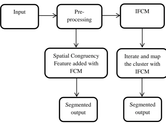
The detection of brain tumor in MRI Brain Image is done with the help of data base which already have the information about MRI tissue. The Training Input has some noise. The input is pre-processed to remove the noise. The median filter is used to remove the noise and it help full to spatial detection. Most of our training input is segmented with spatial feature. The median filter gives the mean value among the neighborhood pixels. It Give better result than other filtering method. For better result the combined form median filter is composed of a median filter and a second median
filter that filters the error signal.
The proposed segmentation method gives the additional iteration and clustering and maps the best matching. This will gives improvement in the segmentation using Improved Fuzzy C Means clustering technique. The segmented image using proposed algorithm then compared with the data base system. By using the morphological operation the data base system is used for detecting the tumor in the segmented output.
Figure 1Block Diagram of Proposed Method
IV. PREPROCESSING
The key function of pre-processing is to improve the image in ways that increase the chances for success of the other processes. The next stage deals with image segmentation. The double median filter is composed of a median filter and a second median filter that filters the error signal. The error signal is the difference between the input signal and the filtered signal after the first median filter. The traditional median filter is used. The median filter is a nonlinear filter often used to remove noise. Such noise reduction is a typical pre-processing step to improve the results of later processing. It preserves edges while removing noise.
V. SEGMENTATION
Image segmentation is an essential procedure in many applications of image processing. Brain image segmentation is a complex and challenging part in the Medical Image Processing. The new approach for detecting tumor in MRI Brain done with modified FCM iterative process. A new level of region growing algorithm has come up that
Input
Pre-processing
IFCM
Spatial Congruency Feature added with
FCM
Iterate and map the cluster with
IFCM
Segmented output
overcomes severe limitations of older approaches. This will extend those ideas by putting them in a general framework, which allows for the combination of a variety of segmentation algorithms, resulting in great potential to optimize all aspects of the segmentation process. Segmentation is the process of partitioning an image into different segments. In medical imaging, these segments often correspond to different tissue classes, organs, pathologies, or other biologically relevant structures. Medical image segmentation is made difficult by low contrast, noise, and other imaging ambiguities. Although there are many computer vision techniques for image segmentation, some have been adapted specifically for medical image computing. This project is focused on MRI Brain images segmentation and analyse about the brain tumor. These MRI Brain images are segmented and the results of segmentation are used for detecting the brain tumor.
SEGMENTATION USING IMPROVED FUZZY C MEANS CLUSTERING:
Clustering is a process for classifying objects or patterns in such a way that the samples of the same cluster are more similar to one another than samples belonging to different clusters. Fuzzy clustering is a soft segmentation method. Fuzzy c-means (FCM) algorithm is the most popular method used in image segmentation because it has robust characteristics for ambiguity and can retain much more information than hard segmentation methods.
Step1.
1) Set the number c of the cluster prototypes change from 2 to cmax.
2) Initialize randomly those prototypes and set ε>0 to a very small value.
Step2.
Compute the local similarity measures sij for all neighbor windows over the image using
𝑠𝑖𝑗 =�
𝑠𝑠−𝑖𝑗∗ 𝑠𝑔−𝑖𝑗, 𝑗 ≠0
0, 𝑗= 0
Step3.
Compute linearly-weighted summed image ξ in terms of ξi.
ξ𝑖=∑𝑗𝜖𝑁i𝑠𝑖𝑗
x
j ∑𝑗𝜖𝑁i𝑠𝑖𝑗 Step4.
Update the partition matrix using
u𝑖𝑙 = (ξ𝑖− 𝑣𝑖) 2 𝑚−1
∑𝑐
𝑗=1 (ξ𝑖− 𝑣𝑗) 2 𝑚−1
Step5.
Update the prototypes using
𝑣𝑖=∑ 𝛾𝑙
𝑞
𝑙=1 𝑢𝑖𝑙𝑚ξ𝑙
∑𝑞𝑙=1𝛾𝑙𝑢𝑖𝑙𝑚
Repeat Steps 4-5 until the following termination criterion is satisfied: |Vnew–Vold|<ε Where V= [v1, v2…vc] are the vectors of cluster prototypes.
SEGMENTATION USING MAPPING THE ITERATIVE OF IFCM CLUSTERING (proposed method1 PM1):
• Concatenate the IFCM clustering output. • Initialize the cluster pixel values.
• Obtained the repmatrix with respect to the output of IFCM Clustering.
• Now get Concatenation of rep matrix • Find the distance between the
Concatenation of input and the Concatenation cluster pixel.
• Update the distance with respect to the rep matrix.
• Get the partition matrix with respect to the updating of distance.
• Update the cluster pixel value with respect to the partition matrix.
• Find out the temporary matrix from the partition matrix.
• Map the values of temporary matrix into the output of IFCM clustering.
• Repeat this still max (temporary matrix) <.00001.
SEGMENTATION USING PHASE CONGRUENCY FEATURES WITH FCM (proposed 2):
Step1:
Set the number of clusters(C) , degree of fuzziness ,stop criterion and neighborhood size.
Step2:
Calculate phase congruency features and define the neighborhood configuration for each pixel.
Step3:
Initialize the center of the clusters v𝑖|I =1,2,…..C. Step4:
Calculate the new similarity measure using,
𝐷𝑖j= 𝑤𝑖𝑗��𝑥𝑗− 𝑣𝑖�� 2
=(1-α𝑠𝑖𝑗)��𝑥𝑗− 𝑣𝑖��2
Step5:
Calculate the membership value,
𝑢𝑖𝑗 = [∑ (𝐷k𝑗𝐷𝑖𝑗) 1 𝑚−1 𝑐
𝑘=1 ]−1
Step6:
𝑢𝑖𝑗−𝑛𝑒w= 𝑢𝑖𝑗𝑀𝑖𝑗 𝛽
∑𝑐𝑘=1𝑢𝑘𝑗𝑀𝑖𝑗𝛽
Step7:
Update 𝑣𝑖using𝑢𝑖𝑗−𝑛𝑒w
𝑣𝑖=
∑𝑁𝑗=1𝑢𝑖𝑗𝑚𝑥𝑗 ∑𝑁𝑗=1𝑢𝑖𝑗𝑚
Repeat the steps from 4 to 7 until it reaches the stopping criteria,
𝑚𝑎𝑥𝑖∈[1,𝑐]||𝑣𝑖𝑙− 𝑣𝑖𝑙+1||∞<∈.
VI. RESULTS
Figure 2 segmented output (IFCM)
Figure 3 segmented output (proposed method 1)
Figure 4 segmented output (proposed method 2)
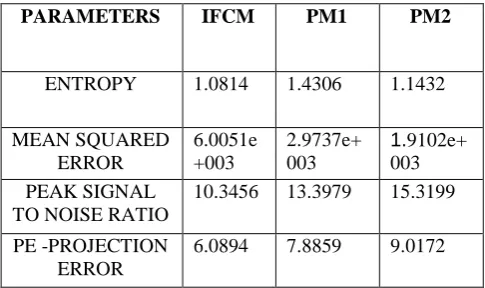
PARAMETERS IFCM PM1 PM2
ENTROPY 1.0814 1.4306 1.1432
MEAN SQUARED ERROR
6.0051e +003
2.9737e+ 003
1.9102e+ 003 PEAK SIGNAL
TO NOISE RATIO
10.3456 13.3979 15.3199
PE -PROJECTION ERROR
6.0894 7.8859 9.0172
Table 1 Comparison of Parameters
VII. CONCLUSION
Thus the image enhancement is done using the median filtering which is often used to remove noise. Median filtering is very widely used in digital image processing because, under certain conditions, it preserves edges while removing noise. The median filtered image is well suited for MRI brain image segmentation using clustering. The MRI brain image segmentation is done by improved fuzzy c means clustering. And the parameters Entropy, Mean square error, Peak signal to noise ratio, Re-projection error have been evaluated. For better performance of image segmentation the Entropy should be small and the other parameters should be improved. The Improved FCM algorithm attempts to partition a finite collection of Fuzzy C clusters with respect to some given criterion. The outcome will be used to further analysis of MRI image more accurately.
References:
[1] Colm Elliott, Douglas L. Arnold, D. Louis Collins, and Tal Arbel, “Temporally Consistent Probabilistic Detection of New Multiple Sclerosis Lesions in Brain MRI”, IEEE Transactions On medical imaging, Vol. 32, No. 8, August 2013.
[2] Ivana Despotović, Student Member, IEEE, Ewout Vansteenkiste, and Wilfried Philips, Member, IEEE,“ Spatially Coherent Fuzzy Clustering for Accurate and Noise-Robust Image Segmentation”, IEEE Transactions On Image Processing, Vol. 22, No. 2, APRIL 2013.
[3] Maoguo Gong, Member, IEEE, Yan Liang, Jiao Shi, Wenping Ma, and Jingjing Ma,“Fuzzy C-Means Clustering With Local Information and Kernel Metric for Image Segmentation”, IEEE Transactions On Image Processing, Vol. 22, No. 2, February 2013.
Minh Nguyen, “A Robust Fuzzy Algorithm Based on Student’s t-Distribution and Mean Template for Image Segmentation Application”, IEEE Signal Processing Letters, FEBRUARY 2013.
[5] D. Salas-Gonzalez a,n, J.M.Go´ rriz a, J.Ramı´rez a, M.Schloegl b, E.W.Lang b, A.Ortiz, “Parameterizationofthedistributionofwhiteandgrey matterinMRI using the a-stable distribution”, Elsevier JAN 2013.
[6] Zexuan Ji, Yong Xia, Member, IEEE, Quansen Sun, Qiang Chen, Member, IEEE, Deshen Xia,and David Dagan Feng, Fellow, IEEE, “Fuzzy Local Gaussian Mixture Model for Brain MR Image Segmentation” IEEE Trans, May 2012.
[7] Stefan Bauer*, Student Member, IEEE, Christian May, Dimitra Dionysiou, Georgios Stamatakos, Member, IEEE, Philippe B¨uchler, and Mauricio Reyes, Member, IEEE “Multiscale Modeling for Image Analysis of Brain Tumor Studies” IEEE Trans, JAN. 2012.
[8] Chunming Li, Rui Huang, Zhaohua Ding, J. Chris Gatenby, Dimitris N. Metaxas, Member, IEEE, and John C. Gore, “A Level Set Method for Image Segmentation in the Presence of Intensity Inhomogeneities With Application to MRI” IEEE Trans, July 2011.
[9] Stelios Krinidis and Vassilios Chatzis, “A Robust Fuzzy Local Information C-Means Clustering Algorithm” IEEE Transactions On Image Processing, Vol. 19, No. 5, May 2010.
[10] Shan Shen, William Sandham, Malcolm Granat, and Annette Sterr, “MRI Fuzzy Segmentation of Brain Tissue Using Neighborhood Attraction with Neural-Network Optimization” IEEE Transactions On Information Technology In Biomedicine, Vol. 9, No. 3, September 2005.