INTRODUCTION
β-Thalassemia is one of the commonest single gene disorders resulting from mutations in and around the α-globin gene located as a cluster on the short arm of chromosome number 11 i.e.11p15(Lin et al., 1985) causing severe anemia. In this disorder, there is relative excess of insoluble β-globin chains that precipitates within the red cells causing ineffective erythropoiesis and severe anemia (Weatherall and Clegg, 1981; Susanne et al., 2000).
In the Jammu region of the J&K state, no high risk community for β-thalassemia is known as there is no previous record of screening for β-thalassemia carrier detection. The present study is an account of the carr ier screening for β-thalassemia trait in Jammu region of the J&K state. The aim of the study was to provide genetic counselling to the carriers thus detected and their
Detection of carriers for
βββββ
-thalassemia:
A case study of Jammu (J&K)
VIJAY BHARAT*, SUBASH GUPTA and RACHNA GUPTA
P.G. Department of Zoology, University of Jammu, Jammu- 180 006 (India).
(Received: July 12, 2009; Accepted: August 04, 2009)
ABSTRACT
The present work embodies screening of about 450 individuals for β-thalassemia carrier detection through NESTROFT and CBC. Of 450 individuals, 125 individuals were found to be NESTROFT positive and 325 were found negative. The sensitivity, specificity, positive predictive and negative predictive values were found to be 100%, 98.5%, 96% and 100% respectively. The efficiency of the NESTROFT was found to be 98.8%. Of the 125 NESTROFT positive individuals, 50 individuals were subjected to CBC analysis and it was found that the hematological parameters like Hb level, red cell count, PCV/HCT, MCV, MCH and MCHC levels varied from 8.1-14.4 g/dl with a mean of 10.63±0.21 g/dl, 2.57-9.80 × 10¹²/litre with a mean of 4.49±1.13 × 10¹²/litre, 25.4-39.0% with a mean of 33.5±3.49%, 53.3-101 fl with a mean of 72.64±1.34 fl, 16.5-31.3 pg with a mean of 22.7±0.49 pg and 29.4-33.4 g/dl with a mean value of 30.8±0.86 g/dl respectively.
Key words: β-thalassemia, NESTROFT, CBC and carrier detection.
relatives that would help in preventing the transmission of the β-thalassemia trait. A number of authors have already studied this aspect viz., Weatherall and Clegg, 1981; Kattamis et al., 1981; Varawalla et al., 1991; Balgir, 1999; Balgir, 2000; Susanne et al., 2000; Singh and Gupta, 2008 and Wiwanitkit. (2009).
MATERIAL AND METHODS
During the present study, screening for the carrier detection was carried out by NESTROFT (Naked Eye Single Tube Red cell Osmotic Fragility Test) and Complete Blood Count (CBC).
A total of 450 individuals were screened and the screening included two categories of people: ´ Individuals belonging to the families having the history of b-thalassemia i.e. the families where at least one β-thalassemic child was born, and,
NESTROFT
NESTROFT was performed using 0.36% buffered saline solution following the method of Singh and Gupta (2008) with least modifications. Based on the observations, sensitivity, specificity, positive and negative predictive values and efficiency of the test were calculated by following the formulae given below (Singh and Gupta, 2008):
Sensitivity 100×TP/ (TP+FN)
Specificity 100×TN/ (TN+FP)
Positive predictive value 100×TP/ (TP+FP) Negative predictive value 100×TN/ (TN+FN)
Efficiency of test 100× (TN+TP)/
(TP+FP+TN+FN)
CBC (Complete Blood Count)
Of the 125 NESTROFT positive samples, only 50 samples were put to CBC analysis because of latter being expensive and cumbersome. CBC was carried out by using the ERMA PC 604 Cell counter within 2-4 hours after collection of the blood samples.
RESULTS NESTROFT
Out of the 450 individuals screened through NESTROFT, 125 individuals were found positive with respect to NESTROFT while the remaining 325 individuals were found negative. Fur ther, of these 125 NESTROFT positive individuals, 61 were females and 64 were males. The nature and number of the individuals with respect to NESTROFT is shown in the table 1.
Table 1: Showing the number of True Positive, False Negative, False Positive
and True Negative individuals Nature of Individuals with Number of respect to NESTROFT Individuals
True Positive (TP) 120
False Negative (FN) 0
False Positive (FP) 5
True Negative (TN) 325
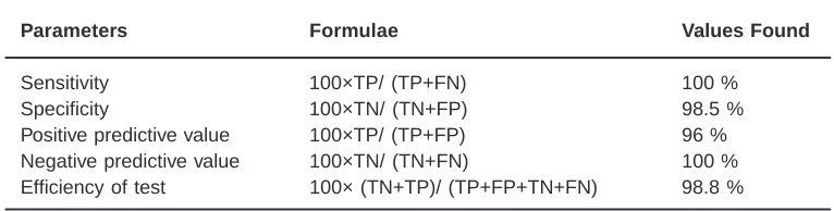
Table 2: Showing the Sensitivity, Specificity, Positive predictive value, Negative predictive value and the Efficiency of NESTROFT
Parameters Formulae Values Found
Sensitivity 100×TP/ (TP+FN) 100 %
Specificity 100×TN/ (TN+FP) 98.5 %
Positive predictive value 100×TP/ (TP+FP) 96 %
Negative predictive value 100×TN/ (TN+FN) 100 %
Efficiency of test 100× (TN+TP)/ (TP+FP+TN+FN) 98.8 %
Table 3: Ranges and Means of different hematological parameters of βββββ-thalassemia carriers
S. No. Hematological Parameters Range Mean Standard Deviation
1. Hb (g/dl) 8.1-14.4 10.63 0.21
2. Red cell count (-×1012/litre) 2.57-9.80 4.49 1.13
3. PCV/HCT (%) 25.4-39.0 33.5 3.49
4. MCV (fl) 53.3-101 72.64 1.34
5. MCH (pg) 16.5-31.3 22.7 0.49
Fig. 1:
Fig. 2(a):
Fig. 2(b):
Fig. 3(a):
Fig. 3(b):
Fig. 3(c):
T
a
b
le 4:
Usefulness of NESTR
OFT in the detection of
βββββ
-thalassemia carrier
s:
a Comparative anal
ysis of data with earlier studies
P arameter Present Kattamis Mehta Gorakshaker R a gha v a n Thomas Manglani Maheshwari Siric hotiy Singh Study et al . et al . et al et al . et al . et al . et al . a kul (2006) (1981) (1988) (1990) (1991) (1996) (1997) (1999) et al . (2004) Sensitivity 100% 98.4% 95.0% 98-100% 95.5% 98.4% 94.4% 91.0% 97.6% 97.7% Specificity 98. 5% 91.0% 82.1% 82-84% 87.0% 66.6% 64.2% 95.0% 72.9% 83.3% P ositiv e 9 6 % 91. 3% 73.1% 50.0% 70.5% 81.5% 97.6% 55.0% 33.6% 95.5% Predictiv e V alue Negativ e 100% 98.35 97.0% 99-100% 98.3% 96.5% 35.3% 99.0% 99.5% 90.9% Predictiv e V alue
The graphical representation of the number of True Positive, False Negative, False Positive and True Negative individuals in Figure 1.
In detecting the heterozygous β -thalassemia cases, the sensitivity of NESTROFT was 100%, specificity 98.5% and the positive and negative predictive values were 96% and 100% respectively. The efficiency of the test was 100%. (Table 2 and Figure 2a & 2b).
The values found for the var ious parameters of NESTROFT can also be shown in a graphical representation in figure 2a and 2b.
CBC
The values of different hematological parameters like Hemoglobin, Red Cell Count, Mean Corpuscular Volume (MCV), Plasma Corpuscular Volume (PCV), Mean Corpuscular Hemoglobin (MCH) and Mean Cor puscular Hemoglobin Concentration (MCHC)of 50 samples in which CBC was done are depicted in the form of graphs shown in the figures 3a,3b,3c,3d,3e and 3f respectively.
In the present study, most of the carriers were diagnosed on the basis of their hematology with special emphasis on complete blood count (CBC). The Hb level in carriers varied from 8.1-14.4 g/dl with a mean of 10.63±0.21 g/dl. The red cell count was relatively high and ranged from 2.57-9.80 × 10¹²/litre with a mean value of 4.49±1.13 × 10¹²/ litre. The MCV and MCH were markedly reduced and ranged from 53.3-101 fl and 16.5-31.3 pg with mean values of 72.64±1.34 fl and 16.5-31.3±0.49 pg respectively.
Ranges and mean values of different hematological parameters of the β-thalassemia carriers are shown in the table 3 and graphically in the figure 4.
DISCUSSION
1.4-7.96%. It is probably the commonest inherited hemoglobin disorder in India, with an uneven distribution amongst the different endogenous populations (Sukumaran, 1975; Varawalla et al., 1991a, b; Venkatesan et al., 1992; Verma, 1994; Verma et al., 1997, Balgir, 1999; Balgir, 2000).
The test is very sensitive and specific. A comparison of the present findings with those of the earlier reports has been shown in the table is shown in the table 4.
The sensitivity of NESTROFT in the present study was found to be 100% which is comparable with sensitivity repor ted by Gorakshaker et al. (1990) and to some extent with the reports of Thomas et al. (1996). The specificity in the present study has been found to be 98.5%. Although the specificity as reported by majority of the workers is less than the one found in the present study, yet, the value of specificity as recorded in the present study (98.5 %) is more or less comparable with the findings of Maheshwari et al. (1999) who reported the value of specificity to be 95.0%.
Similarly, the positive predictive value in the present study has been found to be 96% and it is quite high quite high but the positive predictive value is comparable with the studies made by Kattamis et al. (1981), Manglani et al. (1997) and Singh (2006). The negative predictive value of the test in the detection of β-thalassemia carriers in the present study has been found to be 100%. The same is well comparable with the studies made by Kattamis et al. (1981), Mehta et al. (1988), Gorakshaker et al. (1990), Maheshwari et al. (1999) and Sirichotiyakul et al. (2004) respectively. Thus, a comparison of different parameters has largely been found to be more or less similar to the earlier reports. This suggests that these parameters have their practical significance in the screening of β-thalassemia trait.
The red cell indices have been found as a valuable diagnostic tool for the detection of â-thalassemia carriers and have also been used in the present study. In the carriers detected during the present study, there was a relatively high red cell count, however, the red cells were microcytic and hypochromic having markedly reduced MCV and MCH with averages of 72.64 fl and 22.7 pg respectively.
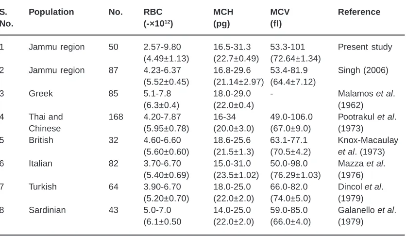
Table 5: Hematological values in βββββ-thalassemia carriers in different populations of the world
S. Population No. RBC MCH MCV Reference
No. (-×1012) (pg) (fl)
1 Jammu region 50 2.57-9.80 16.5-31.3 53.3-101 Present study
(4.49±1.13) (22.7±0.49) (72.64±1.34)
2 Jammu region 87 4.23-6.37 16.8-29.6 53.4-81.9 Singh (2006)
(5.52±0.45) (21.14±2.97) (64.4±7.12)
3 Greek 85 5.1-7.8 18.0-29.0 - Malamos et al.
(6.3±0.4) (22.0±0.4) (1962)
4 Thai and 168 4.20-7.87 16-34 49.0-106.0 Pootrakul et al.
Chinese (5.95±0.78) (20.0±3.0) (67.0±9.0) (1973)
5 British 32 4.60-6.60 18.6-25.6 63.1-77.1 Knox-Macaulay
(5.60±0.60) (21.5±1.3) (70.5±4.2) et al. (1973)
6 Italian 82 3.70-6.70 15.0-31.0 50.0-98.0 Mazza et al.
(5.40±0.69) (23.5±1.02) (76.29±1.03) (1976)
7 Turkish 64 3.90-6.70 18.0-25.0 66.0-82.0 Dincol et al.
(5.20±0.70) (22.0±2.0) (74.0±5.0) (1979)
8 Sardinian 43 5.0-7.0 14.0-25.0 59.0-85.0 Galanello et al.
Carriers for β-thalassemia trait, in general have reduced hemoglobin, reduced MCV (</= 80 fl) and reduced MCH (< 27 pg) (Karimi and Rasekhi, 2002; Leung et al., 2005; Sirichotiyakul et al. 2005; Al-Suliman, 2006). They have relatively high red cell count (>4.5×10¹²/liter) (Gomber et al., 1997), but with pronounced microcytosis and hypochromia. Reticulocyte count is slightly increased around twice the normal value.
A comparison of the data obtained on red blood indices during the present work with other workers is given in the table-5.
The findings of reduced MCV and MCH values in majority of the cases has been used as a basis of population screening for â-thalassemia trait (Pearson et al., 1973 & 1974; Klee, 1980). In the present study also, similar approach was used for the diagnosis of carriers and this approach worked well in the screening programme.
1. Lin, C. C., Draper, P. N. and De Braekeleer, M., High-Resolution Chromosomal Localization of the β-Gene of the Human â-Globin Gene Complex by in situ Hybridization. Cytogenetics, 39: 269-274 (1985).
2. Weatherall, D. J. and Clegg, J. B., In: The thalassemia syndromes. Blackwell Scientific, Oxford (1981).
3. Susanne, R., Christoph, B., Gunter, H., Dieter, O., Michael, M., Pranav, S. andreas, K., Christian, M., Barbara, V. and Andreas, E. K.. Oral isobutyramide reduces transfusion requirements in some patients with homozygous β-thalassemia. Blood, 96(10): 3357-3363 (2000).
4. Balgir, R. S., The burden of
hemoglobinopathies in India and the challenges ahead. Current Science, 79: 1536-1547 (2000).
5. Varawalla, N. Y., Old, J. M., Sarkar, R., Venkatesan, R. and Weatherall, D. J., The spectrum of β-thalassemia mutations on the Indian subcontinent: The basis for prenatal diagnosis. British Journal of Haematology, 78(2): 242-247 (1991a).
6. Higgs, D. R., Thein, S. L. and Wood, W. G., Distribution and population genetics of the thalassemias. In: Weatherall DJ, Clegg JB, eds. The Thalassemia Syndromes. 4th edn. Blackwell Science, Oxford: 237-284 (2001). 7. Singh, S. P. and Gupta, S. C., Effectiveness of red cell osmotic fragility test with varying degrees of saline concentration in detecting beta-thalassemia trait. Singapore Medical
REFERENCES
Journal, 49(10): 823-826 (2008).
8. Khattak, I., Khattak, S. T. and Khan, J., Heterozygous beta-thalassemia in parents of children with â-thalassemia major. Gomal Journal of Medical Sciences, 4(20): 52-56 (2006).
9. Weatherall, D. J. and Clegg, J. B., Inherited hemoglobin disorders: an increasing global health problem. In: Bulletin of the World Health Organization, 79(8): 704-712 (2001b). 10. Sukumaran, P. K., Abnormal hemoglobins in India. Trends in Hematology (eds N.N. Sen & A.K. Basu). Sree Saraswaty Press Ltd., Calcutta, 225 (1975).
11. Venkatesan, R., Sarkar, R. and Old, J. M., β -thalassemia mutations and their linkage to â-haplotypes in Tamil Nadu in southern India. Clinical Genetics, 42: 251-256 (1992). 12. Ver ma, I.C., The challenge of genetic
disorders in India. In: Molecular genetics and gene therapy-the new frontier; Scientific Communications, Amesterdam: 11-20 (1994).
13. Verma, I. C., Saxena, R., Thomas, E. and Jain, P. K., Regional distribution of â-thalassemia in India. Human Genetics, 100: 109-113 (1997).
14. Balgir, R. S., Control and prevention of the genetic load of hemoglobinopathies in India. Natl. Med. J. India, 12: 234-238 (1999). 15. Kattamis, C., Efremov, G. and Pootrakul, S.,
(1981).
16. Mehta, B. C., Gandhi, S. and Mehta, J. B., Naked eye single tube red cell osmotic fragility test for beta-thalassemia: population survey. Indian Journal of Haematology, 6: 187-190 (1988).
17. Gorakshaker, A. C., Colah, R., Desai, S. and Nadkarni, A., Evaluation of the single tube osmotic fragility test in detection of â-thalassemia trait. Natl. Med. J. India, 3: 171-173 (1990).
18. Raghavan, K., Lokeshwar, M. R., Birewar, N., Nigam, V., Manglani, M. V. and Raju, N. B. (1991). Evaluation of naked eye single tube red cell osmotic fragility test in detecting beta-thalassemia trait. Indian Pediatrics, 28(5): 469-472.
19. Thomas, S., Srivastava, A., Jeyaseelan, L., Dennison, D. and Chandy, M., NESTROFT as a screening test for the detection of thalassemia & common haemoglobinopathies—an evaluation against a high performance liquid chromatographic method. Indian Journal of Medical Research, 104: 194-197 (1996).
20. Sharma, U., Agrawal, A. K., Choudhary, A., Sitaraman, S. and Sharma, M. L., Nestroft Screening Tool for â-Thalassemia Trait. Indian Pediatrics, 33: 788 (1996).
21. Manglani, M., Lokeshwar, M. R., Vani, V. G., Bhatia, N. and Mhaskar, V., NESTROFT’— an effective screening test for beta thalassemia trait. Indian Pediatrics, 34(8): 702-707 (1997).
22. Gomber, S., Sanjeev and Madan, N., Validity of Nestroft in screening and diagnosis of beta-thalassemia trait. Journal of Tropical Pediatrics, 43(6): 363-366 (1997).
23. Thool, A. A., Walde, M. S., Shrikhande, A. V. and Talib, V. H., A simple screening test for the detection of heterozygous â-thalassemia. Indian Journal of Pathology & Microbiology, 41(4): 423-426 (1998).
24. Maheshwari, M., Arora, S., Kabra, M. and Menon, P. S. N., Carrier screening and prenatal diagnosis of â-thalassemia. Indian Pediatrics, 36: 1119-1125 (1999).
25. Sir ichotiyakul, S., Tantipalakor n, C., Sanguansermsri, T., Wanapirak, C. and Tongsong, T., Erythrocyte osmotic fragility
test for screening of alpha-thalassemia-1 and beta-thalassemia trait in pregnancy. International Journal of Gynaecology & Obstetrics, 86(3): 347-350 (2004).
26. Singh, S. P., Screening and Molecular character ization of beta-thalassemia Mutations in different populations of Jammu region, J&K State. Ph.D. Thesis, Department of Zoology, University of Jammu (2006). 27. Karimi, M. and Rasekhi, A. R., Efficiency of
premarital screening of beta-thalassemia trait using MCH rather than MCV in the population of Fars Province, Iran. Haematologia (Budap), 32(2): 129-133 (2002).
28. Leung, N. T., Lau, T. K. and Chung, T. K. H., Thalassemia screening in pregnancy. Curr. Opinion in Obstetric & Gynaecology, 17: 129-134 (2005).
29. Sirichotiyakul, S., Maneerat, J., Sa-nguansermsri, T., Dhananjayanonda, P. and Tongsong, T., Sensitivity and specificity of mean cor puscular volume testing for screening for alpha-thalassemia-1 and beta-thalassemia traits. Journal of Obstetric Gynaecology & Research, 31(3): 198-201 (2005).
30. Al-Suliman, A., Prevalence of β-thalassemia trait in premarital screening in Al-Hassa, Saudi Arabia. Ann. Saudi Med., 26(1): 14-16 (2006).
31. Pearson, H. A., McPhedran, P., O’Brien, R. T., Aspner, G. T., Mclntosh, S. and Guiliotis, D. K., Comprehensive testing for thalassemia trait. Ann. N.Y. Acad. Sci., 232: 135 (1974). 32. Pearson, H. A., O’Brein, R. T. and Mclntosh,
S., Screening for thalassemia trait by electronic measurement of mean corpuscular volume. New England Journal of Medicine, 288: 351 (1973).
33. Klee, G. G., Role of mor phology, and er ythrocyte indices in screening and diagnosis. The automated hematology group (CBC) as a screen for thalassemia and hemoglobinopathies. In: Hemoglobinopathies and Thalassemia- Laboratory Methods and Clinical Cases (ed. V. F. Fairbanks), Decker, New York: 38 (1980).
Hypotheses Ideas, 3: 1 (2009).
35. MAZZAG, U., Sagliof,. Caligarisc Appioc, Camaschella, G. Nerettaond E. Gallo., Clinical and Haematological Data in 254 Cases of Beta-Thalassaemia Trait in Italy. British Joumal of Haematology, 33: 91 (1976). 36. MALAMOS, B., FESSAS, P. STAMATOYANNOPOULOS, G., Types of thalassaemia-trait carriers as revealed by a study of their incidence in Greece. British Journal of Haematology, 8: 5 (1962).
37. PO OT R A K U L, P., WA S I, P. NA-KA KO R N, S.,
Haematological data in 312 cases of â-thalassaemia trait in Thailand. British Journal of Haematology, 24: 703 (1973).
38. KNOX-MACAULAY, H.H.M., WEATHERALL, D.J., CL E G G, J.B. PE M B R E Y, M.E. (1973). Thalassaemia in the British. British Medical Journal, iii, 150.