EVALUATIO OF WOUD HEALIG EFFECT OF A POLYHERBAL FORMULATIO BY DIFFERET CUTAEOUS WOUD MODELS
Swapnil Goyal1*, Shruti Sureka1, Kratika Daniel1, Vivek Daniel1, Rahul Trivedi2, Pradeep Deshmukh2, Navin R. Sheth3
1. Department of Pharmacology, Mandsaur Institute of Pharmacy, Mandsaur, M.P., 458001, INDIA
2. Diabetes Research Division, Department of Pharmacology and Toxicology, B.R. Nahata College of Pharmacy, Mandsaur, M.P., 458001, INDIA
3. Department of Pharmaceutical Sciences, Saurashtra University, Rajkot, Guj., INDIA
*Author for Correspondence Swapnil Goyal
Lecturer, Department of Pharmacology
Mandsaur Institute of Pharmacy, Mandsaur, M.P., 458001, INDIA Email- [email protected]
Contact no. 09907860790
Summary
In present study, a Polyherbal formulation (PHF) containing the hydro alcoholic extracts of root bark of Calotropis gigentea Linn., leaves of yctanthes arbortristis Linn., and flower of Tridax procumbens in an optimized ratio (5:3:2) was evaluated in different cutaneous wound models. An Ointment of PHF (10%w/w) was prepared and was applied topically in excision wound model in rats. In dead space and incision wound model oral suspension of PHF at doses of 200 and 400 mg/kg was given orally to rats. In excision wound model, PHF ointment showed a significant (p< 0.05) wound contraction rate compared with normal control and standard group i.e. nitrofurazone ointment applied topically (0.2%w/w) on 10th, 15th, 20th day of experiments. In incision model, PHF treated groups showed significant increase (p< 0.05) in tensile strength at 200 mg/kg and at 400 mg/kg compared with normal groups. In dead space model, there was increased weight of granuloma tissue in drug treated groups and significant increase in hydroxyproline contents. Increased hydroxyproline contents showed greater synthesis of collagen.
Introduction
Healing is a normal physiological process that proceeds through a series of co-ordinate cellular and cytokine mediated events, culminating in the restoration of functional integrity of tissues.[1] Wound repair in healthy individuals depends on several interrelated processes, including the migration of inflammatory cells into the wound space to colonize the provisional matrix, proliferation of fibroblasts and vascular cells, apoptosis, and synthesis of extracellular matrix proteins to reconstitute dermal architecture.[2] Wound healing occurs in three interrelated and interdependent phases: inflammation, granulation tissue formation, and remodeling. The inflammatory phase begins immediately after injury. Tissue injury causes the disruption of blood vessels and extravasations of blood constituents that lead to clot formation. It provides a provisional extracellular matrix, formed mainly by fibrin and fibronectin that allow cell migration. During granulation tissue formation, fibroblasts and endothelial cells proliferate and move into the wound space leading to extracellular matrix deposition and angiogenesis, which are typical features of granulation tissue formation.[3] The remodeling, involves the formation and maturation of extracellular matrix. Fibrin, but also fibronectin and thrombospondin-1 and other (glyco-) proteins are replaced step by step by collagen.[4] The factors involved in the modulation of myofibroblastic differentiation include cytokines and growth factors such as Transforming Growth Factor, platelet-derived growth factor, granulocyte macrophage- colony stimulating factor, fibroblastic growth factors (FGFs), tumor necrosis factor and interferon. Impaired wound healing may be consequences of pathologic states associated with diabetes, immune disorders, ischemia, and in injuries such as burns, frostbite and gunshot wounds. Decreased wound healing may be due to decreased synthesis of collagen, increases levels of proteases and defective macrophages function. [3]
In traditional system of medicines leaves of yctanthes arbortristis [5] Linn.flowers of Tridax procumbens [6] Linn. and root bark of Calotropis gigentea Linn.[7] have been used as a natural wound healing remedy. Keeping in view the complication and multi etiological factors related to acute and chronic wounds, a Polyherbal formulation (PHF) was prepared by combining the hydro alcoholic extract of root bark of Calotropis gigentea Linn., leaves of yctanthes arbortristis Linn., and flower of Tridax procumbens in an optimized ratio (5:3:2). The main aim of present study and to make a Polyherbal formulation was that all the selected plants for investigation have potent wound healing activity so we prepared a topical formulation according to potency and evaluate for its wound healing property by using different wound models in rats.
Materials and Methods
Plant Materials Collection
Preparation of Extract
Extractions were made by process of maceration. The fixed amount of dried coarse powders were weighed of all three dried coarse powders were kept in a separate three an iodine flask with alcohol and water in a ratio of 70:30 for 72 hrs. Continuous shaking was maintained by electrical shaker. After 72hrs. extracts were filtered out using muslin cloth. The filtrates were evaporated on water bath until it becomes solid mass. The dried extracts were used for the evaluation of wound healing activity in rats.
Animals
Wistar albino rats of either sex weighing between 180 and 200 g were procured from animal house of B.R. Nahata college of Pharmacy, Mandsaur. The Institutional animal Ethical Committee (IEAC) for animal experimentation of the institute approved the study protocol. These animals were used for the wound healing activity studies. The animals were stabilized for 1 week. They were maintained in standard conditions at room temperature, 60±5% relative humidity and 12 hrs. light dark cycle. They had been given standard pellet diet supplied by Hindustan Lever Co., Mumbai and water ad libitum throughout the course of the study.
Preparation of an Ointment
The dried extracts of all three plants (root bark of Calotropis gigentea Linn., leaves of yctanthes arbortristis Linn., and flower of Tridax procumbens) in a ratio of (5:3:2) were triturated with the minimum quantity (20 gm.) of soft white petroleum jelly I.P. to avoid any microbial contamination of growth. Then, the remaining quantity (70 gm.) of soft white petroleum jelly I.P. was added in the above ointment and triturated for 30-40 minutes to form a homogenous ointment. This ointment was packed in air tight umber coloured bottle to avoid any contamination and microbial growth.
Preparation of Oral Suspension-
Calotropis gigantea, yctanthes arbortristis, Tridax procumbens: were weighed in a ratio of 5:3:2 and suspended in a 50 ml of pyrogen free distilled water (Parenteral drugs Pvt. Ltd., Indore, India). This suspension was given in a dose of 200 mg/kg and 400 mg/kg.
Treatment Schedule
Wound healing activity was studied using excision, incision, and dead space wound model. The test suspension was given in a dose of 200 and 400 mg/kg in dead space and incision models. The effect of extracts were seen on various physical parameters like wound breaking strength, wet and dry granulation tissue weight, and biochemical parameters like hydroxyproline. In excision wound model, 10 % w/w ointments were made and applied topically once daily. After 0, 5, 10, 15 and 20 days interval wound area were measured using transparent sheet.
Experimental Procedure
1. Excision Wound Model
The animals were divided into three groups of six rats in each group and kept in separate cages. Animals were anaesthetized with diethyl ether shaved on back side using scalp blade. The area of wound to be created was outlined using methylene blue using circular stenless steel stencils. A full thickness wound of square area of 1 cm2 was created along with markings. The entire wound left open. After making wounds in animals, all animals were kept in a separate cage to avoid any infections. Animals were observed closely for the any infection.
Normal control without any treatment (group 1) was applied petroleum jelly topically, positive control (group 2) was applied with nitrofurazone and experimental control (group 3) was applied with test drug extract. Treatment was done topically in all cases and once daily. Drug extracts were applied topically in a dose 10% w/w ointment for 20 days. Wound areas were measured on 0, 5, 10, 15, 20 days using transparent sheet with permanent markers. Recording of wound areas were measured on graph paper. [8, 9, 10]
2. Dead Space Wound Model
The animals were divided into three groups of six rats each and kept in separate cages. Implanting subcutaneously sterile cotton 10 mg each in the lumbar region on dorsal side created the dead space wound. Animals received test extracts from 0 day to 9th post-wounding day. On 10th post-wounding day, the granulation tissue harvested on each implanted tube was carefully dissected out along with the tube and employed for determination of breaking strength and the estimation of hydroxyproline content. Experimental control (group 2 & 3) received test drug extracts in a dose of 200 mg/kg and 400 mg/kg. [8, 9, 10]
3. Incision Wound Model
The animals were divided into three groups of six rats each and kept in separate cages. Two Para-vertebral straight incisions of 5 cm length each were made through the entire thickness of the skin, on either side of the vertebral column with the help of a sharp scalpel. After complete homeostasis the wounds were closed by means of interrupted sutures placed at equidistance points about 1 cm apart. On the 7th days sutures were removed and on the 10th post-wounding days tensile strength was measured by continuous water flow technique using tensiometer. Experimental control (groups 2 & 3) received test drug extracts in a dose of 200 and 400 mg/kg. [8, 9, 10]
After noting the weight of the wet granulation tissue, the tissue was dried at 60°C for 12 hrs and the dry tissue weight was recorded on next day.
4. Determination of Hydroxyproline
The dry granulation tissue was kept in 6N HCL for 24 hrs. Then the hydroxyproline content was determined using UV spectrophotometer.
Hydroxrproline is a post transitional product of praline hydroxylation catalyzed by an enzyme polyhydroxylase. The occurrence of this amino acid is confined to the connective tissue collagen.[11]
6. Statistical Analysis-
The results were expressed in mean±SEM and statistical significance by means of ANOVA followed by Dunnet’s test. P < 0.05 was considered significant.
Results
1. Excision Wound Model
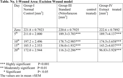
Topical application of PHF increased the percentage of wound contraction and completed wound healing by 20th day, which indicates rapid epithelization and collagenization. In fact, topical application of PHF accelerated the progression of wound healing by 10th day, i.e. (174.5±3.658***) p < 0.001 compared with control (197.2 ± 2.496). There was complete healing on 20th day of PHF treated groups, i.e. (96.83±3.928***) compared with control group, i.e. (172.0 ± 2.944). Nitrofurazone treated groups showed significant increase in wound contraction rate on 20th day, i.e. (116.2±2.286***) when compared with normal i.e. (172.0 ± 2.944) and soframycin treated groups i.e. (153.8±1.869***). (Table 1)
Table. o. 1-Wound Area: Excision Wound model
Day Group-I Normal Control [mm2]
Group-III
Standard control (Nitrofurazon treated) [mm2]
Group-IV
Extract treated [mm2]
Zero 221.8 ± 0.7923 220.6 ± 0.7925 222.6 ± 0.7892 5th 211.0 ± 2.000 189.3±3.703*** 189.7±4.137***
10th 197.2 ± 2.496 178.7±2.985*** 174.5±3.658*** 15th 185.5 ± 2.553 156.0±1.932*** 143.2±4.037*** 20th 172.0 ± 2.944 116.2±2.286*** 96.83±3.928***
*** Highly significant P<0.001 ** Moderately significant P<0.01 * Significant P< 0.05 The values are in mean ±SEM
2. Incision Wound Model
Table. o. 2- Tensile Strength Measurement: Incision Wound model –
3. Dead Space Wound Model
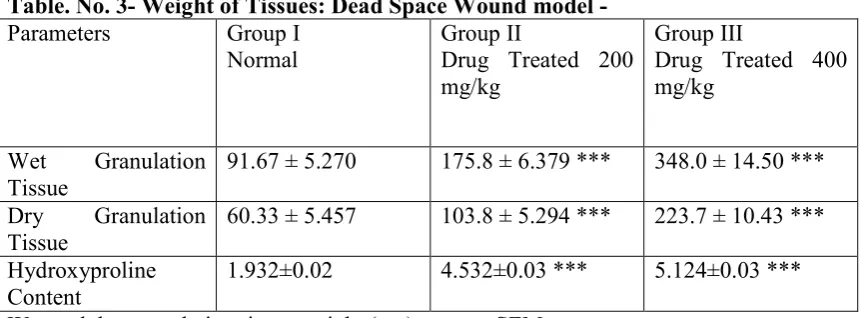
In the dead space wound study, there was a significant increase in granuloma breaking strength in extract treated groups at 200, and 400mg/kg doses when compared to control (Table 3). There was significant increase in hydroxyproline content in extract treated groups at 200 and 400mg/kg doses.
Table. o. 3- Weight of Tissues: Dead Space Wound model -
Parameters Group I Normal
Group II
Drug Treated 200 mg/kg
Group III
Drug Treated 400 mg/kg
Wet Granulation Tissue
91.67 ± 5.270 175.8 ± 6.379 *** 348.0 ± 14.50 ***
Dry Granulation Tissue
60.33 ± 5.457 103.8 ± 5.294 *** 223.7 ± 10.43 ***
Hydroxyproline Content
1.932±0.02 4.532±0.03 *** 5.124±0.03 ***
Wet and dry granulation tissue weight (mg) mean ± SEM
Discussion
In the ancient time, the combination of classical phytotherapy using herbal drug combination with superior efficacy and lesser side effects than a single plant extract or constituents has been frequently tried pharmacologically. Wound healing is a complex multifactorial process that can be managed by more effectively through use of PHF containing a number of bioactive substances. [13]
In present study, PHF was found to increase rate of wound contraction in PHF treated groups and this may be due to increased proliferation and transformation of fibroblast cells. The faster wound contraction might be due to increased keratinocyte proliferation, and their migration into wound surface.
Wound healing process consists of different phases such as granulation, collagenization, collagen maturation and scar maturation which are concurrent but independent to each other. Hence this study uses three different models to assess the effect of PHF on various phases of wound healing process. The results of the present study showed that PHF possesses a definite prohealing action. In excision wound healing model, the PHF showed significant increase in percentage closure of excision wounds by enhanced epithelization.
S.No. Normal Drug Treated 200
mg/kg
Drug Treated 400 mg/kg
wound healing activity as compared to drug treated groups i.e. nitrofurazone. Since in experiments we used a Polyherbal formulation and the formulation contained various bioactive phytoconstituents i.e. Flavanoids, volatile oils, alkaloids and tenpins that may contribute to progressive wound healing. Similarly, the breaking strength of the incision wounds was increased in extract treated groups in incision wound healing model. The increased breaking strength may be due to increased production of hydroxyproline which is responsible for collagen synthesis and provide hardness to skin. Deposition of newly synthesized collagens at the wound site increases the collagen concentration per unit area and hence the tissue tensile strength increases at treated groups. The higher breaking strength indicates better healing of wounds. Higher hydroxyproline content was seen with extract treatment. The increased amount of hydroxyproline in test groups underlines increased collagen content, since hydroxyproline is the direct estimate of collagen synthesis it supports the wound healing activity of PHF. In dead space model, there was a significant increase in weight of granuloma tissue in extract treated groups in dead space wound model. Recent studies have shown the use of many Polyherbal formulations in promoting the wound healing.
Earlier studies have shown that antimicrobial activities of selected plants which supports our findings since microbial infection is one of the factors for poor healing of Cutaneous wounds.[14,15] Hence present research supports traditional claims of the plants in PHF in wound healing.
Conclusion
Recent studies have shown that phytochemical constituents like flavonoids [16] and triterpenoids [17] are known to promote the wound healing process mainly due to their astringent and antimicrobial properties, which appear to be responsible for wound contraction and increased rate of epithelialization.
The preliminary phytochemical analysis of the all selected medicinal plants extract showed the presence of tannins, flavonoids, triterpenoids, and alkaloids. Any one of the observed phytochemical constituents present in selected plants may be responsible for the wound healing activity. Postoperative wounds are commonly known to be complicated by infection. Earlier studies have shown that antimicrobial activity of various plants supports the wound healing.[14] Further the plants have been evaluated for antimicrobial activity by previous researchers, hence present research supports traditional claims of the plant in wound healing.
Acknowledgement
The author and co-authors are greatly thankful to Dr. Gyanendra tiwari for identification and authentification of selected medicinal plants. The author is also thankful to Mr. Pradeep Desmukh, head deptt. Of pharmacology, B.R. Nahata college of pharmacy, mandsaur for providing all the facility regarding to research work and approval of animal experiments from institutional animal ethical committee (IAEC).
References
2. Nirendra K, Kamlakar T, Deborshi S, Vijay S. Apoptosis: A basic pathologic process in wound healing. Lower Extremity Wounds.2005; 4: 138-144.
3. Thais PA, Bernard C, Alexis D, Andrea M. Cutaneous Wound Healing:s Myofibroblastic Differentiation & in vitro models. Lower Extremity Wounds.2003;2: 60-68
4. Michael S, Maria W. Horst Dieter Becker, Models to Study Ischemia in Chronic Wounds. Lower Extremity Wounds. 2002; 1: 104-111.
5. Sharma PC, Yelne MB, Dennis TJ. Database on medicinal plants used in ayurveda. Vol.-4. New Delhi: Central Council for Research in Ayurveda & Siddha; 2002.
6. The Wealth of India, A Dictionary of Indian Raw Materials & Industrial Products, First Supplement Series (raw material). Vol.-VI1. New Delhi: NISCAIR; 1996. 7. Nadkarni KM. Indian Materia Medica. 1st edition. Bombay: Popular Prakashan;
1908.
8. Swamya HM, Krishna V, Shankarmurthy K, Abdul Rahimana KL, Mahadevan KM, Harish BG, Raja Naika H. Wound Healing Activity of Embelin Isolated From the Ethanol Extract of Leaves of Embelia ribes Burm. Journal of
Ethnopharmacology. 2007; 109: 529–534
9. Pulok KM, Rob Verpoorte B, Suresh B. Evaluation of in-vivo Wound Healing Activity of Hypericum patulum (Family: Hypericaceae) leaf Extract on Different Wound Model in Rats. Journal of Ethnopharmacology. 2000;70: 315–321
10.Pradeep TD, Jennifer F, Atul A, Emmanuel T. Wound Healing Activity of Calotropis gigantea Root Bark in Rats. Journal of Ethnopharmacology. 2009;125:178–181
11.Woessner JF. The Determination of Hydroxyproline in Tissue and Protein Samples Containing Small Proportion of this Imino acid. Archives of Biochemistry and Biophysics.1961; 93:440–447.
12.Lee KH. Studies on Mechanism of Action of Salicylates, Retardation of Wound Healing by Aspirin. Journal of Pharmaceutical Sciences.1968; 57:1042–1043. 13. Asheesh G, Nitin U, Sawhney RC, Ratan K. A Poly-herbal Formulation Accelerates Normal and Impaired Diabetic Wound Healing. Wound Rep Reg. 2008; 16:784–790
14.Ashraful MA, Rowshanul MH, Nikkon F, Rahman M, Karim MR. Antimicrobial activity of Akanda (Calotropis gigantea L.) on some Pathogenic Bacteria. Bangladesh Journal of Scientific and Industrial Research.2008; 43: 397–404. 15. Rasik AM, Ram R, Gupta A, Shukla MP, Srivastava S, Jain HK, Kulshrestha DK. Healing Potential of Calotropis procera on Dermal Wounds in Guinea pigs. Journal of Ethnopharmacology. 1998; 68:261–266
16.Tsuchiya H, Sato M, Miyazaki T, Fujiwara S, Tanigaki S, Ohyama M, Tnanka T, Linuma M. Comparative study on the antibacterial activity of phytochemical flavanones against methicillin resistant Staphylococcus aureus. Journal of Ethnopharmacology.1996; 50:27–34.