Research Paper
MORPHOLOGICAL STUDY OF
Echinococcus
WORM ISOLATED FROM
DOGS IN ALBIDA CITY -LIBYA
Ahmad. M. H. Ekhnefer, Abdulwahed B. Hador and Salem M. Bowashia
Department of Zoology, Omar AL Mukhtar University,
Albida, Libya.
Abstract
Genus Echinococcus has been complicated by attempts to assign species, subspecies status but their some different between strains of the parasite. The genus has Only four species of Echinococcus are accepted at the present time these are E. granulosus, E. oligarthus, E. vogeli, E. multiocularis. The adult Echinococcus possesses a specialized attachment organ, the scolex, which has four muscular suckers and two rows of hooks, one large and one small, on the rostellum. Strobili, is segmented and consists of a number of reproductive units (proglottids), which may vary in number from two to six. Like all tapeworms, Echinococcus has no gut and all metabolic interchange takes place across the syncytial outer covering, the tegument, For the isolation sample of parasite Echinococcus from dogs, In the present study,30 dogs from Albida city in Libya were examined. By tow method the 1st dogs were using a dose of 1:2 mg / kg of arcoline hydrobromide and the second by dissecting the dead dogs from anywhere in the Albida city, then empty contained intestinal and where samples of faeces were collected and washed with saline solution (0.6 – 0.9) several times to get out the cestode worms to be examined Light microscopy fresh, some worms were fixed in 2.5% glutaraldehyde for the electron microscopy. The adult worm consists of the head and 3 segments. It is flattened conical-shaped and its length ranges from 5.6-6.2mm. On the head there are four suckers and two parallel circles of hooks: an outer and an inner one. The number of hooks ranges from 28-38 distributed equally between the two circles and the length of large hooks measures 25-49μm while that of the small ones’ ranges from 16-32μm. The hooks are oriented posteriorly towards the peduncle. Scanning electron microscopy of the head revealed that the hooks in each circle originated alternatively from the places around the rostellum. The second segment (mature) of the adult worm contained the testes, which range in number from 26-79. The surface ultrastructure illustrated the dense covering of the protrusions giving it the velvety appearance. The
Journal of Global Biosciences
ISSN 2320-1355local isolates of Echinococcus has been identified according to morphology and parasitological standards and was found to belong to Echinococcus granulosus.
Key words: Morphology; Echinococcus.
INTRODUCTION
The tapeworm (Echinococcus.sp) spends most of its adult life in the intestine of its definitive host, (canids) and in particular the dogs and wolves. The tapeworm eggs become voided in the canid’s faeces and as a result of ingesting the eggs, infection passes to the intermediate host, commonly herbivores while grazing. However, humans can become accidentally infected and hydatid cysts may develop throughout the body. Therefore, cystic echinococcosis (CE) or hydatidosis is a disease caused by the metacestode stage of Echinococcus. The disease is not apparent to farmers but is of considerable economic and public health importance [1,2]. In farm animals, it causes considerable economic loss due to condemnation of edible organs, decreased meat and milk production, reduced hide and fleece value and decrease in fecundity [3,4]. The incidence of human hydatid disease in any country is closely related to the prevalence of the disease in domestic animals and is highest where there is a large dog population and high sheep production [5]. Speciation in the genus Echinococcus has been complicated by attempts to assign species, subspecies status or the different strains of the parasite. Only four species of the genus Echinococcus are accepted at the present time these are E. granulosus, E. oligarthus, E. vogeli, E. multiocularis, [6,7,8]. The adult Echinococcus possesses a specialized attachment organ, the scolex, which has four muscular suckers and two rows of hooks, one large and one small, on the rostellum. Strobila, is segmented and consists of a number of reproductive units (proglottids), which may vary in number from two to six [9]. Like all tapeworms, Echinococcus has no gut and all metabolic interchange takes place across the syncytial outer covering, the tegument [10].
2.MATERIALS AND METHODS
microscopy[13] Light microscopy fresh. The adult worms were fixed between a slide and cover glass for a bout 3h using drops of 70% alcohol as a fixative. The specimens were washed by distilled water, stained with eosin dehydrated, cleared and mounted with Canada balsam. The specimens were put in an oven at 36 C0 for at least one night. Then they were examined under the microscope. The total length of the worm and hooks was measured and the segments and hooks were enumerated [11].
Fig (1): Photographs of the worm Echinococcus in the Petri dish under the dissection microscope 20x, A- sample taken from the feces of an infected dog B-The worm Echinococcus in the Petri dish, a sample taken from the feces of a dog infected by camera
Canon 14-megapixel full zoom.
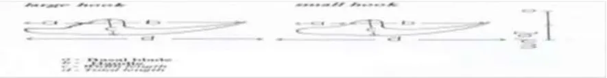
Fig. (2): diagrams showing the method of measuring the length and width of hooks by [2].
2.2Electron microscopy:
3. RESULTS
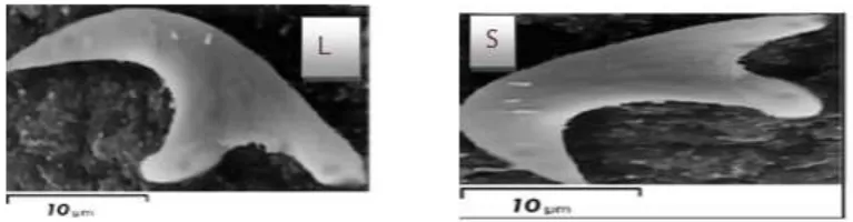
3.1 Description of Parasites: The adult worm consists of the head 3 segments. It is flattened conical-shaped and its length ranges from 5.6-6.2mm. On the head there are four suckers and two parallel circles of hooks: an outer and an inner one. The number of hooks ranges from 28-38 distributed equally between the two circles and the length of large hooks measures 25-49μm while that of the small ones’ ranges from 16-32μm(Fig.5). The hooks are oriented posteriorly towards the peduncle. Scanning electron microscopy of the head revealed that the hooks in each circle originated alternatively from the places around the rostellum (Fig.3). The second segment (mature) of the adult worm contained the testes, which range in number from 26-79 (Figs 3 and 4 and Table 1). The surface ultrastructure illustrated the dense covering of the protrusions giving it the velvety appearance (Fig, 4 B).
Fig. (3):A: a photograph showing the adult worm(10X), B: photograph showing R- Rostellum and H-Hooks (400X).
Table (1): Measurements of morphometric for adult worm (Echinococcus).
Parameters Range Mean ±SD
Length mm 5.8-6.1 6±0.2
No. of hooks 28-38 28±2.5
No. of Suckers 4 4±0
No. of segments 3 3±1
No. of testes 27-75 50±10
No. of Large Hooks 16-19 16±2.5
No. of Small Hooks 16-19 16±2.5
Length of Large Hooks
µm 26-48 38.7±4
Arrangement of hooks One small hook between two large hooks.
Fig. (4): Scanning electron micrographs E.granulosusA: Adult worm, Sg = Segments, 1=Immature, 2=Mature, 3=Gravid, G=Genital pore He=head. B: Magnification of the head
of worm, R=Rostellum, H=Hooks, S=Sucker.
Fig. (5): Scanning electron microscope showing of large(L) and small(S) hooks.
DISCUSSION
for E. granulosus studies.
ACKNOWLEDGEMENTS
THANKS OT ALL THE STAFF OF ZOOLOGY DEPARTMENT IN OMAR AL MUKHTAR UNIVERSITY, ALBIDA, LIBYA.
REFERENCES
[1]- P.R. Torgerson, &C.M Budke, : Echinococcosis – an international public health challenge. Res. Vet. Sci., 74, 191–202 .2003.
[2]- Ahmadi, N.A. & Meshkehkar, M. (2011): An abattoir-based study on prevalence and economic losses due to cystic Echinococcosis in slaughtered herbivores in Ahwaz, south-western Iran. J. Helminthology. 85, 33–39.2011.
[3]- Polydorou, K. : Animal health and economics. Case study: echinococcosis with reference to Cyprus. Bul. Int. Epz., 93, 981-992.1981.
[4]- Romazanov, V.T., : Evaluation of economic losses due to echinococcosis. In: LysendoA, editor. Zoonosis control: colle- ction of teaching aids for international training course vol. II. Moscow: Centre of International Projects GKNT, pp 283– 85.1983.
[5]- M.S Khuroo,. Hydatid disease: current status and recent advances. Ann. Saudi Med. 122, 56-64.2002
[6]- D.J. Jenkins, ; T. Romig,; R.C Thompson, : Emergence /reemer-gnce of Echinococcus spp. a global update. Int. J. Parasitol. 35:1205.2005.
[7]- N. Xiao; J. Qiu, ;M. Nakao, ;T. Li, ; W. Yang, ;X. Chen , ;P.M Schantz ;P.S Craig, &A. Ito:. Echinococcus shiquicus, a new species from the Qinghai -Tibet plateau region of China: Discovery and epidemiological implications Parasit. Int., 55, S233–236.2006 [8]- S. Mojtaba,; H.Hosein; S. Mansour; R. Ali, &B. Ali ,: Molecular characterization of human and animal Echinococcus granulosus isolates in Ispahan, Iran . journal homepage: www. elsevier. Com /locate /actatropica.2010.
[9]- Klaus Rohde.: Tapeworms Cestoda ,http: //knol .google .com/k /-/-/1bbsle13m97c0/431.2011
[11]- Smyth, J.D. & Barrett, N.J. (1980): Procedures for testing the viability of human hydatid cysts following surgical removal, especially after chemo therapy. Tran. Roy. Soci . Trop. Med. Hyg. 74: 649-652.1980.
[12]- Smith, S.A & Richards, K.S. (1991): Ultrastructure and microanalyses of the protoscolex hooks of Echinococcus granulosus. Parasitology, vol.103, pp.267-274.1991.
[13]- Ekhnefr, A.M.: Biological Studies on Hydatid Cysts Infesting Sheep in Libya, Ph.D. Zoology Department, Faculty of Science, Mansoura University, 2012.
[14]. Mansour, M.F.A. :Light and scanning electron microscope studies on the cestode nematotaenia kashmerensis fotedar, 1966(nematotaeniidaei cyclophyllidea)infecting the toad bufo regularis in dakahlyia gorernorate, Egypt. Egypt.j.zool.,45:155-168.2005.
[15]. M. Galindo, ; M.J Gonzalez,& N.Galanti: Echinococcus granulosus protoscolex formation in natural infections. Biomedical Research,vol.35, pp.365-371.Khuroo,2002.
[16].F.B Almeida; Rodrigues-Silva, R.; Neves, R.H.; Gonçalves, M.M.L.; Romani, E.L. & Machado-Silva, J.R.: morphological and morphometric studies on protoscolec- es rostellar hooks of Echinococcus granulosus from peru visualized by several microscopic techniques asociación peruana de helmintología invertebr-ados afines (aphia). Neotropical helminthology, vol. 3, nº 2, pp. 65-72.2009.
[17]. L.M Kumaratilake,; R.C Thompson &J. Eckert: Echinococcus granulosus of equine origin from different countries possess uniform morphological characteristics. Int. J. Parasitol. 16, 529–540.1986.
[18]. S.A Smith, & K.S Richards : Ultrastructure and microanalyses of the protoscolex hooks of Echinococcus granulosus. Parasitology, vol.103, pp.267-274.1991.
[20]. P. Dubinský,; A. StefancÌková,; L. Turceková,; J.Macko, J & Soltýs: Development and morphological variability of Echinococcus granulosus. Parasitology Research, vol.84, pp.221-229.1989.