Abstract— Laserpuncture exposure effectively stimulates gonad maturation and spawning, but the associated gonadotropin release mechanism is still unknown. Our study was aimed to investigate the release of gonadotropin hormones in the catfish (Clarias sp.) after laserpuncture exposure at the reproduction acupoint. The test fish consisted of 54 males and 54 females aged 8-9 months from the F1 hybrid S angkuriang type female and Paiton type male parents. Our study employed an experimental method with completely randomized design. The treatment comprised 6 levels with 9 repetitions.
We were to observe gonadotropin (GtH-I and GtH-II) hormone concentrations pre-spawn, spawn, post-spawning in the laser exposed group. As a comparison investigations were also conducted to untreated group (control). Blood sampling was performed six hours post exposure. Hormone level test was performed using Elisa test kits. The test results showed a significant effect of laser exposure on the increase of gonadotropin hormone levels pre-spawn, spawn and post- spawning. This suggests that laser exposure at reproduction acupoint can stimulate the release of gonadotropin hormones which affect the acceleration of gonad maturation and spawning.
Index Term— Laserpuncture, catfish (Clarias sp.), reproduction acupoints, gonadotropin hormones
I. INT RODUCT ION
ENVIRONMENT AL factors such as photoperiod and water temperature give a signal to initiate a series of oocyte development processes. In response, the hypothalamus releases gonadotropin-releasing hormone (GnRH), which in turn stimulates the pituitary to release gonadotropin hormones
P.S.W. Kusuma is with the Postgraduate Program, Faculty of Fishery and Marine Science, Brawijaya University, Jl. Veteran 2, Malang 65145, East Java, Indonesia (corresponding author, phone: +62-85655085599;
e-mail: [email protected]).
A.P.W. Marhendra is with the Dept. of Biology, Faculty of Mathematics and Natural Sciences, Brawijaya University, Jl. Veteran 2,
Malang 65145, East Java, Indonesia.
Aulanni’am is with Dept. of Chemistry, Faculty of Mathematics and Natural Sciences, Brawijaya University, Jl. Veteran 2, Malang 65145,
East Java, Indonesia.
Marsoedi is with the Faculty of Fishery and Marine Science, Brawijaya University, Jl. Veteran 2, Malang 65145, East Java, Indonesia.
(GtH-I and GtH-II). During the spawning season, gonadotropin hormone GtH-II increases sharply in blood serum [1, 2]. GtH-I involves in the process of vitellogenesis and GtH-II plays a role in final oocyte maturation and ovulation [3-5]. Gonad development and spawning are controlled through regulation of the hypothalamic-pituitary-gonad and liver axis [6, 7].
Various exogenous hormones have been used to induce spawning, such as Ovaprim® injections. These efforts are made to elevate the concentration of gonadotropin hormones in blood so as to induce egg development and spawning [8]. Laser excitatory technology as a reproduction biostimulation is a relatively new approach that has been shown to s timulate gonad maturation and spawning of tilapia [9].
Laser is electromagnetic waves that may cause inhibition and biostimulation in biological tissues [10]. Low-power laser may give a biological stimulus such as changing the cell membrane potential and membrane permeability to sodium, potassium and calcium ions that increases cellular activity such as enzyme activity, nerve regeneration, either central or peripheral, and is able to stimulate the production of hormones [11-13]. The effect caused by laser beam is electrobioluminance, i.e. if the laser beam irradiates the tissue it will stimulates the cells and produce electrical signals. The 4-5 mW laser beam (He-Ne) that was exposed to the skin may penetrate the epidermis and dermis which leads to stimulation [14].
In previous studies [9, 15] we have managed to locate the reproduction acupoints and reproduction cycle on female black tilapias variety GIFT (Genetic Improvement Farmer Tilapia) that were spawn for the first time. Laser exposures on the fish for 6 seconds to reproduction acupoint exactly at 2/6 ventral body (governor vessel) optimally influenced the gonad maturity stage (GMS), namely the GMS IV. The GMS value indicates that the fish are ready for spawning. With this technique the tilapia can spawn three times in 30 days, whereas in a normal environment the tilapia spawns only once every 30 days [16]. In a study on catfish (Clarias sp.) the control group were still at GMS I within 10 weeks, which indicates that the catfish are not ready for spawning, while in the 15-second-laser-exposed group at 2/3 the ventral body (governor vessel) optimally influenced the GMS to reach GMS IV 15 days after exposure laser [17]. Thus, either in tilapia or catfish laser exposure at
Mechanism of Gonadotropin Hormone Release in
Catfish (Clarias Sp.) upon Laserpuncture
Exposure to Reproduction Acupoint
reproduction acupoint will give similar effect in the maturation of the gonads. This indicates that laser exposure can improve the hormone performance that plays an important role in the reproduction control system.
There is a presumption that laser exposure on the tissue influences the increase of gonadotropin releasing hormone (GnRH) released by the hypothalamus. GnRH will stimulate the anterior pituitary to secrete gonadotropin hormones (GtH-I and GtH-II). The gonadotropin produced will work on theca cells in the gonads to produce testosterone [18]. Testosterone will then enter the granulosa layer and converted to estradiol-17 β with the help of aromatase enzyme [19]. Estradiol-17 β plays a role in vitellogenin biosynthesis in the liver. In addition, estradiol-17 β in the blood provides feedback to the hypothalamus to produce GnRH that stimulates the pituitary gland to release gonadotropin. This hormonal control continues to run during the process vitellogenesis [18-20].
Given that the mechanism of gonadotropin hormones (GtH-I and GtH-II) release in catfish (Clarias sp.) after being exposed to the laser at the reproduction acupoint is still unknown, this paper presents the results of our study to prove that s uch laser exposure to the reproduction acupoint can stimulate the release of gonadotropin hormones (GtH-I and GtH-II), which will control gonad maturation and spawning in catfish.
II. MAT ERIALS AND MET HODS
Our study was conducted in January to March 2012 in Freshwater Aquaculture Management Unit (UPBAT) Kepanjen Malang. Catfish blood samples were prepared in the Organic Chemistry Laboratory of Brawijaya University to analyze the hormone level profile by Elisa method.
Test Animals and Experiment Matters
Animals used in the study were prospective catfish (Clarias sp.) parent with matured gonads and have never spawn ed, about 8-9 months old. Animal samples were F1 hybrids of Sangkuriang female and Paiton male parents obtained from UPBAT Kepanjen Malang. The number of sample was 54 males and 54 females. The fish weighted ranged from 1000-1700 g for the females and between 1150-1750 g for the males.
Parents Keeping
The selected male and female catfish (Clarias sp.) parent candidates with mature gonads and never spawn were adapted separately in a tarp pool for 14 days to avoid spawning before treatment. During maintenance of the fish were fed with commercial fish food Pokphan 781-3 with 36% protein content produced by CP Prima and was administered daily in the morning and evening as much as 5% of body weight. The parent candidates in the control group and laser induced groups were kept in the coded tarp ponds of 2×2×1 m3 in size. Each pond was filled with a pair of fish.
Laser Exposures
Our study employed an experimental method with completely randomized design (CRD). The treatments consisted of six levels and were repeated nine times. The treatments included one control group and laserpuncture exposed groups (pre-spawn, spawn, and post-spawning). The treatment groups were exposed to the Helium-Neon (He-Ne) soft laser of 5 mW maximum power and a wavelength of 632.8 nm. The laser beams were exposed at the reproduction acupoints exactly at 2/3 of ventral body (governor vessel) for 15 seconds each. The dose was the optimal dose obtained in previous researches [17]. The control and treatment groups consisted of 27 fish pairs .
Observed Parameters
The parameter observed in this study was the profile of gonadotropin hormones (GtH-I and GtH-II) in blood serum of catfish (Clarias sp.) after being exposed to laser beam at the reproduction acupoint pre-spawn, spawn, and post-spawning. Measurements of the hormone level profile were conducted using Elisa test kits (Cusabio Biotech Co. Ltd.) with cat. no. CSB-E15790Fh (967) for GtH-I and cat. no. CSB-E15791Fh for GtH-II.
Data Analysis
The data were analyzed using SPSS software ver. 15.0 for windows. Tests on the differences in response between treatments were performed by analysis of varian ce (ANOVA) in both directions. Gonadotropin hormone (GtH-I and GtH-II) profiles were observed in pre-spawn, spawn, and post-spawning in both animal groups. To the significantly different groups the test was continued using LSD test at 95% confidence level.
III. RESULT S GtH-I Profile
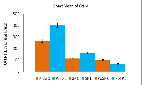
The average values of GtH-I profile of the control group and the treatment groups are displayed based on the conditions of pre-spawn, spawn, and post-spawning. We can see from the values that the GtH-I profile from the pre-spawn to spawning conditions decreases, where the average values for the treatment groups are higher than the control. This is different from the situation after the spawn which shows that the average GtH-I profiles of the two groups drop, but in the control group it is higher than those of the treatment groups (see Table I).
TABLEI
VALUES OF GTH-I P ROFILE IN CATFISH (CLARIAS SP.) BLOOD SERUM OF CONTROL AND TREATMENT GROUP S
Group GtH-I Profile N Average ± SD
Control PrSPC 9 270 ± 60a
SPC 9 110 ± 10b
PoSPC 9 100 ± 10c
Laser exposed PrSPL 9 400 ± 100a
SPL 9 160 ± 20b
PoSPL 9 70 ± 10c
Remarks: Values in the column followed by different superscript indicates significantly different (p < 0.05). PrSPC/PrSPL: pre-spawn, SPC/SPL: spawn, PoSPC/PoSPL: post -spawning.
Fig. 1. GtH-I profile for the control and treatment groups in pre-, during, and post -spawning).
GtH-II Profile
The GtH-II profile of the control and the treatment groups are displayed based on the conditions pre-spawn, spawn, and post-spawning. It appears that the GtH-II profile in the condition pre-spawn to spawn decreases, where the average values for the treatment groups are higher than in the control group. This is different from the post-spawning situation which shows that the GtH-II profiles of both groups decrease but in the control group it is higher than those of the treatment groups (see Table II).
TABLEII
VALUES OF GTH-II P ROFILE IN CATFISH (CLARIAS SP.) BLOOD SERUM OF CONTROL AND TREATMENT GROUP S
Group GtH-II Profile N Average ± SD
Control PrSPC 9 11.9 ± 1.7a
SPC 9 4.6 ± 1.2b
PoSPC 9 2.4 ± 1.1c
Laser exposed PrSPL 9 14 ± 3a
SPL 9 6.1 ± 1.6b
PoSPL 9 1.3 ± 1.2c
Remarks: Values in the column followed by different superscript indicates significantly different (p < 0.05). PrSPC/PrSPL: pre-spawn, SPC/SPL: spawn, PoSPC/PoSPL: post -spawning.
The profiles of gonadotropin hormone GtH-II level in the treatment groups at pre-spawn condition (PrSPL) as well as of spawn (SPL) are higher than the control. However, it is
different in conditions post spawning (PoSPL), where the value of the treatment groups is lower than that of the control group (Fig. 2).
Fig. 2. GtH-II profile for the control and treatment groups in pre-, during, and post -spawning).
IV. DISCUSSION
The profile of gonadotropin hormones (GtH-I and GtH-II) levels in conditions from pre-spawn to spawn in the treated group is higher than the control group. While in condition after spawning, the profile in the treatment group was lower than the control group. The increase and decrease of gonadotropin hormones are affected by GnRH in the hypothalamus, where GnRH is influenced by environmental factors through the central nervous system. This gives an indication that laser exposure at the reproduction acupoint stimulates GnRH through nervous pathways.
A 15-second exposure of 5 MW/cm2 low power 632.8-nm laser (soft laser) Helium-Neon (He-Ne) to the reproduction acupoint releases an equivalent energy of 0.375 joule/cm2. This energy is capable of inducing the release of gonadotropin hormones from the hypothalamus. This is possible because the laser exposure stimulates the release of neurotransmitters. The laser beam will penetrate and hit the peripheral nerve ends situated between the epidermis and dermis of the skin tissue. The laser beam will further transduced into chemical signals to be received by various ion channels, such as G-proteins (GTP-binding protein)-coupled receptors subunit α and VGCC (voltage-gated Ca2+ channels), or through calcium receptors, such as calcium sensing receptor (CaSR), located in the nervous membrane cells. Bonding of ligands to specific receptors will trigger the release of second messengers that will cause a chain reaction and bring about a change in the cell.
propagated to the brain. Intracellular and extracellular Ca2+ ions mediated through spontaneous membrane potential changes will play an important role in stimulating the release of GnRH from the hypothalamus which in turn stimulates the pituitary to release GtH-I and GtH-II in carp, African catfish and tilapia [23, 24].
The release of neurotransmitters such as GABA (gamma-aminobutyric acid) from GABAergic neurons depend on nerve cell membrane depolarization, action potential, calcium ions, CCP, decarboxylation of glutamate and GAD (glutamic acid decarboxylase). GAD is a catalytic enzyme expressed abundantly in telencephalon and hypothalamus mediobasal of trout that have an important role in GABA synthesis [25]. Synthesis of GABA starts from glutamate decarboxylation catalyzed by GAD that is generally located at the tip end of the nerve [26-28]. There are two kinds of GAD known in fish, namely GAD-65 and GAD-67, where GAD-65 is more pronounce due to its sensitivity to changes in circulating sex steroids and is able to modulate the synthesis an d release of neurotransmitters. The results of our previous studies showed that GAD-65 is abundant after 6 hours. The activity of GAD-65 determines the synthesis of GABA in GABAergic neurons [28]. Later, the released GABA convey messages directly through synaptic contacts between GABAergic nerve terminals and GnRH neurons in which these GABAergic neurons are directly innervated by the anterior pituitary [26, 29], so it has a stimulatory effect on the release of GtH-II [26-28, 30, 31].
The results showed that laser exposure raised the level of gonadotropin hormones (GtH-I and GtH-II) in blood serum 6 hours before spawning compared with the control group, because the laser exposure on reproduction acupoint of catfish (Clarias sp.) can stimulate GABAergic neurons to synthesize GABA, which will immediately convey the message through the synaptic contacts between nerve terminals of GABAergic and GnRH neurons [32]. GABA released by GABAergic neurons will further stimulate the hypothalamus to release GnRH. The released GnRH will stimulate pituitary neurons to release gonadotropin hormones (GtH-I and GtH-II). Gonadotropin hormones (GtH-I and GtH-II) are then channeled into the bloodstream towards the gonads (ovaries) that enables a wide range of activities. Gonadotropin hormone GtH-I will be stimulating follicle development through secretion of estradiol-17 β in the ovaries that plays a role in vitellogenin biosynthesis in the liver. The content of estradiol-17 β is in accordance with the development of the egg [19, 33]. Likewise, gonadotropin hormone GtH-II will be required in the final oocyte maturation and stimulate ovulation and spawning [3, 5]. This process repeats when there is nerve cell membrane depolarization [25]. The results are consistent with the finding of Trudeau et al. [28] that stimulation of GABA can influence the increase in gonadotropin hormone release in the hypothalamus and the decrease in dopaminergic activity.
On condition after spawning the profile of gonadotropin
hormones (GtH-I and GtH-II) was higher in the control group compared with the treatment groups. This was due to the release of gonadotropin hormones (GtH-I and GtH-II) in some control group fish was incomplete because some fish were yet to spawn. While in the treatment group all fish had completed the spawn that made the profile of gonadotropin hormones (GtH-I and GtH-II) in the blood serum was detected low. Naturally, many physiological factors affect the GnRH release in the hypothalamus, such as irradiation, sexual behavior, stress and gonadal maturation. Besides, there are many mediator substances in regulating the synthesis and release of GnRH, such as glutamate and GABA [34], dopamine (DA) [35], serotonin [36, 37] and neuropeptide Y (NPY) [38].
Our research suggests that the release of GABA from GABAergic neurons can stimulate the hypothalamus to release gonadotropin hormones which in turn will stimulate the pituitary to release GtH-I and GtH-II even without laser stimulation. This is because GABAergic neurons can regulate its own expression in the brain over a period of 24 hours [39] and as a result the profile of gonadotropin hormones (GtH-I and GtH-II) in the control group was higher than in the treatment groups after spawning. Levels of the hormones (GtH-I and GtH-(GtH-I(GtH-I) in both the control and the treatment groups should decrease after spawning, but the decline in the treatment groups was accelerated. Another possibility is that the level of GABA released by GABAergic neurons was not sufficient to stimulate the hypothalamus to release gonadotropin hormones.
V. CONCLUSION
Our study shows that laserpuncture exposure to reproduction acupoint in skin tissue of catfish (Clarias sp.) was capable of raising the profile of gonadotropin hormones (GtH-I and GtH-II) either at the time of pre-spawn and spawn the time of spawning. Laserpuncture exposure to reproduction acupoint in skin tissue can accelerate the maturation of the gonads and stimulate spawning.
Laserpuncture exposure to reproduction acupoint in skin tissue of catfish (Clarias sp.) can be used to speed up the production of hatchlings. Our results can be used as a reference for further research on the possible side effects such as genetic abnormalities in the offspring.
ACKNOWLEDGMENT
REFERENCES
[1] R. W. Schulz, L. R. d. França, J.-J. Lareyre, F. LeGac, H. Chiarini-Garcia, R. H. Nobrega, and T . Miurae, "Spermatogenesis in fish," Gen Com par Endocrin, vol. 165, pp. 390-411, 2010.
[2] R. W. Schulz and T . Miura, "Spermatogenesis and it s endocrine regulation," Fish Physiol Biochem , vol. 26, pp. 43-56, 2002.
[3] Y. Nagahama, "Endocrine regulation of gametogenesis in fish," Int J Dev Biol, vol. 38, pp. 217-229, 1994
[4] Y. Nagahama and M. Yamashita, "Regulation of oocyte maturation in fish," Dev Growth Diff, vol. 50, pp. S195-S219, 2008.
[5] H. Q. Pham, A. T . Nguyen, M. D. Nguyen, and A. Arukwe, "Sex steroid levels, oocyte maturation and spawning performance in Waigieu seaperch (Psam m operca waigiensis) exposed to thyroxin, human chorionic gonadotropin, luteinizing hormone releasing hormone and carp pituitary extract," Com par Biochem Physiol A, vol. 155, pp. 223-230, 2010.
[6] O. Kah and S. Dufour, "Conserved and divergent features of reproductive neuroendocrinology in teleost fishes," in
Horm ones and Reproduction of Vertebrates. Vol. 1 Fishes, D.
O. Norris and K. H. Lopez, Eds.: Academic Press, 2010, pp. 15-42.
[7] R. Urbatzka, M. J. Rocha, and E. Rocha, "Regulation of ovarian development and function in teleosts," in Horm ones and Reproduction of Vertebrates. Vol. 1 Fishes, D. O. Norris and K. H. Lopez, Eds.: Academic Press, 2010, pp. 65 -82. [8] A. Hardjamulia and S. Atmawinata, "T eknik Hipofisasi
beberapa Jenis Ikan Air T awar," in Prosiding Lokakarya Nasional Teknologi Tepat Guna Bagi Pengem bangan
Perikanan Budidaya Air Tawar, Bogor, 1980 (in Indonesian),
pp. 1-16.
[9] P. S. W. Kusuma, "Optimalisasi Letak T itik dan Frekuensi Penembakan Laserpuncture (He-Ne) terhadap GSI Ikan Nila
(Oreochrom is sp)," 1999 (unpublished, in Indonesian).
[10] A. N. D. Chester, S. Martelucci, and A. M. Scheggi, Laser
System for Photobiology and Photom edicine. New York:
Plenum Press, 1991.
[11] T . I. Karu, "Cellular Mechanisms of Low Power Laser T herapy: New Questions," in Lasers in Medicine and Dentistry. vol. 3, Z. Simunovic, Ed. Rieka: Vitgraf, 2003, pp. 79-100.
[12] J. Kert and L. Rose, Clinical Laser Therapy. London: Scandinavian Medical Laser T herapy, 1989.
[13] M. Koutna, R. Janisch, and R. Veselka, "Effects of Low-Power Laser Irradiation on Cell Proliferation," Scripta Medica
(BRNO), vol. 76 pp. 163-172, 2003.
[14] Sukarto, "Penggunaan Laser untuk Akupunktur," J. Akupunk., vol. 1, pp. 49-54, 1992 (in Indonesian).
[15] P. S. W. Kusuma, Pengaruh Penem bakan Soft Laser He-Ne
terhadap Siklus Reproduksi Ikan Nila. Masters T hesis.
Program Pascasarjana, Airlangga University. Surabaya. 2000 (in Indonesian).
[16] L. L. Loshin and H. H. Ibrahim, "Effects of broodstock exchange on Oreochromis niloticus egg and fry production in net enclosures," in The Second International Sym posium on
Tilapia in Aquaculture. ICLARM Conference Proceeding,
1988, p. 623.
[17] P. S. W. Kusuma, D. Hariani, A. T . Mukti, and W. A. Satyantini, "Aplikasi T eknologi Laser untuk Peningkatan Produksi Lele dalam Rangka Pengembangan Ekonomi Masyarakat Ekonomi Masyarakat Desa di Kabupaten Boyolali
Jawa T engah," LP3K Kabupaten Boyolali, Boyolali. 2007 (in Indonesian).
[18] Y. Nagahama, "T he Functional Morphology of T eleost Gonads," in Fish Physiology IX B, W. S. Hoar, D. J. Randall, and E. M. Donaldson, Eds. New York: Academic Press, 1983, pp. 223 -275.
[19] Z. Yaron, "Endocrine control of gametogenesis and spawning induction in the carp," Aquaculture, vol. 129, pp. 49-73, 1995.
[20] A. J. Matty, Fish Endocrinology. Portland: T imber Press, 1985.
[21] I. A. Graef, F. Chen, L. Chen, A. Kuo, and G. R. Crabtree, "Signals T ransduced by Ca2+/Calcineurin and NFAT c3/c4 Pattern the Developing Vasculature," Cell, vol. 105, pp. 863-875, 2001.
[22] P. M. Stemmer and C. B. Klee, "Dual Calcium Ion Regulation of Calcineurin by Calmodulin and Calcineurin B," Biochem , vol. 33, pp. 6859-6866, 1994.
[23] J. P. Chang, J. D. Johnson, F. V. Goor, C. J. H. Wong, W. K. Yunker, A. D. Uretsky, D. T aylor, R. M. Jobin, A. O. L. Wong, and J. I. Goldberg, "Signal transduction mechanisms mediating secretion in goldfish gonadotropes and somatotropes," Biochem Cell Biol, vol. 78, pp. 139-153, 2000.
[24] J. P. Chang, F. Van Goor, R. M. Jobin, and A. Lo, "GnRH Signaling in Goldfish Pituitary Cells," Neurosignals, vol. 5, pp. 70-80, 1996.
[25] I. Anglade, D. Mazurais, V. Douard, C. Le Jossic-Corcos, E. L. Mañanos, D. Michel, and O. Kah, "Distribution of glutamic acid decarboxylase mRNA in the forebrain of the rainbow trout as studied by in situ hybridization," J Com par Neurol, vol. 410, pp. 277-289, 1999.
[26] O. Kah, V. L. T rudeau, B. D. Sloley, J. P. Chang, P. Dubourg, K. L. Yu, and R. E. Peter, "Influence of GABA on Gonadotrophin Release in the Goldfish," Neuroendocrinology vol. 55, pp. 396-404, 1992.
[27] V. L. T rudeau, B. D. Sloley, and R. E. Peter, "T estosterone Enhances GABA and T aurine but not N-Methyl-D,L-Aspartate Stimulation of Gonadotropin Secretion in the Goldfish: Possible Sex Steroid Feedback Mechanisms," J
Neuroendocrinol, vol. 5, pp. 129-136, 1993.
[28] V. L. T rudeau, B. D. Sloley, and R. E. Peter, "GABA stimulation of gonadotropin-II release in goldfish: involvement of GABAA receptors, dopamine, and sex steroids," Am J Physiol, vol. 265, pp. R348-R355, 1993. [29] M.-G. Martinoli, P. Dubourg, M. Geffard, A. Calas, and O.
Kah, "Distribution of GABA-immunoreactive neurons in the forebrain of the goldfish, Carassius auratus," Cell Tissue Res, vol. 260, pp. 77-84, 1990.
[30] E. L. Mañanos, I. Anglade, J. Chyb, C. Saligaut, B. Breton, and O. Kah, "Involvement of gamma-aminobutyric acid in the control of GT H-1 and GT H-2 secretion in male and female rainbow trout," Neuroendocrinology, vol. 69, pp. 269-280, 1999.
[31] B. D. Sloley, V. L. T rudeau, J. G. Dulka, and R. E. Peter, "Selective depletion of dopamine in the goldfish pituitary caused by domperidone," Can J Physiol Pharm acol, vol. 69, pp. 776-781, 1991.
[32] C. Leranth, N. J. MacLusky, H. Sakamoto, M. Shanabrough, and F. Naftolin, "Glutamic Acid Decarboxylase-Containing Axons Synapse on LHRH Neurons in the Rat Medial Preoptic Area," Neuroendocrinology, vol. 40, pp. 536-539, 1985. [33] J. D. T an-Fermin, S. Ijiri, H. Ueda, S. Adachi, and K.
macrocephalus (Gunther) during an Annual Reproductive Cycle," Fisheries Sci, vol. 63, pp. 867-872, 1997.
[34] V. L. T rudeau, D. Spanswick, E. J. Fraser, K. Larivière, D. Crump, S. Chiu, M. MacMillan, and R. W. Schulz, "T he role of amino acid neurotransmitters in the regulation of pituitary gonadotropin release in fish," Biochem Cell Biol, vol. 78, pp. 241-259, 2000.
[35] S. Dufour, F. A. Weltzien, M. E. Sebert, N. Le Belle, B. Vidal, P. Vernier, and C. Pasqualini, "Dopaminergic Inhibition of Reproduction in T eleost Fishes: Ecophysiological and Evolutionary Implications," Ann NY Acad Sci, vol. 1040, pp. 9-21, 2005.
[36] I. A. Khan and P. T homas, "Immunocytochemical Localization of Serotonin and Gonadotropin -Releasing Hormone in the Brain and Pituitary Gland of the Atlantic Croaker Micropogonias undulatus," Gen Com par Endocr, vol. 91, pp. 167-180, 1993.
[37] B. Senthilkumaran, K. Okuzawa, K. Gen, and H. Kagawa, "Effects of Serotonin, GABA and Neuropeptide Y on Seabream Gonadotropin Releasing Hormone Release In Vitro from Preoptic-Anterior Hypothalamus and Pituitary of Red Seabream, Pagrus major," J Neuroendocrinol, vol. 13, pp. 395-400, 2001.
[38] C. Li, P. Chen, and M. S. Smith, "Morphological Evidence for Direct Interaction between Arcuate Nucleus Neuropeptide Y (NPY) Neurons and Gonadotropin-Releasing Hormone Neurons and the Possible Involvement of NPY Y1 Receptors," Endocrinology, vol. 140, pp. 5382-5390, November 1, 1999 1999.